


August 2, 2026 Cross-Polarization: The Filter Trick That Kills Glare and Reveals a Tooth’s True Structure
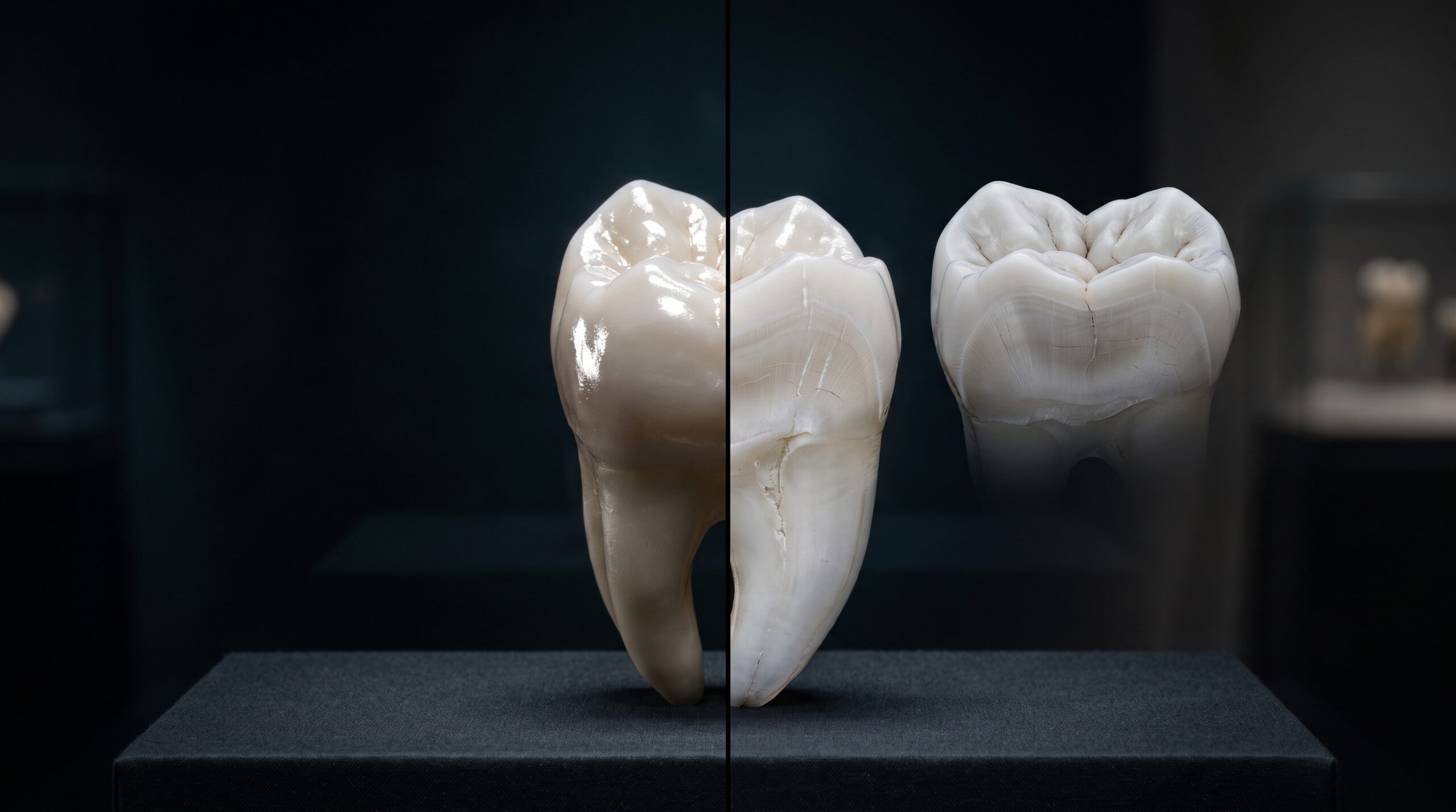
Photograph a polished tooth under a bright flash and the first thing the sensor records is a lie of light. Across the wet, curved enamel sits a scatter of brilliant white highlights, the specular reflection of the flash bouncing straight back into the lens. Those hot spots are beautiful in a clinical portrait and useless in a diagnostic one. They sit precisely where the surface is most convex, obscuring craze lines, early decalcifications, the delicate boundary between enamel and dentin, and the very chroma a laboratory needs to match. Cross-polarization is the technique that removes them, and in doing so it changes what a dental photograph is for.
The idea is borrowed from optics and mineralogy, where crossed polarizers have long been used to see through surface reflection into structure. Applied to the mouth, it turns a glossy, highlight-strewn image into a matte, even one in which the tooth reveals its interior. This is imaging as investigation rather than flattery, and understanding how it works is the difference between using it as a gimmick and using it as an instrument.
What glare actually is, and why it hides so much
Light reflecting from a tooth arrives in two distinct populations. The first is specular reflection: light that bounces cleanly off the outer wet surface of the enamel, unchanged, producing those mirror-bright highlights. The second is diffuse reflection: light that penetrates the translucent enamel, scatters through the underlying dentin, and returns carrying information about color, opacity, and internal structure. The diagnostic value lives almost entirely in that second population.
The trouble is that the specular component is far more intense and sits on top of everything, drowning out the subtle diffuse signal exactly where the surface curves toward the camera. The clinician wants the second population and is handed the first. To see structure, you have to remove the surface bounce without disturbing the light that came from within.
The crossed-filter trick
Polarization makes that separation possible. A polarizing filter placed over the flash forces the outgoing light to vibrate in a single plane. When that polarized light reflects specularly off the smooth enamel surface, it keeps its orientation. But when it penetrates the tooth and scatters through dentin, the internal scattering randomizes its polarization entirely.

Now place a second polarizing filter over the lens, rotated ninety degrees to the first. This crossed filter blocks any light still traveling in the original plane, which means it eliminates the specular highlights that preserved their polarization. The diffuse light returning from inside the tooth, its polarization already scrambled, passes through freely. The result is an image stripped of glare, showing only the light that actually interrogated the tooth’s internal structure. Two cheap pieces of optical film, oriented against each other, accomplish what no amount of post-processing can genuinely recover.
What the glare-free image gives back
Remove the highlights and the tooth appears almost matte, and that flatness is the point. Details that were sitting under the reflections become plainly visible. Mamelons and the fine internal gradient where translucent incisal enamel gives way to more opaque cervical dentin read clearly. Early white-spot lesions and decalcifications, so easily lost in glare, stand out. Craze lines, micro-cracks, and the margins of existing restorations gain definition.
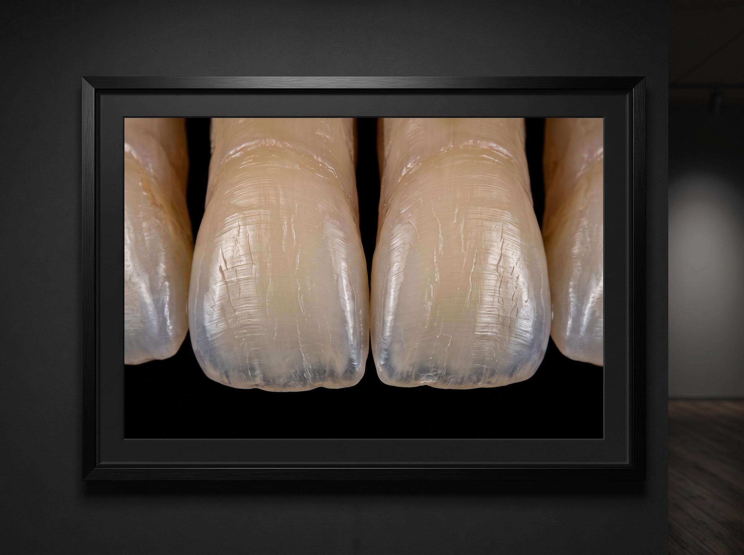
Just as important is color. Because the recorded light has genuinely traveled through the tooth rather than skidding off its surface, the chroma and value are far more faithful to the real internal shade. This is why cross-polarization has become a favored tool for communicating with a ceramist: the image carries the tooth’s true color information instead of a surface reflection of the flash. What the laboratory receives is closer to the tooth as it is, not as the lighting flattered it to be.
The technique, and its honest trade-offs
In practice, the method requires matched polarizing filters, one on the light source and one on the lens, with the lens filter crossed against the flash. Many clinicians use a dedicated ring or twin flash fitted with a polarizing sleeve, paired with a rotating polarizer on a macro lens so the crossed position can be dialed in precisely.

Nothing is free, optically. Crossing two polarizers discards a large fraction of the light, commonly two stops or more, so exposures must compensate with more flash power, a wider aperture, or higher sensitivity, each with its own cost to depth of field or noise. The resulting image is also, by design, matte and slightly flat, which is diagnostically honest but less immediately flattering than a glossy clinical portrait. For that reason many practitioners capture both a conventional and a cross-polarized frame of the same view: one for the patient’s eye, one for the clinician’s and the laboratory’s. Used well, the technique is not a replacement for standard photography but a second, more revealing pass at the same subject.
Future Developments
Cross-polarization is quietly moving from a specialist trick toward a standard layer of dental documentation. Purpose-built polarizing flash systems continue to shrink and simplify, lowering the barrier to routine use, and calibrated cross-polarized capture is increasingly paired with software that reads shade values directly from the glare-free image, turning a photograph into quantitative color data. The most interesting frontier is analytical: because the glare-free frame exposes internal structure so cleanly, it is fertile input for algorithms trained to flag early demineralization, cracks, and shade mismatches that the unaided eye skims past. As those tools mature, the crossed filter may come to be seen not as an aesthetic refinement but as the moment a clinical photograph stopped merely depicting the tooth and started measuring it.
Sorry, the comment form is closed at this time.