


September 24, 2026 Transillumination: Photographing Cracks and Craze Lines With Light Instead of Radiation
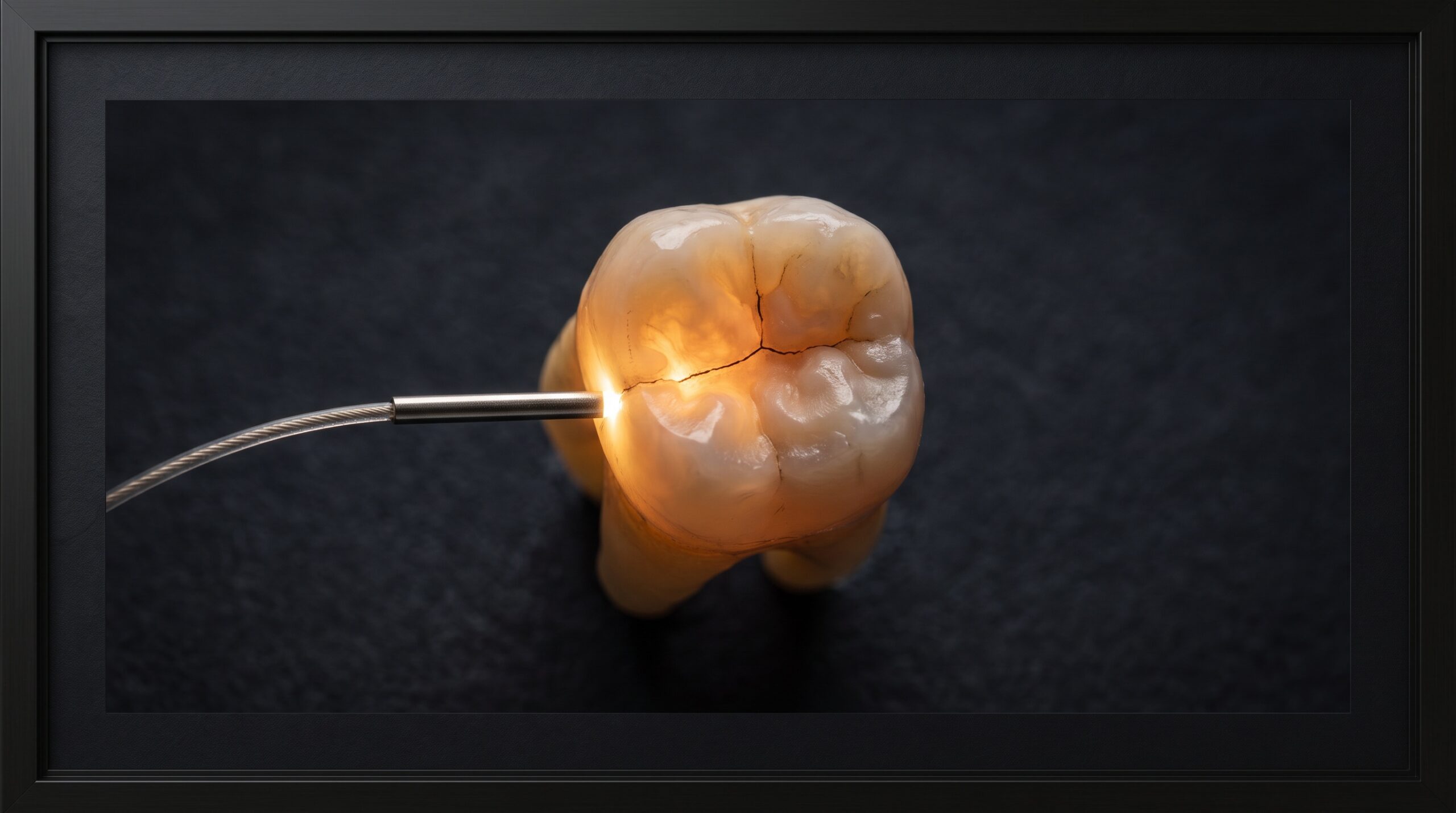
Ask a periapical radiograph to show you a hairline crack and it will often shrug. A fracture that runs buccolingually — with the X-ray beam rather than across it — casts almost no shadow, and the tooth reads as sound on film even as the patient winces on release. This is the blind spot transillumination was made for. Instead of casting a beam through the tooth and reading the shadow on a sensor, we press a bright, cold point of light against the tooth and photograph what the enamel does with it.
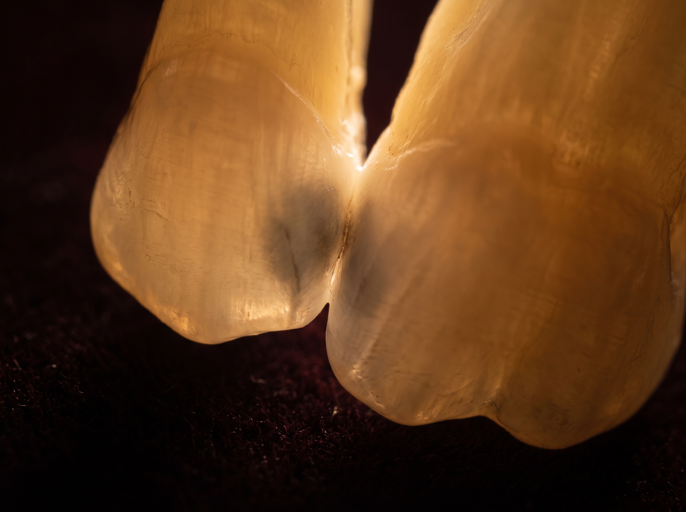
It is one of the oldest ideas in imaging and one of the most quietly beautiful. Sound enamel is remarkably translucent; it carries light the way frosted glass does. A crack, a caries lesion, or a band of demineralization interrupts that light path, scattering and absorbing it. The result is a glowing tooth with a dark line or shadow drawn precisely where the trouble lives — a picture no other modality composes quite the same way.
Why light sees what the X-ray misses
Radiography and transillumination answer different questions. An X-ray maps density along the beam’s path, which is why it excels at bone level, root anatomy, and lesions with a mineral-loss footprint broad enough to attenuate the beam. But a clean fracture is a plane, not a volume — it removes almost no mineral, so it barely dents the beam unless the geometry is perfect. Light, by contrast, is exquisitely sensitive to that plane: the fracture surface acts like an internal mirror, reflecting light back and starving everything behind it. The crack becomes a shadow.
The same physics makes transillumination a gentle screening tool for early interproximal decay, where a demineralized zone scatters light and prints as a soft grey shadow long before it has cavitated into something a probe can catch. None of this replaces the radiograph — it complements it, and photographing both gives the clinical record two independent witnesses. For teeth already under scrutiny, pairing this with the detail that cone-beam CT reveals that a flat radiograph cannot builds a fuller picture of what is happening inside the crown and root.
The instrument and the setup
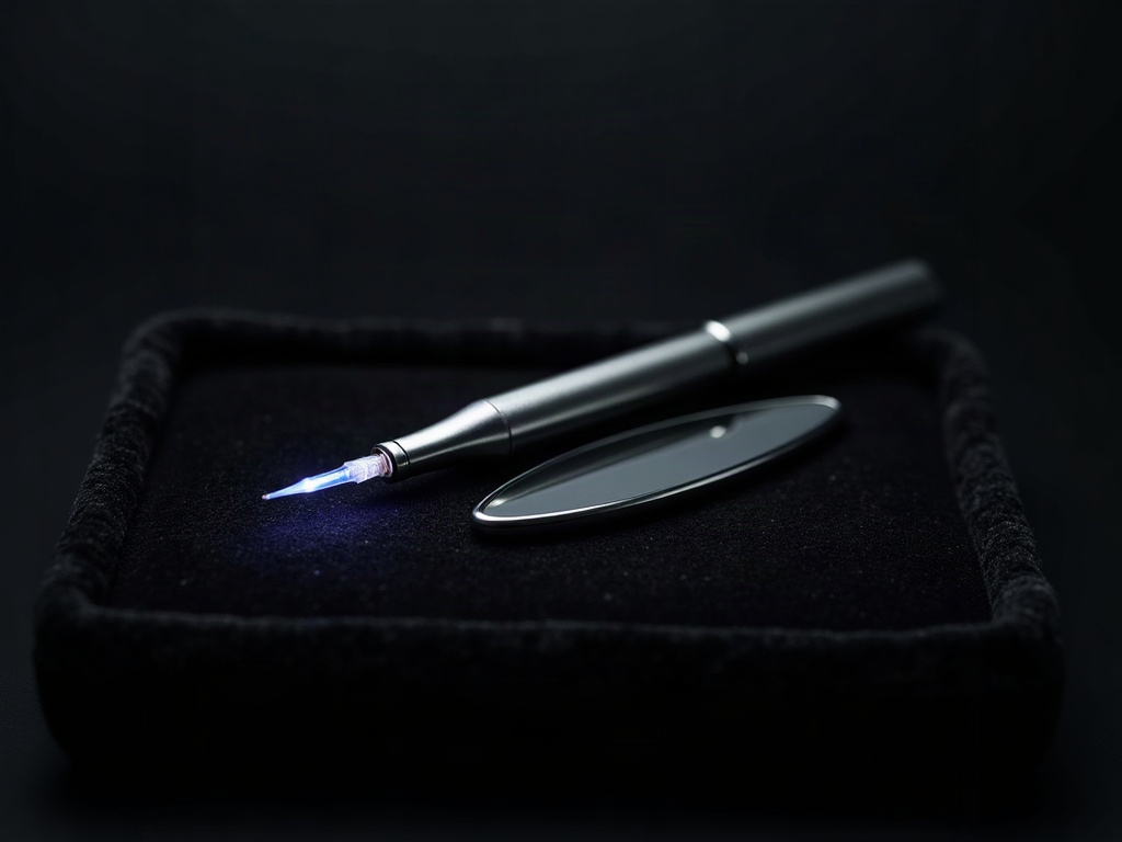
The light source can be as simple as a fiber-optic curing-light guide with the tip shielded, a dedicated transillumination wand, or a purpose-built near-infrared unit. What matters photographically is three things: the light must be bright, cool in temperature, and tightly focused so it enters the tooth rather than washing across the whole field. A broad, warm light floods the frame and erases the very contrast you are hunting.
- Placement. The light goes behind or to the side of the tooth relative to your lens — lingual light for a facial photograph, or interproximal placement to throw a contact-point shadow. You are always trying to put the tooth between the light and the camera.
- Darkness. Kill the overhead and operatory lights. Transillumination is a low-light craft; ambient light competes with the glow and flattens the shadow into nothing.
- Isolation. A dry, clean tooth transmits cleanly. Saliva and debris scatter light at the surface and add noise that reads as false shadow.
Photographing the glow
Capturing transillumination is a genuine exposure problem, because you are photographing a dim, self-luminous subject in a dark field. Shooting on full auto will almost always fail — the camera sees black surroundings, opens up, and blows the glowing tooth into a featureless white blob. Manual control is the craft:
- Expose for the glow, not the room. Meter off the lit tooth. You want the enamel bright but not clipped, so the fracture shadow retains its edge.
- Small aperture, patient shutter. Macro depth of field is punishing; stop down for front-to-back sharpness, then let the shutter stay open long enough to gather the faint light. A stabilized hand or a rest matters here.
- Kill the ring flash. The on-lens flash that serves a normal clinical portrait is the enemy of transillumination — it lights the surface and destroys the internal glow. This is one of the few dental shots taken with the flash off.
- Watch your white balance. Fiber-optic light skews warm and orange; a near-infrared unit skews the other way. Set white balance deliberately so the shadow reads as shadow, not as a color cast.
The discipline is the same one that governs every serious clinical capture — controlling light rather than accepting it. The principles that make a well-composed clinical portrait repeatable apply here too: standardize the setup so today’s image can be honestly compared with next year’s.
Reading and documenting what you see
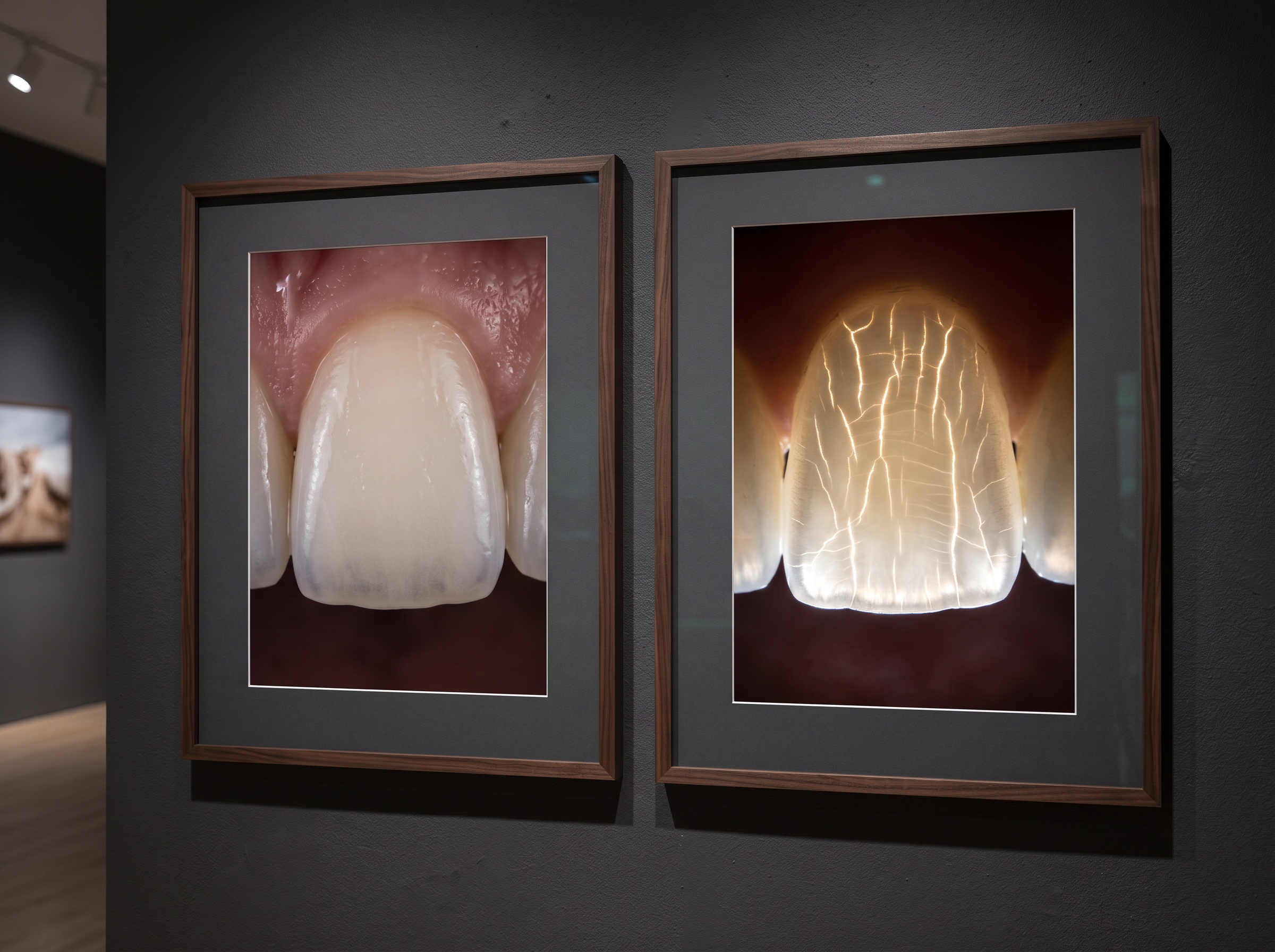
A transillumination image is diagnostic only if it is interpreted with care. A bright, uninterrupted glow suggests intact structure. A sharp dark line that stops at the light and does not cross a marginal ridge points toward a crack. A diffuse grey cloud at a contact point suggests demineralization. But shadows can deceive — a restoration margin, a stain, or a thick area of dentin can all mimic pathology. The photograph is evidence to be weighed against the radiograph, the probe, and the patient’s symptoms, never a verdict on its own.
Where the technique earns its keep is documentation over time. Because it uses no radiation, you can re-photograph a suspicious tooth as often as clinical judgment warrants, building a dated visual record of whether a craze line is stable or a crack is propagating. That serial record — standardized lighting, standardized angle, standardized exposure — is the difference between an anecdote and evidence.
Where transillumination fits in the imaging library
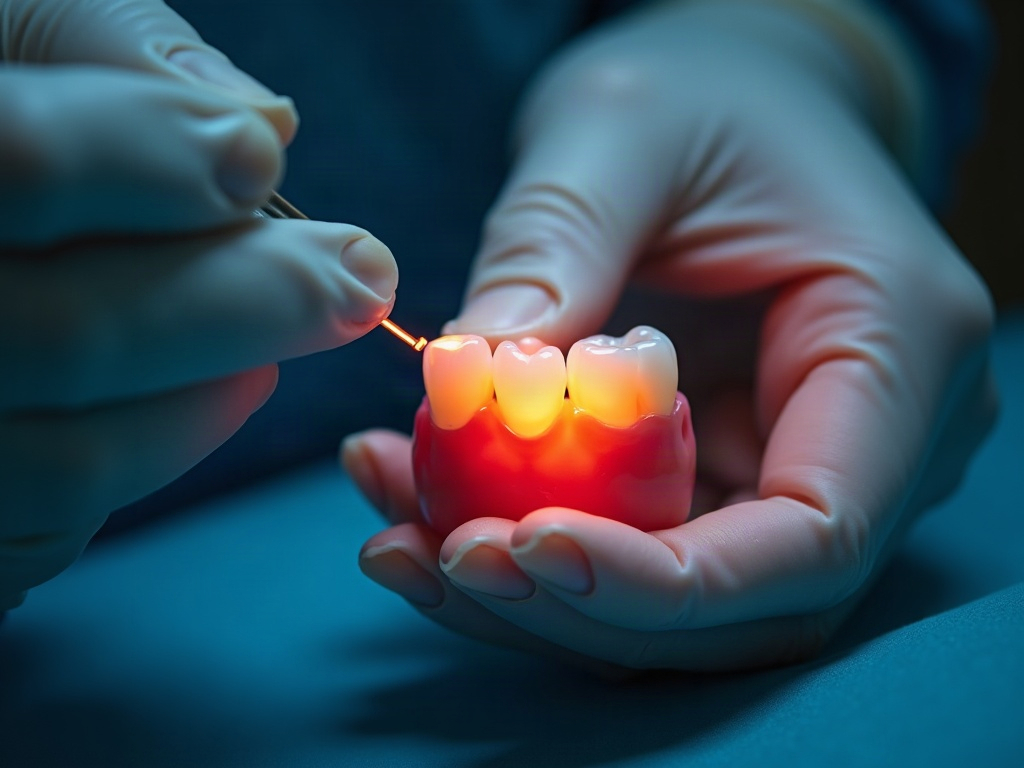
Think of transillumination not as a rival to radiography but as another lens on the same tooth — one tuned to fractures and early surface demineralization that the beam struggles to render. It is fast, comfortable, radiation-free, and, when photographed well, genuinely striking. A gallery of transillumination images alongside the radiographs and clinical portraits gives a case something close to a complete visual account: density from the X-ray, surface and color from the portrait, and internal integrity from the light.
Future developments
The frontier here is near-infrared. Longer wavelengths penetrate enamel more deeply and scatter less, sharpening the contrast between sound and demineralized tissue, and dedicated near-infrared transillumination cameras now feed calibrated digital images straight into the record rather than relying on a clinician’s phone held against a wand. As those sensors improve and as software learns to flag and measure a shadow’s growth from one visit to the next, transillumination is likely to shift from a quick chairside check to a quantified, longitudinal imaging channel — a way to watch a crack or an incipient lesion the way we already watch bone. The craft will remain the same at its heart: put the tooth between the light and the lens, darken the room, and let the enamel tell you where it is broken.
Sorry, the comment form is closed at this time.