


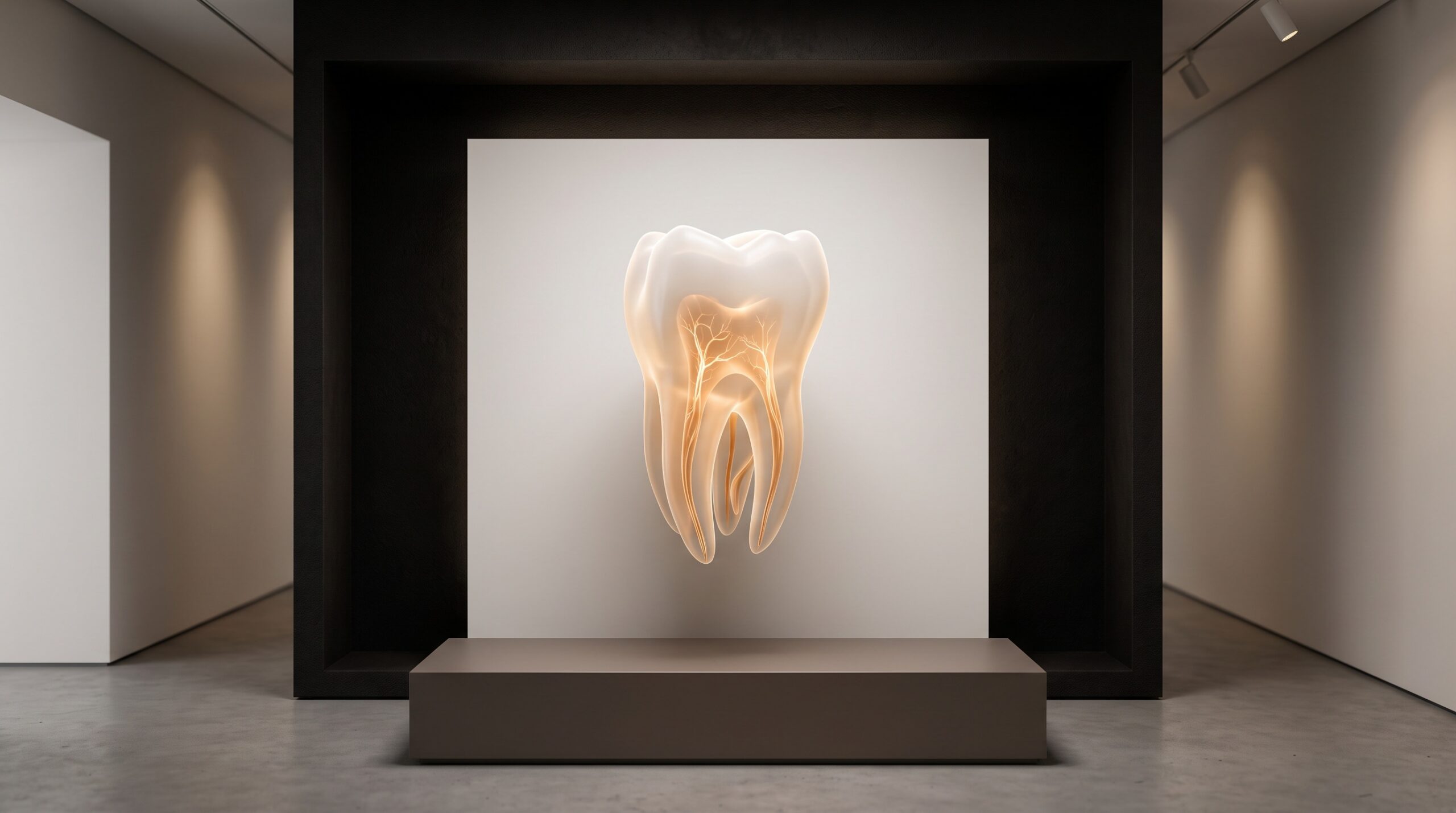
September 14, 2026 The Hidden Architecture of a Tooth: What Cone-Beam CT Reveals That a Flat Radiograph Cannot
For most of a century the dental radiograph told us nearly everything about a tooth, and quietly kept one secret. It is a projection – a shadow cast by X-rays that pass straight through the tooth and strike a sensor behind it – and a shadow, however sharp, is flat. A real root system is not. It curves and divides and rejoins in three dimensions, and when that architecture is pressed onto a single plane, whatever lies directly behind something else simply disappears into it. The classic casualty is the second mesiobuccal canal of an upper molar, the so-called MB2: present in a clear majority of maxillary first molars, and yet routinely invisible on a periapical film because it sits almost exactly buccal to the main mesiobuccal canal, one hiding behind the other like two people photographed in a doorway. The film is not lying. It is only two-dimensional, and the tooth is not.
Cone-beam computed tomography gives the missing dimension back. Instead of one shadow it acquires a true volume – a solid block of data that can be resliced along any plane, axial, sagittal, coronal, and rendered as an object in the round. The overlap that erased the MB2 vanishes the moment you can look down the long axis of the root and see two distinct openings where the flat film showed one. This is the same enlargement of vision that lets a scan reveal so much that lies beyond the teeth themselves – only turned inward, onto the hidden interior of a single root.
The Anatomy a Flat Film Cannot Show
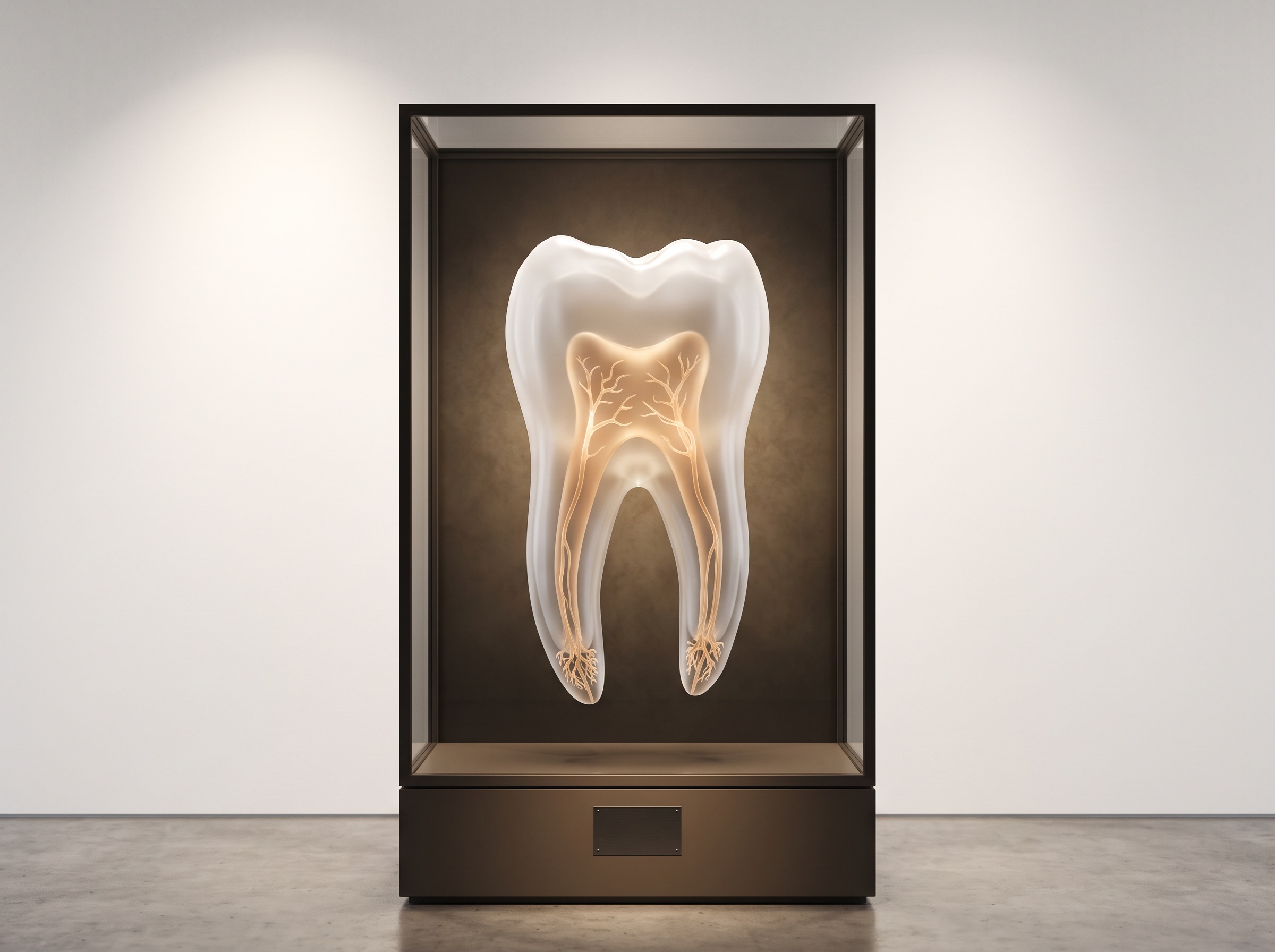
Once you can navigate a tooth in three dimensions, a whole vocabulary of internal form becomes visible that a projection can only hint at. Canals divide and merge along their length. Lateral and accessory canals branch off the main channel toward the surrounding bone. The apex, far from a single neat opening, often frays into an apical delta of several tiny exits. And within a single broad root, two canals may be joined by a narrow ribbon of space called an isthmus – a connection that traps tissue and defies a round instrument, and that no flat image will ever resolve. None of this is exotic; it is simply the ordinary complexity of teeth, made legible.
When Two Roots Become One
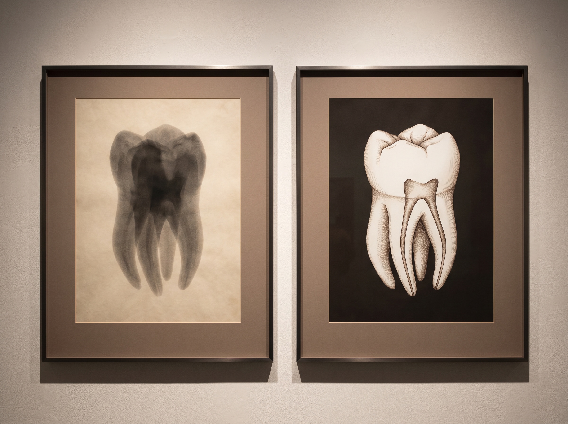
Root fusion is a good example of how three dimensions overturn a flat film’s easy assumptions. On a radiograph, two roots that have merged into one broad structure read as a single simple shadow – deceptively straightforward. In the volume, and confirmed by the micro-CT studies that examine these teeth at the finest scale, the fused root turns out to be anything but simple: its merged canals frequently retain a narrow connecting isthmus and an intricate cross-section that changes shape as it descends. CBCT and in-vivo studies find fused roots and canal merging common enough in maxillary second molars to matter in everyday practice. The lesson is counter-intuitive and worth stating plainly: a fused root is usually a more complicated tooth to treat, not a less complicated one, and only a volumetric view shows why.
Why the Missing Dimension Matters
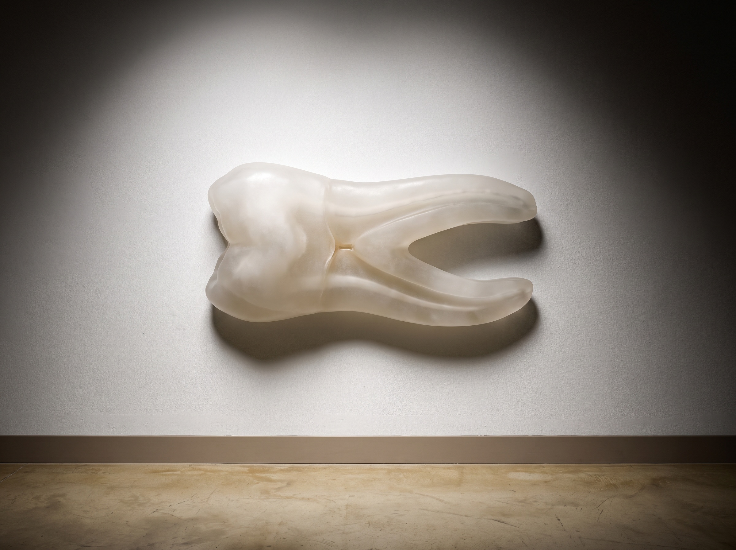
This is not detail for its own sake. A canal that is never found is a canal that is never cleaned or sealed, and a missed canal is among the leading reasons a root canal treatment fails months or years later. Knowing before the first instrument enters the tooth that an MB2 is present, that a root is fused around a hidden isthmus, or that the apex divides into a delta, converts what would have been an intraoperative surprise into a plan. The image does the reconnaissance so the clinician does not have to discover the anatomy the hard way. It is the same principle that runs through all of diagnostic imaging: you cannot treat what you cannot see, and seeing, here, is three-dimensional.
The Honest Limits
None of this makes CBCT a free lunch, and PatientGallery’s admiration for the image has never meant ignoring its costs. A cone-beam scan delivers meaningfully more radiation than a single periapical film, so it is justified case by case rather than taken reflexively – a genuine weighing of benefit against dose that we have explored in the risk of not looking versus the risk of the scan itself. Its resolution, though remarkable, still trails the micro-CT of the research laboratory, so the very finest accessory canals sit at the edge of what a clinical voxel can resolve. And the volume has its own demons: dense restorations and posts throw beam-hardening and metal-streak artefacts across exactly the region an operator most wants to read, occasionally hiding the canal the scan was ordered to find. The three-dimensional image is a better map, not a perfect one.
Reading the Volume Well
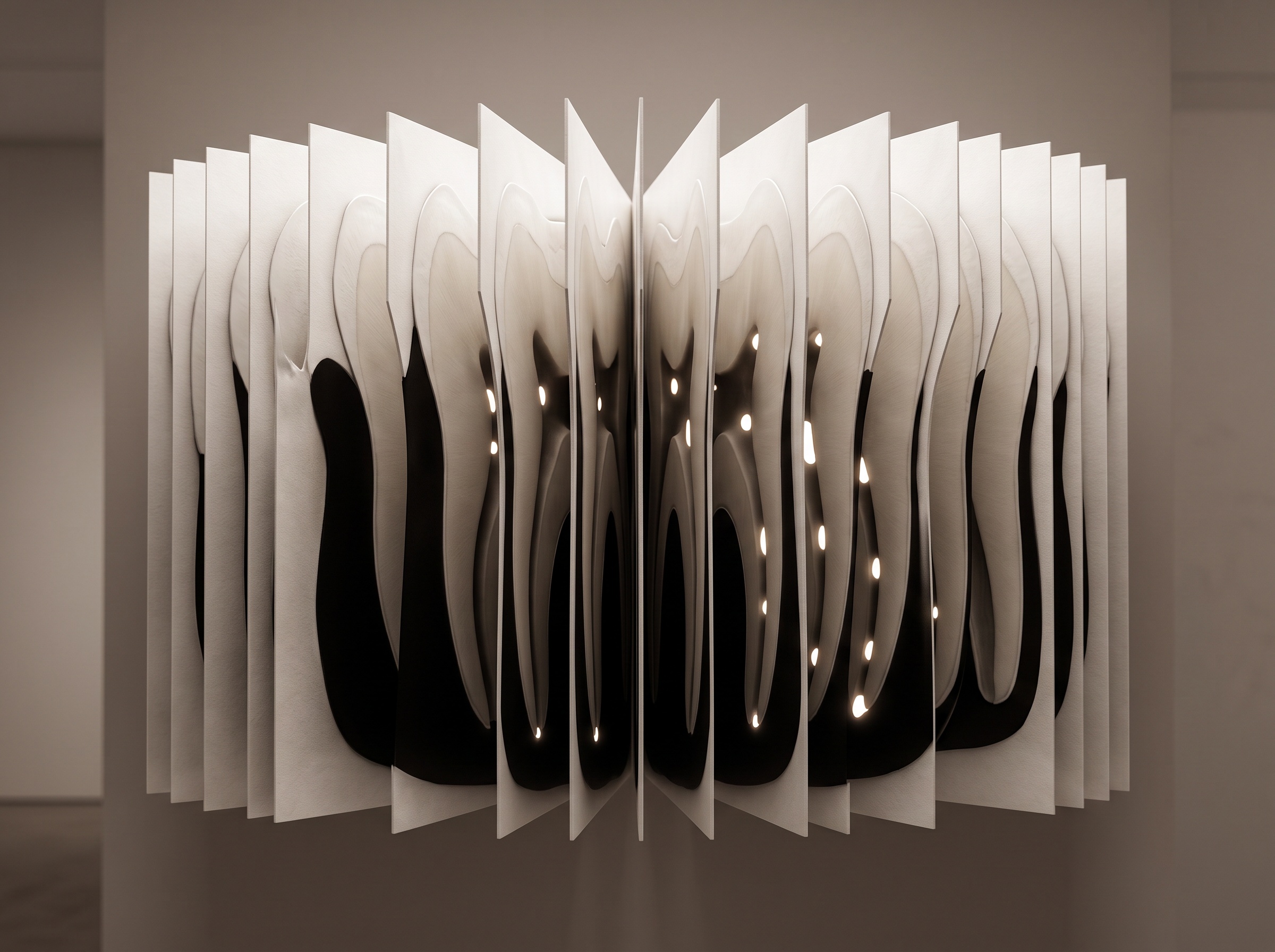
Because the data is a volume rather than a picture, reading it is an active craft – scrolling plane by plane down the root, watching the canal count and cross-section change, mentally reassembling the slices into the branching object they describe. It is a skill, and increasingly a shared one: software now traces the canal system automatically, and the growing ability of AI to segment a CBCT volume and surface exactly what it finds is beginning to turn hours of manual slice-by-slice study into a labelled map delivered in seconds. The machine proposes; the clinician still confirms. But the direction is clear – the volume is not only richer than the flat film, it is becoming easier to read than it once was.
Future Developments
The trajectory is toward a three-dimensional anatomical map that is routine, low-dose, and self-annotating. Detector physics keeps pushing resolution upward while photon-counting and smarter reconstruction pull dose downward, narrowing the gap between the clinic’s CBCT and the laboratory’s micro-CT so that finer accessory anatomy comes into everyday reach. Automated canal-mapping will mature from a helpful overlay into a trusted first read, flagging the extra canal and the hidden isthmus before a human has scrolled a single slice. Further out, the radiation-frugal ideal beckons – the same architectural richness captured with a fraction of today’s dose, or paired with radiation-free modalities for the soft-tissue story the volume cannot tell. What will not change is the essential insight underneath all of it. Cone-beam CT did not so much add detail to the radiograph as give the tooth back a dimension the flat film had quietly taken away – and a tooth seen in the round, at last, is a tooth seen as it truly is.
Sources & further reading:
- Prevalence of Root Fusions and Main Root Canal Merging in Human Upper and Lower Molars: A Cone-beam Computed Tomography In Vivo Study
- Influence of root fusion on canal orifice configuration, lateral anatomy, isthmus formation, and apical morphology in maxillary second molars: a micro-CT analysis
Sorry, the comment form is closed at this time.