23 Mar Periapical Radiography in Endodontics: Mastering the Gold Standard for Root Canal Diagnostics
Periapical radiography remains the cornerstone of endodontic diagnosis and treatment planning, serving as the primary imaging modality for root canal therapy. Understanding proper technique, interpretation, and integration with modern diagnostic tools is essential for successful endodontic outcomes.
The Foundation of Endodontic Imaging
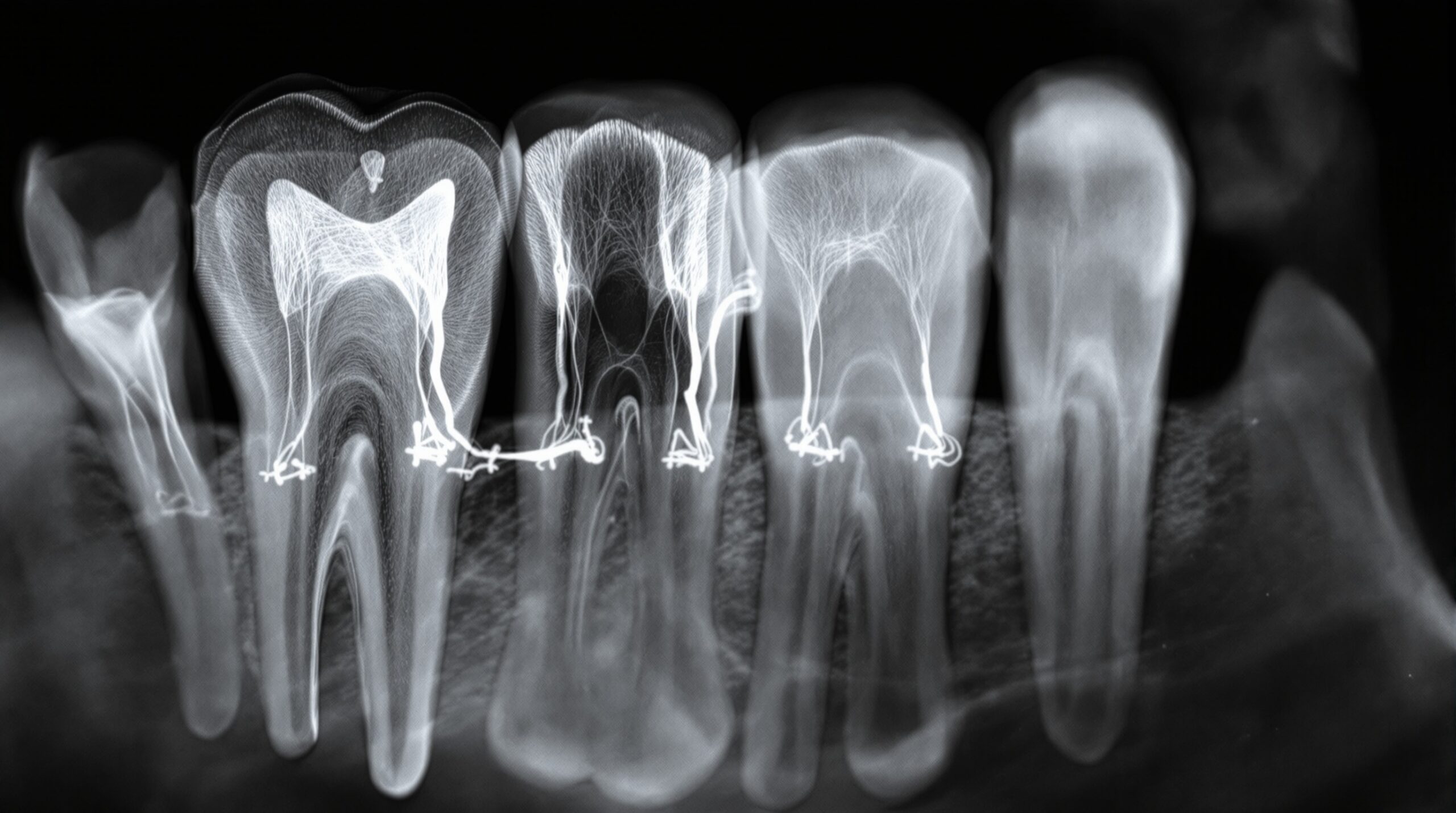
Intraoral periapical (IOPA) radiographs provide essential diagnostic information for endodontic treatment planning. These two-dimensional images offer detailed visualization of root morphology, periapical pathology, and anatomical structures crucial for successful root canal therapy. The 2026 ADA guidelines emphasize using periapical radiographs as the primary imaging modality for initial endodontic evaluations, reserving CBCT for complex cases requiring three-dimensional visualization.
Optimal Technique and Angulation
Achieving diagnostic-quality periapical radiographs requires adherence to standardized positioning techniques. The paralleling technique, using film holders and positioning devices, ensures optimal image geometry and reduces magnification. Proper horizontal and vertical angulation prevents overlapping of adjacent structures and ensures complete visualization of the root apex and surrounding periapical tissues.
Key Technical Considerations:
- Parallel technique – Maintains consistent geometry and reduces distortion
- Complete root coverage – Ensures 2-3mm of periapical tissue is visible beyond the apex
- Proper exposure parameters – Optimizes contrast and detail for endodontic diagnosis
- Multiple angulations – Different projection angles reveal additional diagnostic information
Advanced practitioners often employ eccentric projections, such as mesial and distal tube shifts, to separate superimposed root structures and identify additional canals or complex root morphology that may be missed on standard periapical views.
Integration with Electronic Apex Locators
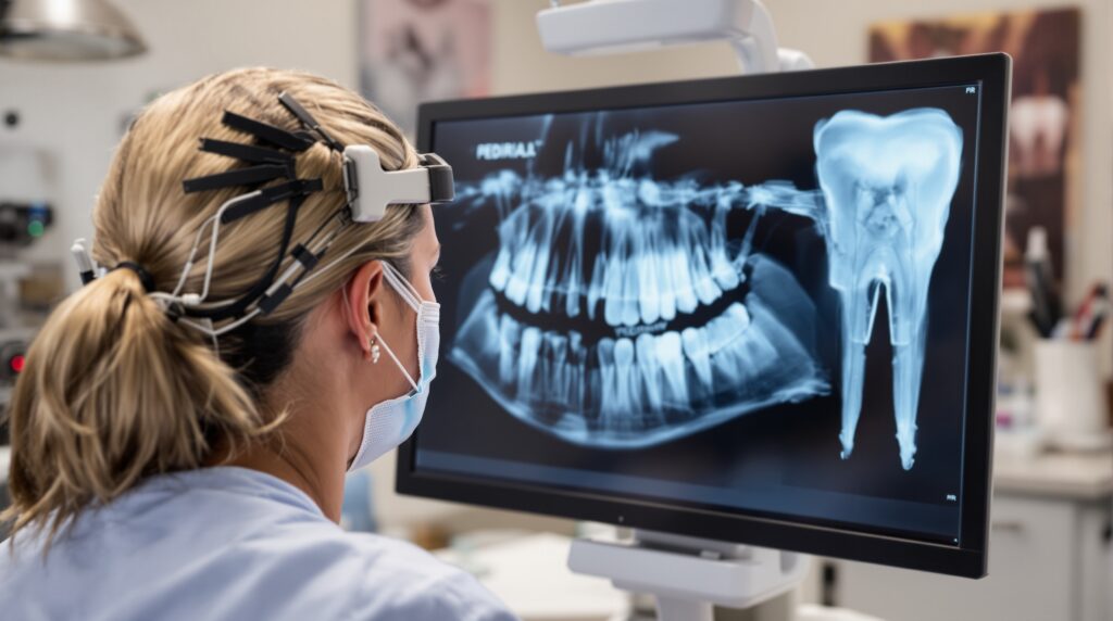
Modern endodontic practice combines periapical radiography with electronic apex locators for enhanced accuracy in working length determination. This dual-modality approach significantly improves treatment outcomes by providing both anatomical visualization and precise electronic measurement of the apical constriction.

Electronic apex locators measure electrical impedance changes as files approach the apical foramen, providing real-time feedback during instrumentation. When combined with periapical radiographs, this technology enables precise working length determination even in challenging cases with complex anatomy or periapical pathology.
Diagnostic Applications in Endodontics
Periapical radiographs serve multiple diagnostic functions throughout endodontic treatment:
Pre-treatment Assessment:
- Identification of periapical pathology and extent of lesions
- Root morphology analysis and canal configuration
- Assessment of root resorption or fractures
- Evaluation of previous endodontic treatment
Intraoperative Guidance:
- Working length verification
- Canal negotiation and instrumentation monitoring
- Master cone fit confirmation
- Obturation quality assessment
Post-treatment Monitoring:
- Healing assessment of periapical tissues
- Long-term treatment outcome evaluation
- Detection of treatment complications
Limitations and When to Consider CBCT
While periapical radiographs excel in routine endodontic diagnosis, certain clinical situations may warrant three-dimensional imaging with cone beam computed tomography (CBCT). The 2026 ADA guidelines recommend considering CBCT after initial clinical examination and 2D imaging assessment for complex cases.
CBCT indications include suspected root fractures, complex root anatomy, retreatment cases with persistent symptoms, and pre-surgical endodontic planning. However, the increased radiation exposure necessitates careful risk-benefit analysis for each patient.
Best Practices for Modern Endodontic Imaging
Contemporary endodontic practice emphasizes evidence-based imaging protocols that maximize diagnostic information while minimizing radiation exposure. Key recommendations include:
- ALARA principle – Use lowest radiation dose necessary for diagnosis
- Selective imaging – Base radiographic frequency on clinical findings
- Digital sensors – Reduce exposure time and enable immediate image review
- Quality assurance – Regular equipment calibration and technique standardization
- Patient positioning – Consistent positioning reduces retakes and exposure
Modern digital radiography systems offer significant advantages including reduced radiation exposure, immediate image availability, and enhanced image manipulation capabilities for improved diagnostic interpretation.
Conclusion
Periapical radiography continues to serve as the gold standard for endodontic imaging, providing essential diagnostic information for successful root canal treatment. Mastery of proper technique, combined with integration of electronic apex locators and judicious use of advanced imaging when indicated, ensures optimal patient outcomes. As endodontic technology continues to evolve, the fundamental principles of quality periapical radiography remain central to exceptional endodontic care.
Sorry, the comment form is closed at this time.