



August 23, 2026 The Risk of Not Looking: Weighing a Cone-Beam Scan’s Radiation Against What It Reveals
Every profession that handles radiation learns to fear it first, and rightly so. We teach dose before we teach diagnosis; we recite ALARA – as low as reasonably achievable – long before we can read a film. That instinct has protected countless patients. But an instinct trained only in one direction can become its own kind of blindness. A cone-beam scan is not a decision about a single risk. It is a decision between two, and the second one is almost never named aloud: the risk of not looking.
A recent commentary in the British Dental Journal put the tension in exactly those terms – radiation risk, or diagnosis risk? – and the framing deserves to travel beyond the letters page. For all our fluency in millisieverts, we are strangely inarticulate about the harm of the image we decline to make.
Two Risks on One Scale
Set the two side by side honestly. On one pan sits the radiation risk of cone-beam computed tomography: real, cumulative, stochastic – a small increment to a lifetime probability, weighted more heavily in children and in tissues like the thyroid and salivary glands. On the other pan sits the diagnostic risk of proceeding without the information a scan would have given: an undetected second canal that dooms a root canal to failure, a root fracture read as periodontal disease, an implant placed a fraction too close to the inferior alveolar nerve.
Neither pan is empty. The error we are prone to is treating only the first as a risk and the second as merely the absence of a benefit. But a missed diagnosis is not a neutral outcome – it is a harm with its own morbidity, its own reoperations, its own dose incurred later when the problem finally forces a scan anyway. Weighed like for like, the question is not “is there radiation?” – there always is – but “which risk is larger for this patient, this question, today?”
The Dose in Proportion
Fear is calibrated by comparison, and here the comparison matters. Effective dose is measured in microsieverts, and a modern, limited-field dental cone-beam scan typically lands in the tens of low hundreds of microsieverts – a fraction of a multi-detector medical CT of the head, and comparable in order of magnitude to the natural background radiation a person absorbs over a span of days to a few weeks simply by living on this planet. It is not nothing. It is also not the catastrophe that reflexive radiophobia imagines.
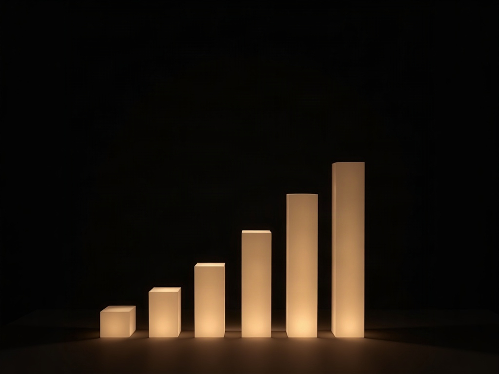
This is why dose belongs on a continuum rather than in a binary of safe versus dangerous. A large field-of-view scan of both jaws is a genuinely larger exposure than a small volume of a single quadrant, and the two should never be spoken of as one thing. The craft lies in matching the exposure to the question – and in refusing to let a vague dread of “CT-level radiation” stand in for the actual, and often modest, number on the console.
Justification Comes Before Optimization
Radiation protection rests on two pillars, and we tend to remember only the second. Optimization – keeping any given exposure as low as reasonably achievable – is the one we drill. Justification – the prior question of whether the exposure should happen at all – is the one that actually governs whether a patient is served. An exquisitely optimized scan of a patient who never needed one is still an unjustified dose. A slightly higher exposure that answers a genuine clinical question is justified radiation, honestly spent.
Justification is where selection criteria earn their keep. A cone-beam scan is not a routine screening tool and was never meant to be; it is indicated when a specific question cannot be answered by two-dimensional imaging – complex endodontic anatomy, impacted teeth in perilous relation to nerves, implant site assessment, certain trauma and pathology. The professional guidelines on CBCT use and ALARA exist precisely to keep the modality tethered to need rather than habit. The discipline is to ask the question first and reach for the volume only when the flat image genuinely cannot reach it.
When the Flat Image Is Not Enough
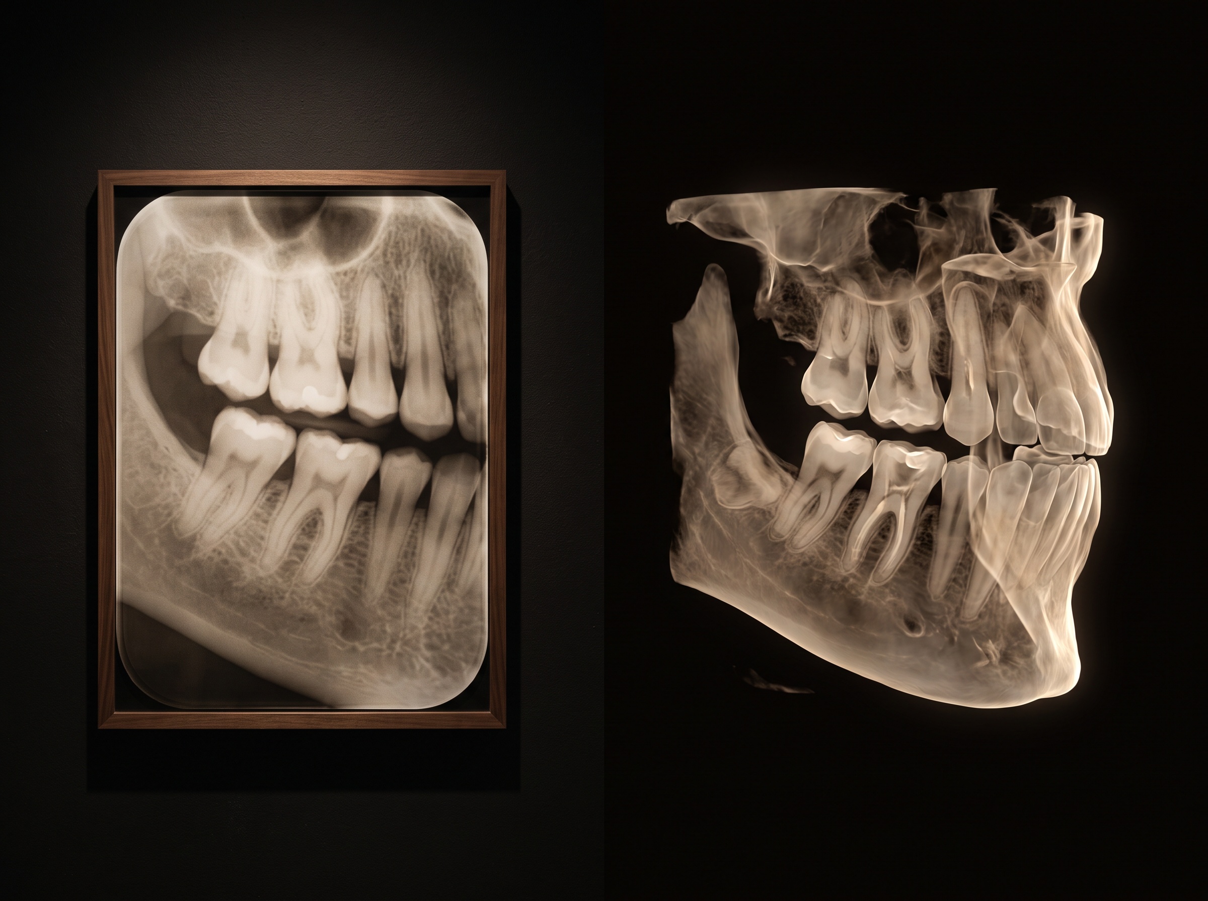
It is worth being concrete about what a volume sees that a plane cannot. A periapical radiograph collapses three dimensions onto one; anatomy stacks on anatomy, and a finding can hide behind the very structure in front of it. A cone-beam acquisition, by contrast, reconstructs true spatial relationships – depth, buccolingual position, the real distance between a root apex and a canal – which is exactly what a cone-beam scan actually captures that a two-dimensional exposure cannot. When the clinical question is spatial, the flat picture is not merely inferior – it is unable to answer, and a confident reading of it is a confident error.
This is the diagnosis risk made vivid. The harm is not hypothetical; it is the treatment planned on incomplete information, the surgery guided by a shadow. To decline the scan in such a case is not caution. It is a different, quieter gamble – one whose stakes simply arrive later.
Restraint as Craft
None of this is a license for volume. The answer to radiophobia is not radio-indifference; it is precision. Once a scan is justified, optimization becomes an act of craftsmanship: collimating to the smallest field of view that contains the question, choosing low-dose acquisition protocols tuned to the diagnostic task, and accepting a little more image noise when noise is the honest price of a lower exposure. A tightly limited scan of one tooth is a categorically different exposure from a full-arch volume, and the operator who reaches instinctively for the smaller one has internalized ALARA as reflex rather than recitation.
There is a bookkeeping dimension to this, too. Just as the exposure index restored accountability for dose in two-dimensional radiography, a mature practice tracks why each volume was acquired – the question it answered – so that justification is a documented judgment rather than a habit that quietly expands. Restraint that is not recorded tends, over months, to erode.
Future Developments
The trajectory of the field is toward dissolving the dilemma rather than merely balancing it. Iterative and AI-based reconstruction already recover diagnostic images from markedly lower exposures, pushing the radiation pan of the scale steadily lighter without surrendering the depth that makes a volume worth acquiring. Smarter selection tools promise to sharpen justification at the point of decision, flagging when a flat image will suffice and when it demonstrably will not. And as effective doses fall toward those of conventional radiography, the old opposition – radiation risk versus diagnosis risk – begins to soften into a single, better question: what is the least exposure that will let us see clearly enough to be right? That is the direction worth advancing toward, because it honors both risks at once – the one we have always feared, and the one we are only now learning to name.
Sources & further reading:
Sorry, the comment form is closed at this time.