


October 16, 2026 Raking Light: Borrowing the Art Conservator’s Trick to Photograph Tooth Wear and Erosion
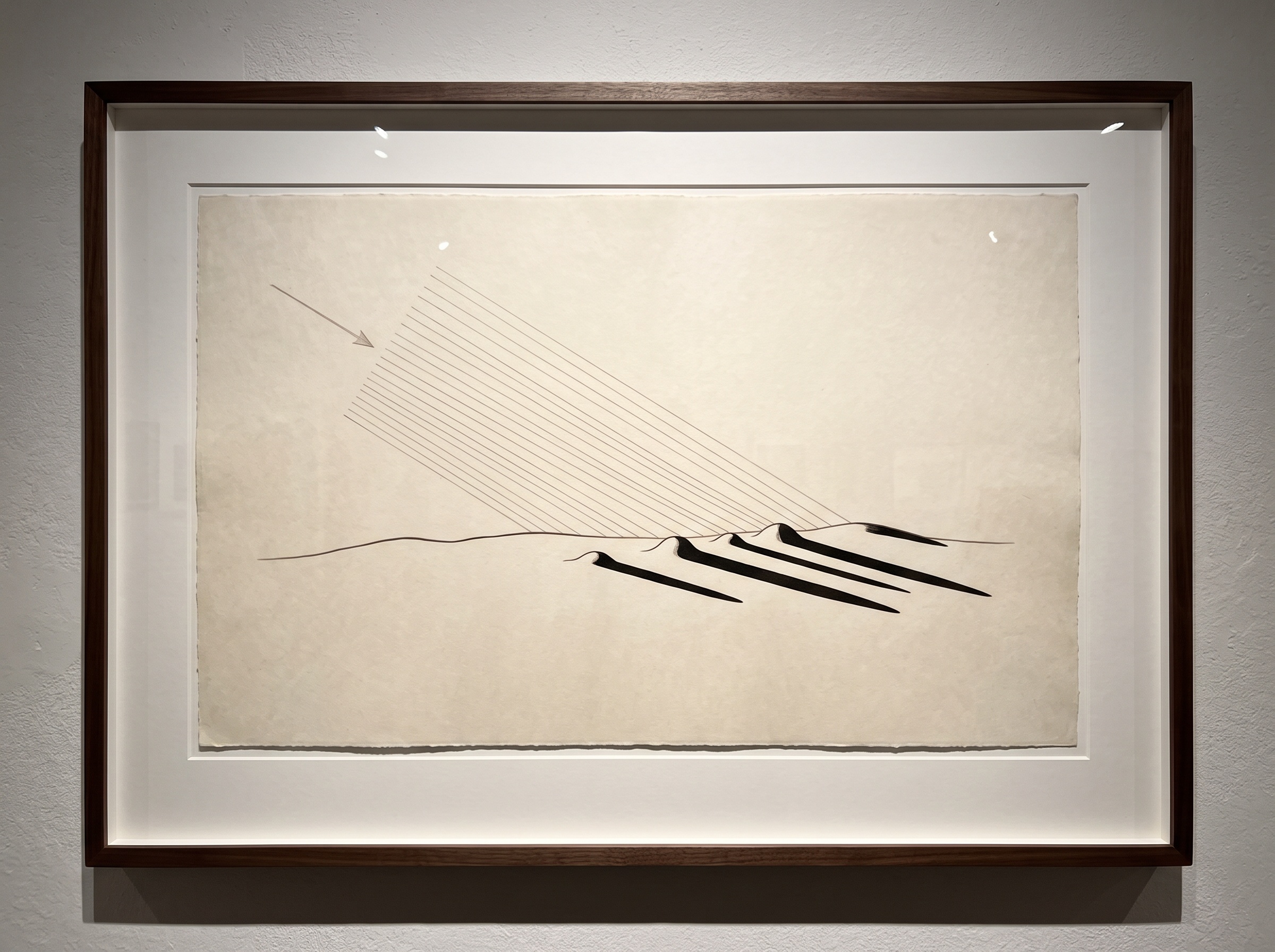
Walk into a painting conservator’s studio and you will eventually see a strange little ritual. The conservator dims the room, takes a single lamp, and holds it almost flat against the surface of a canvas, so the light skims across the paint rather than shining onto it. In that grazing beam the painting transforms. Brushstrokes rise into ridges, the weave of the canvas appears, old cracks open into dark rivers, and the faint warp of a panel that has breathed with a century of humidity becomes suddenly, undeniably visible. The technique is called raking light, and it has been the conservator’s first diagnostic tool for generations. What it does for a painting it will do just as faithfully for a tooth — and most clinical photography, built to chase even, shadowless illumination, throws that gift away.
What raking light actually does
The principle is almost embarrassingly simple, which is part of its elegance. When light strikes a surface head-on, every point is lit roughly equally, and the eye and the sensor record color and reflectance but very little form. Shadows are what tell us about shape, and frontal light casts almost none. Lower the angle of the light, however, and every raised feature begins to throw a shadow across the valley beside it. Push the angle down until the beam is nearly parallel to the surface — raking it — and even a texture measured in fractions of a millimeter casts a shadow long enough to see. A dimple becomes a crater; a ridge becomes an escarpment. The surface has not changed at all, but its relief, previously flattened by generous light, is now exaggerated into legibility. Raking light is, in effect, a way of converting height into contrast, the one quality a camera renders superbly.
The textures a tooth hides from flat light
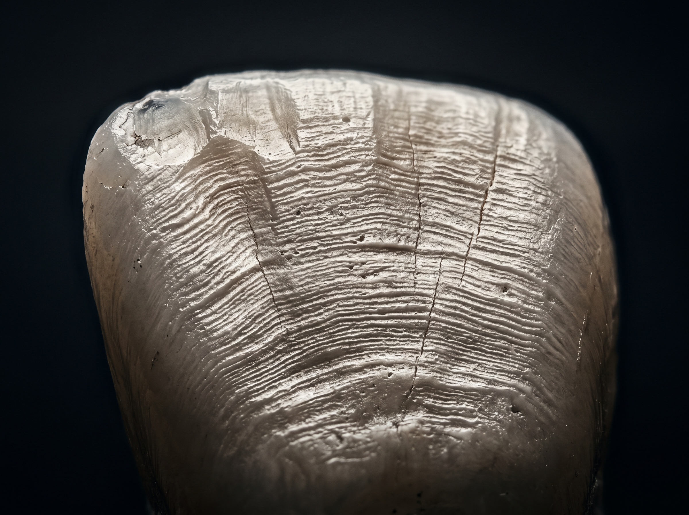
Standard clinical photography is engineered to suppress exactly this effect. Ring flashes and twin flashes with diffusers are chosen precisely because they wrap the tooth in soft, even light that renders color and translucency faithfully and minimizes distracting shadow — the right instinct when the goal is shade matching or a clean record of a restoration. But that same softness erases the surface story. A tooth’s enamel is not a blank porcelain dome; it is a worked surface carrying the record of everything that has acted on it. There are the gentle horizontal waves of perikymata, the growth lines of the enamel itself, most visible on young, unworn teeth. There are wear facets — the flattened, polished planes where opposing teeth have ground against one another, each one angled to tell you how the patient’s jaw actually moves. There is the shallow, cupped, dished-out loss of acid erosion, often maddeningly invisible under flat light because it changes form without changing color. There are the fine vertical craze lines running up from the biting edge, and the wedge-shaped notches of abfraction at the gumline. Every one of these is a change in shape, and shape is exactly what frontal lighting refuses to show.
How to rake light at the chair
Borrowing the conservator’s trick requires no new hardware, only a willingness to break the usual rule. The essential move is to take the light off the lens axis. Instead of the ring flash or a twin flash balanced on either side, use a single light source — one flash head, a small LED panel, even a handheld light — brought around to the side of the tooth and lowered until it skims nearly parallel to the surface being studied. The steeper you want the relief to read, the lower you bring the light. Direction matters as much as angle: light raking from the incisal edge downward will reveal different features than light crossing horizontally, so it is worth making more than one exposure from more than one direction, exactly as a conservator circles a painting with the lamp. Expect the image to look dramatic and even harsh; that is the point, and it is why this is a supplement to, not a replacement for, your routine documentation. Because the technique sacrifices even color rendering, it pairs naturally with the discipline you already bring to setting white balance at the moment of capture on your standard series — the two serve opposite masters, color and form, and a complete record wants both. A polarizing filter can be added to tame the specular hotspots that hard light provokes on wet enamel, the same reflection-control problem that dogs photographing glossy restorations.
Reading what the shadows say
A raking-light image is not merely dramatic; it is diagnostic, and it earns its place in the record by making change measurable. Erosion is the clearest case. Early acid erosion flattens cusps and hollows out shallow cups on occlusal and palatal surfaces — a loss of structure that is notoriously hard to catch early because it alters topography, not shade. Under raking light those cups fill with shadow and announce themselves. The real power, though, is longitudinal. If you photograph the same surface, with the light brought in from the same direction and angle on each visit, you create a series of images that can be laid side by side to track whether a lesion is progressing or holding steady. Consistency of lighting geometry becomes the equivalent of a fixed camera position: it is what makes two images honestly comparable rather than merely similar. This is monitoring by photograph, and for conditions where the decision to intervene hinges on whether wear is active or historical, a shadow that has deepened between two visits can be worth more than any single measurement.
The tooth as relief
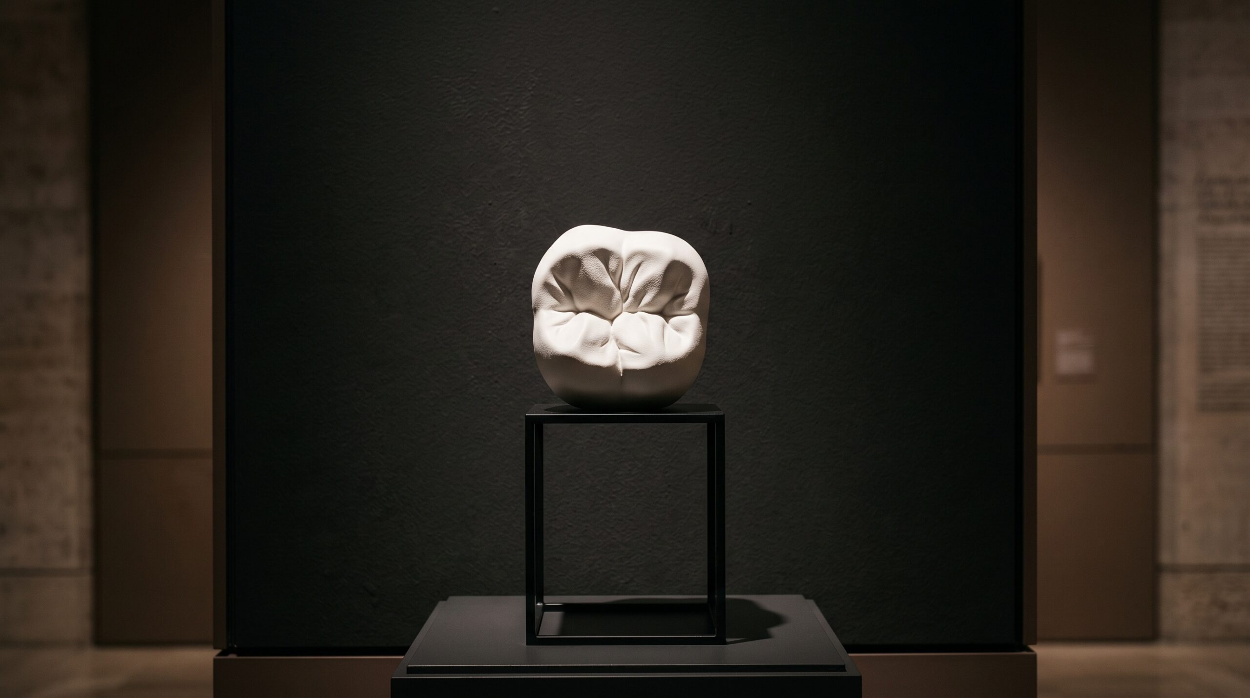
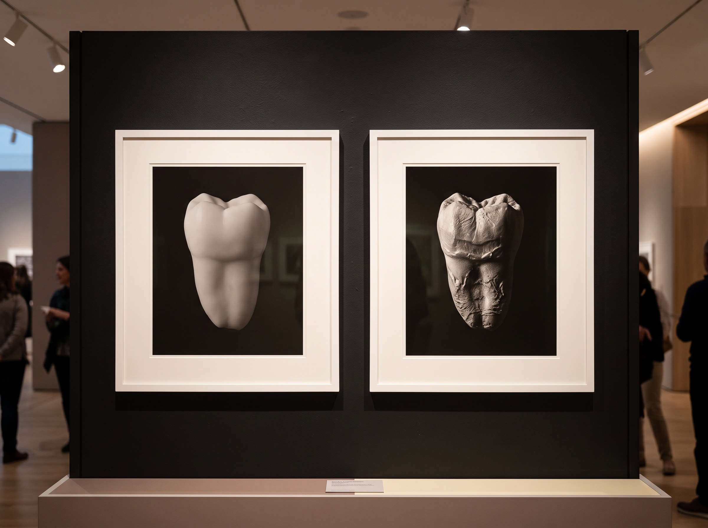
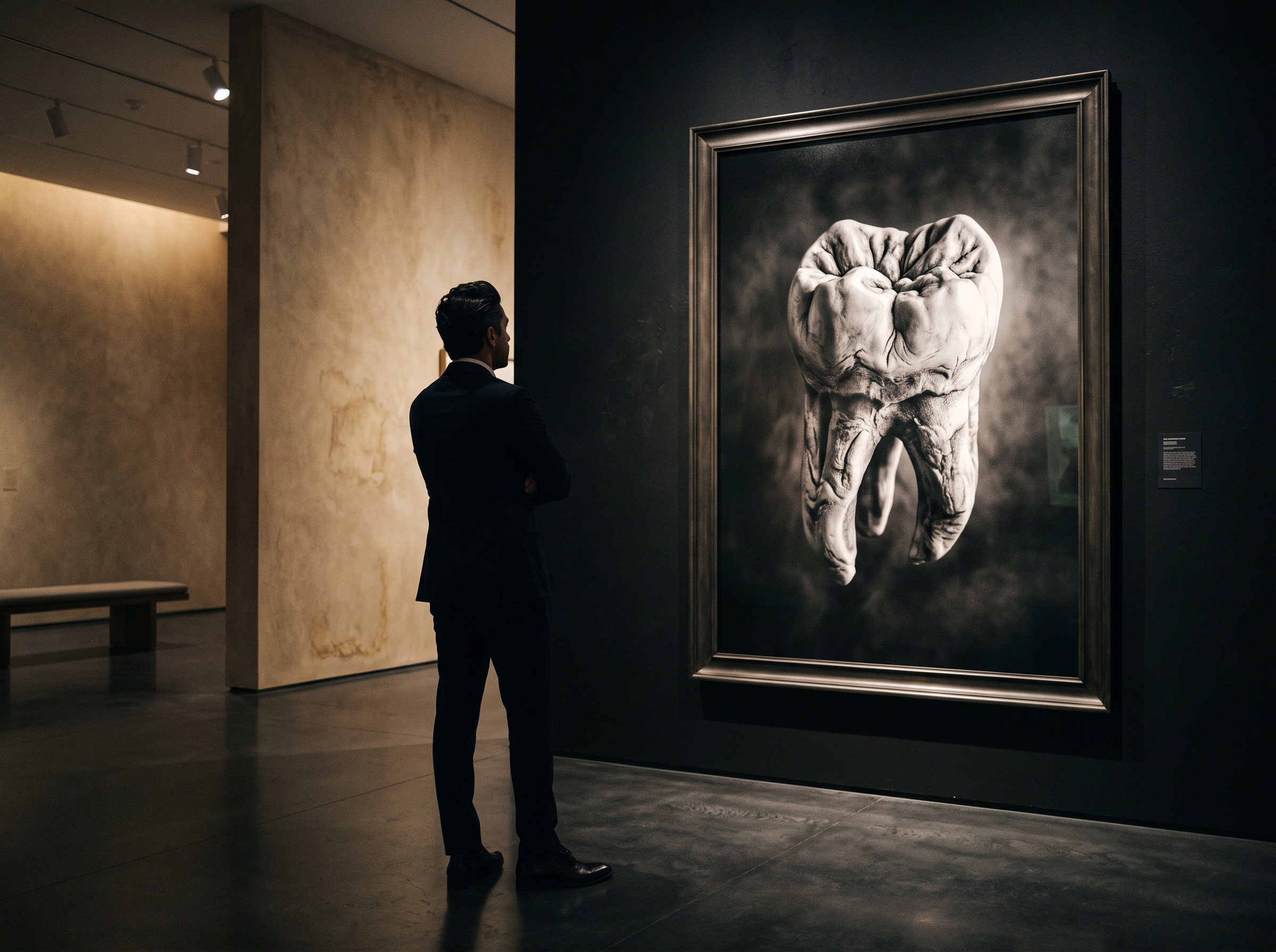
There is an aesthetic dividend here too, and it is not incidental to the craft. Lit this way, a tooth stops looking like a clinical specimen and starts looking like what it is: a small sculpture, worn and shaped by use, bearing a surface as expressive as weathered stone or an old bronze. The art conservators understood long ago that raking light does not only diagnose; it reveals a kind of beauty that even illumination politely hides — the evidence of time and force written into a surface. A dental image made under grazing light carries that same double charge. It is a document a specialist can read and an object that rewards simply looking, the growth waves and wear facets composing themselves into a landscape. That is the sensibility this gallery was built around: that the most honest clinical image and the most arresting one are frequently the same photograph, and that a borrowed trick from another discipline can give us both at once.
Future Developments
Raking light is the analog ancestor of a family of surface-mapping techniques now arriving in dentistry by other means. Intraoral scanners already capture the tooth’s geometry as a dense three-dimensional mesh, and from that mesh a surface can be re-lit from any virtual angle after the fact — a software raking light, consistent to the degree, that could make longitudinal wear comparison fully quantitative rather than visual. Photometric and photogrammetric methods borrowed, once again, from the art-conservation and cultural-heritage world — reflectance transformation imaging among them, where dozens of exposures under light from many directions are fused into a single relightable record — point toward a day when a clinician will capture a tooth once and interrogate its relief endlessly afterward. Until that is routine, the single lamp held low remains the fastest, cheapest, and most quietly revealing instrument we have for seeing a surface as it truly is. The conservators have known it for a century. It is past time the operatory did too.
No Comments