Logo Logo Logo Logo Logo
  • Home
  • Download
  • About
  • Blog
  • Home
  • Download
  • About
  • Blog
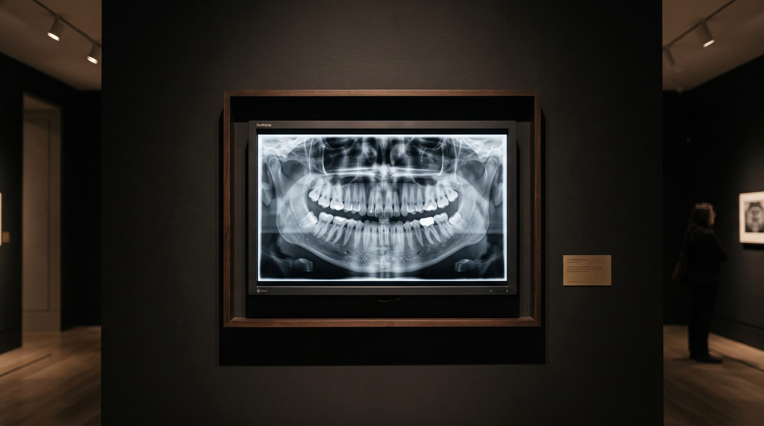
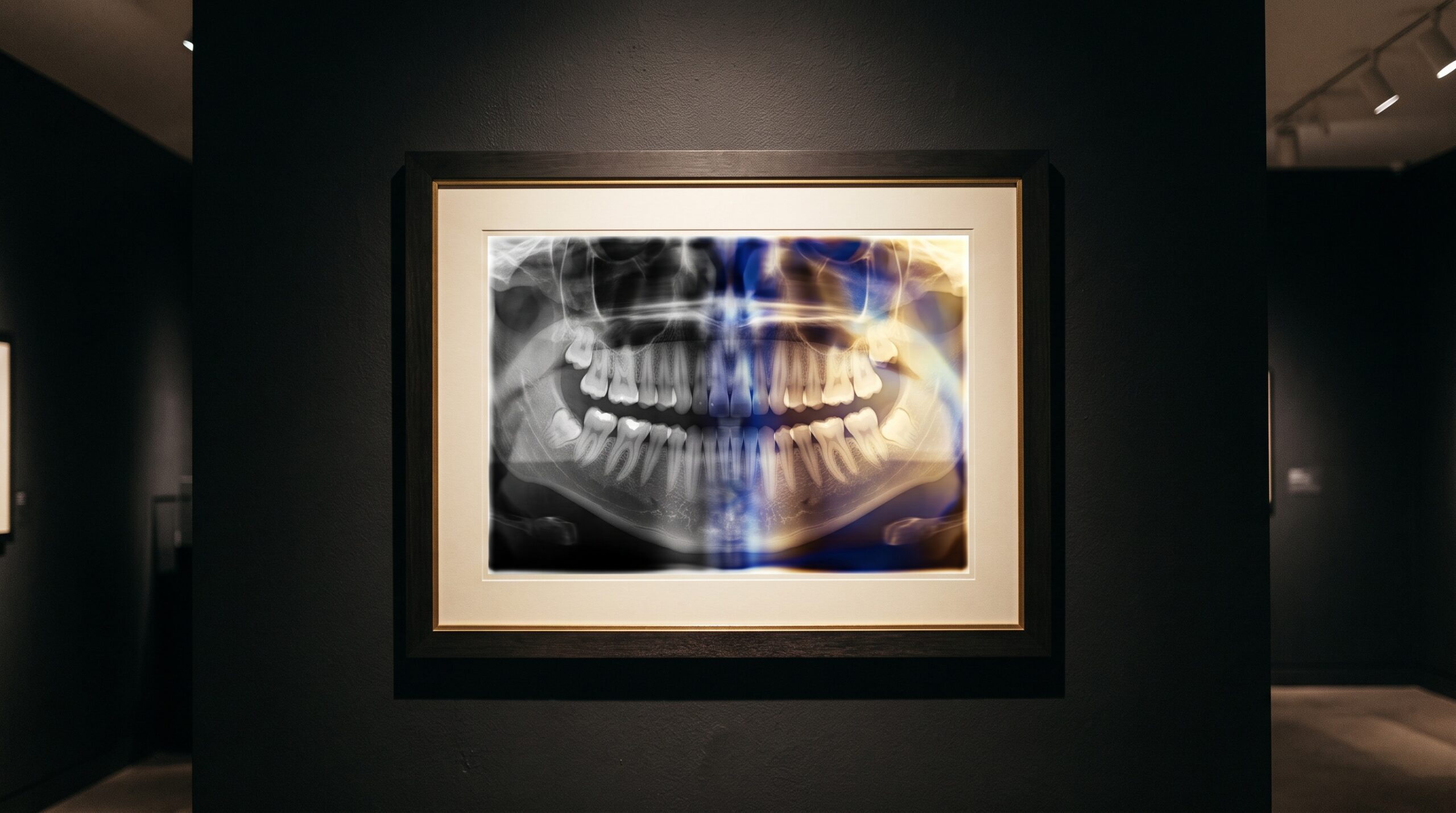
A diagnostic display monitor shown as framed gallery art, its screen holding a dental radiograph in a smooth ramp of grays

17 Aug The Last Instrument: Diagnostic Display Calibration and the DICOM Grayscale Standard for Reading a Radiograph

Posted at 11:38h in Dental Imaging by Raymond 0 Comments
0 Likes

Every radiograph ends its journey on a screen, and the screen is the one instrument in the imaging chain that most practices never calibrate. The DICOM grayscale standard exists because the human eye reads brightness unevenly — and because a monitor that does not account...

Read More
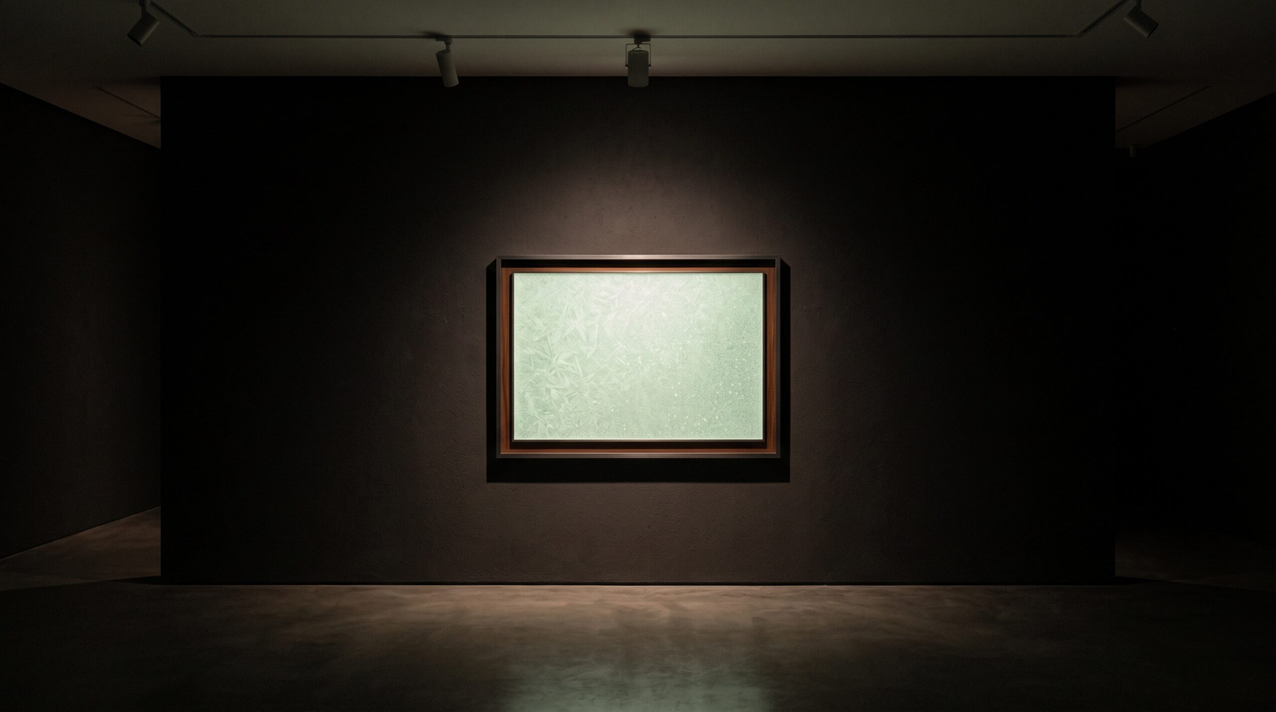
A thin scintillator screen framed as gallery art, faintly glowing with soft internal light

15 Aug The Needle Forest: How Columnar Cesium Iodide and the Scintillator Decide What a Sensor Can See

Posted at 10:37h in Dental Imaging by Raymond 0 Comments
0 Likes

A digital dental sensor does not, in most cases, feel the X-ray at all. It watches a thin crystalline screen flash with light and reads that instead. The material of that screen — whether a forest of light-piping cesium iodide needles or a settled bed...

Read More
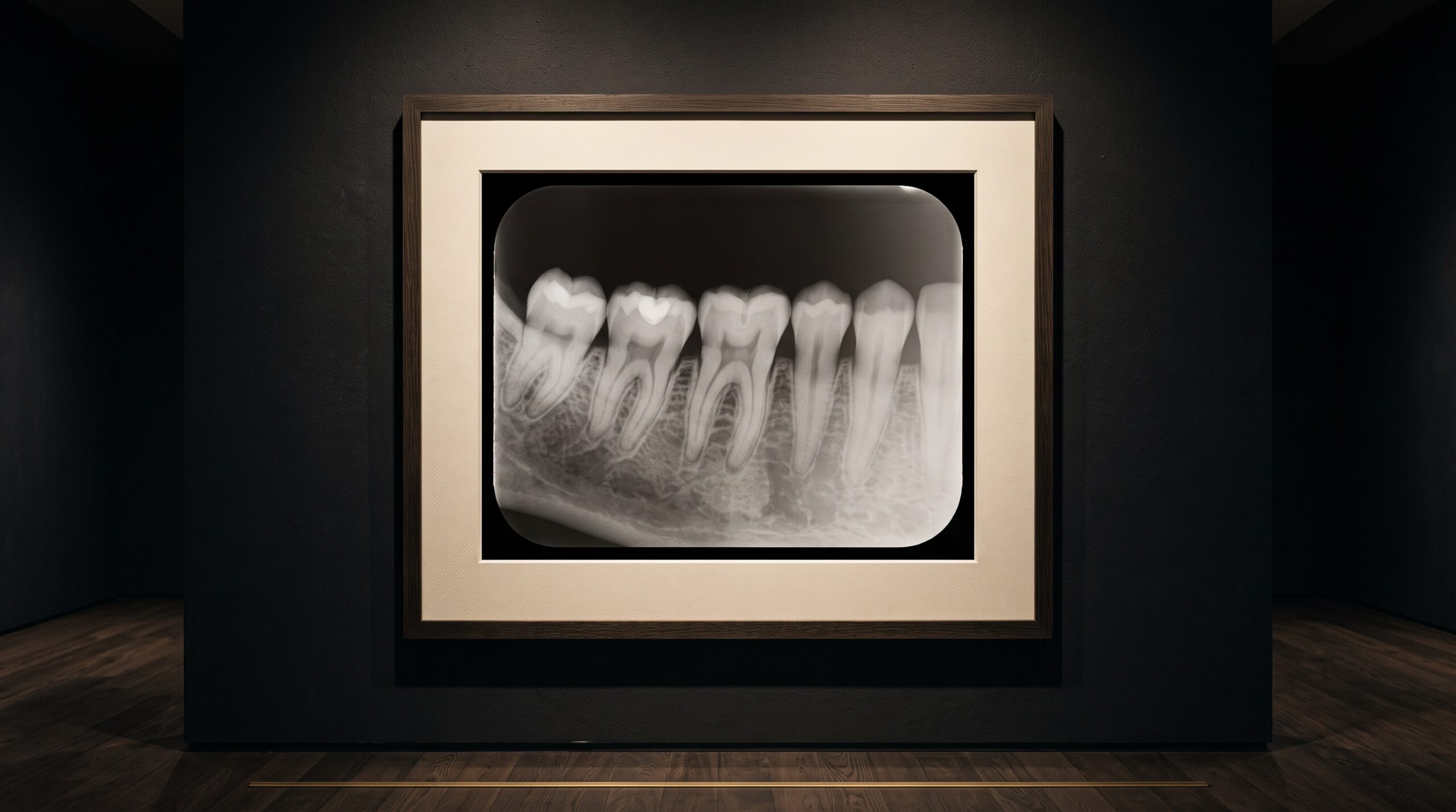
A framed dental radiograph under gallery lighting showing a smooth density gradient from one edge to the other

14 Aug The Anode Heel Effect: The Invisible Gradient Shading Every Radiograph

Posted at 10:38h in Dental Imaging by Raymond 0 Comments
0 Likes

Every X-ray beam leaves the tube already uneven — brighter toward the cathode, dimmer toward the anode. The anode heel effect is the quiet gradient built into the physics of the source itself, and it shades the density of an image before the anatomy is...

Read More
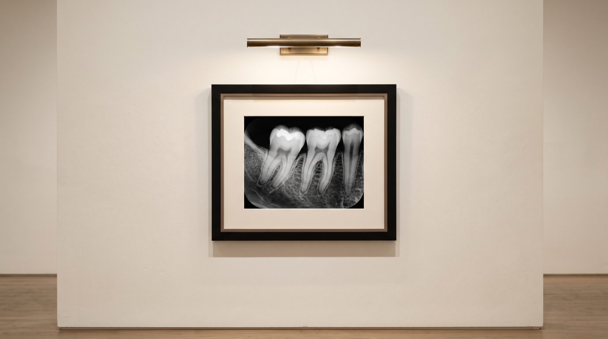
A clean, low-noise dental radiograph presented as framed fine art on a gallery wall

06 Aug Detective Quantum Efficiency: The True Measure of a Dental Sensor, Beyond Resolution

Posted at 10:12h in Dental Imaging by Raymond 0 Comments
0 Likes

Every specification sheet leads with resolution — line pairs per millimetre, the sensor's finest possible line. But two sensors can share a resolution and be worlds apart at the dose you actually use. The number that separates them is detective quantum efficiency, and it is...

Read More
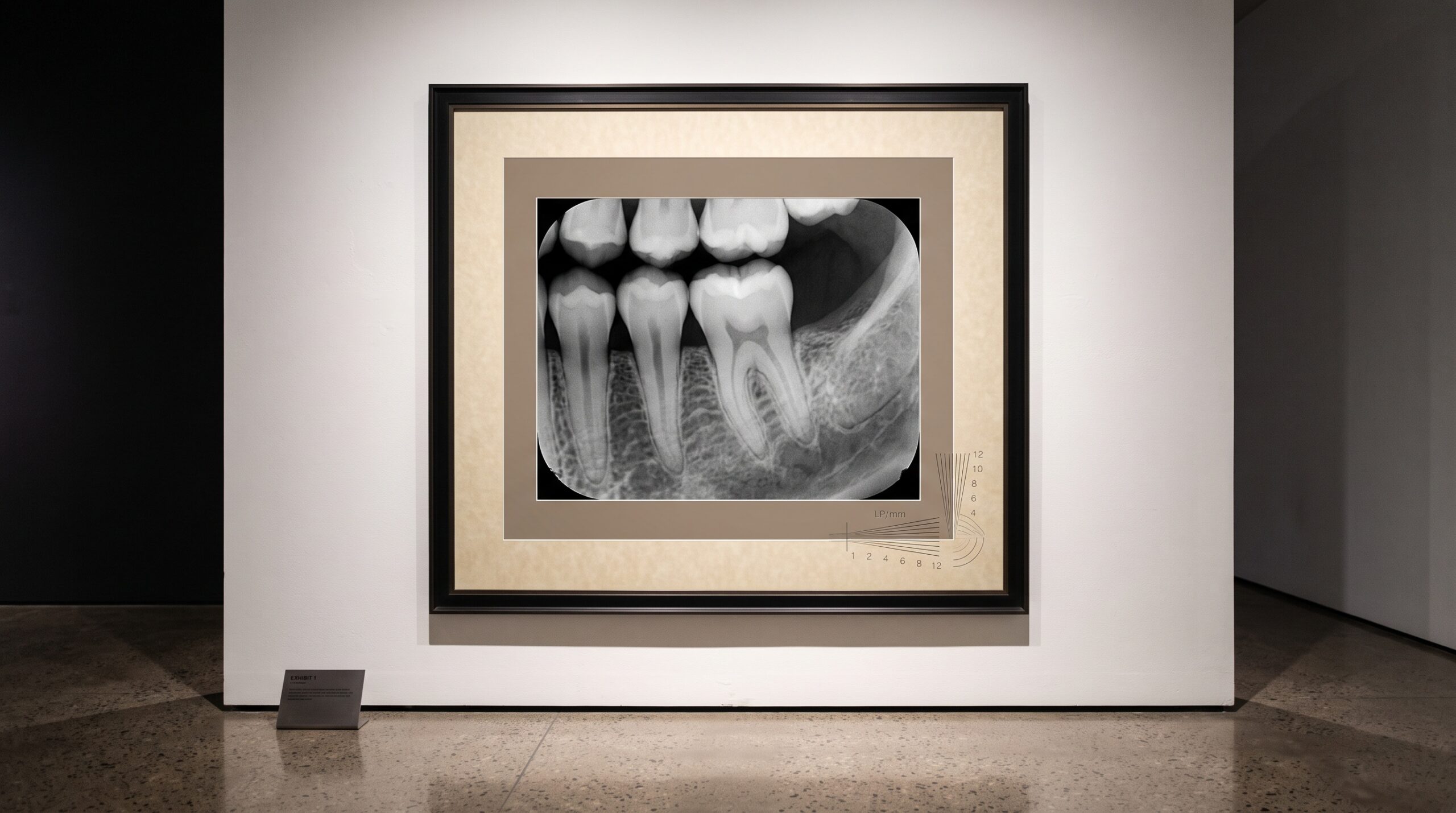
A framed dental radiograph on a gallery wall overlaid with a faint converging line-pair resolution pattern

03 Aug How Sharp Can a Dental Image Truly Be? Pixel Pitch, Line Pairs, and the Limits of Resolution

Posted at 00:39h in Dental Imaging by Raymond 0 Comments
0 Likes

Sharpness in a dental image is not a feeling but a measurable ceiling. A tour of pixel pitch, line pairs per millimeter, and the modulation transfer function that quietly decides what your radiograph can and cannot show....

Read More
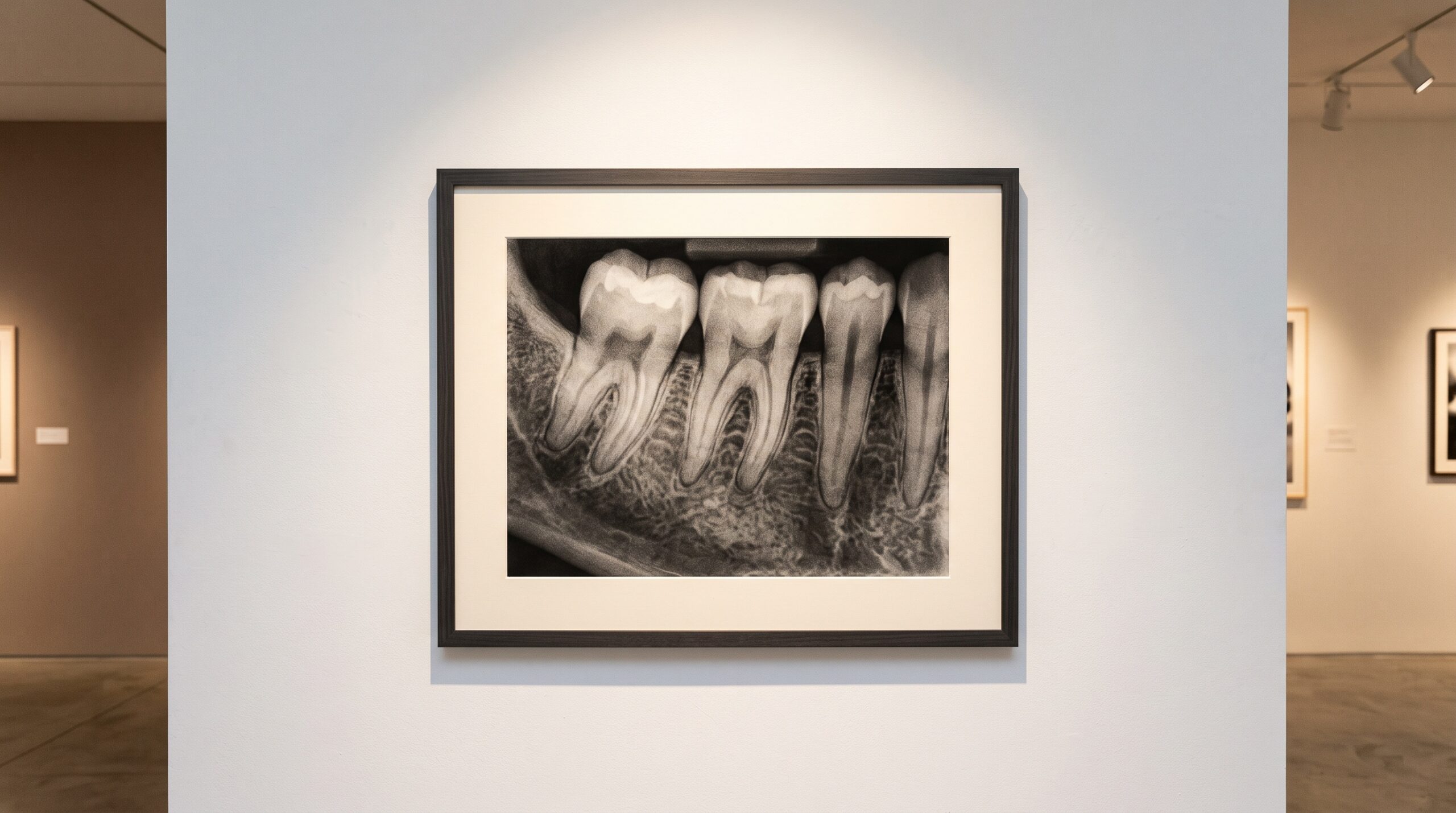
A dental radiograph presented as framed fine art on a white gallery wall under museum lighting

28 Jul Contrast and Density: The Grayscale Craft Behind a Diagnostic Radiograph

Posted at 17:39h in Dental Imaging by Raymond 0 Comments
0 Likes

A radiograph is a painting in gray. Understanding contrast, density and the tonal scale is the quiet craft that separates a diagnostic image from a merely visible one....

Read More
A single dental radiograph framed and lit like a work of art on a gallery wall

20 Jul Before the Patient Stands Up: Judging a Radiograph at the Moment of Capture

Posted at 14:05h in Dental Imaging by Raymond 0 Comments
0 Likes

A flawed radiograph discovered at read-out is a retake that rarely happens. Software that grades technical quality while the sensor is still in the mouth changes the economics of getting it right the first time....

Read More
A framed dental periapical radiograph on a gallery wall beside a glowing panel appraising it with a faint scoring overlay

09 Feb When the Machine Becomes the Critic: Deep Learning Now Grades the Radiograph Itself

Posted at 10:12h in Dental Imaging by Raymond 0 Comments
0 Likes

A neural network can now look at a periapical radiograph and tell you, in the instant it is captured, whether it is diagnostic. What it means when the image acquires its own critic....

Read More
A dental radiograph presented as framed fine art on a gallery wall, its background deep true-black

08 Feb The Photon-Counting Detector: A New Physics Beneath the Radiograph

Posted at 10:12h in Dental Imaging by Raymond 0 Comments
0 Likes

For a hundred years the dental radiograph has been an average — a blur of charge summed across an exposure. The photon-counting detector refuses to average. It counts each X-ray individually, and in doing so quietly rewrites what a diagnostic image can be....

Read More
A dimensionally faithful dental periapical radiograph presented as framed fine art on a gallery wall

07 Feb The Geometry of an Honest Radiograph: Paralleling, the Bisecting Angle, and the Truth About Length

Posted at 10:12h in Dental Imaging by Raymond 0 Comments
0 Likes

A dental radiograph looks like a photograph, but it is really a measurement — a shadow cast by an X-ray beam onto a flat receptor. Whether that shadow reports a root's true length or a convincing lie depends almost entirely on geometry, and on the...

Read More
  • 1
  • 2
  • 3
© Copyright 2020, Raster Builders Inc. All Rights Reserved.