


September 28, 2026 The Weight of the Instrument: The Featherweight Scanner and the Craft of a Scan the Patient Barely Notices
For most of the intraoral scanner’s short history, the argument was about accuracy. Trueness and precision, margin fidelity, how faithfully the point cloud stitched a full arch together — these were the numbers that sold the instrument and filled the literature. They still matter. But a quieter revolution has moved to a different part of the story: not the sensor at the tip, but the mass in the hand. The newest wands weigh about as much as a fountain pen, and that single fact is reshaping both the patient’s experience of a scan and the quality of the image that comes out of it.
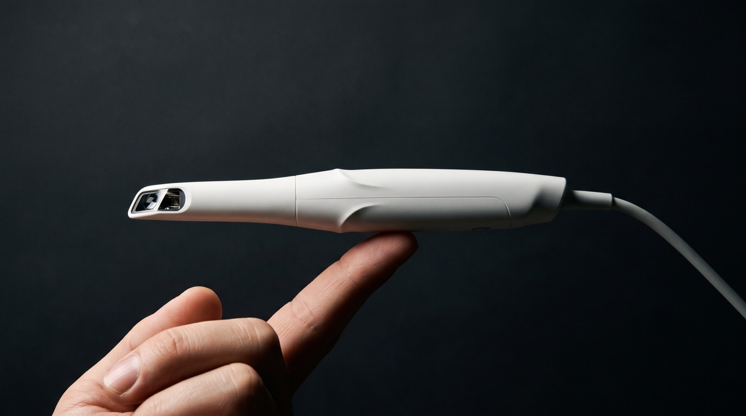
The signal is easy to read in the market. When SHINING 3D introduced the Aoralscan Elf, the headline was not its optics but its heft — roughly 106 grams, among the lightest wands available, connected by a single cable with no bulky power box tethering it to a cart. Competitors are racing down the same curve, and new entrants are staking their pitch on ergonomics rather than raw resolution. The instrument is disappearing into the hand, and an instrument you barely feel is one you can carry with a craftsman’s steadiness.
Why grams became the headline
An intraoral scan is a sustained act of fine motor control. The clinician holds the wand at the end of a braced arm and glides it through a warm, wet, curved, softly moving space, keeping the optical window at a near-constant distance from tissue while the software knits thousands of frames into one surface. Do that for ninety seconds with a heavy wand and a stiff cable and the forearm fatigues, the hand develops a fine tremor, and the path through the arch grows less even. Every one of those faults is written into the data as noise, drift, or a gap the software must guess across.
Weight, then, is not a comfort feature bolted onto a clinical tool — it is a determinant of image quality. A lighter, better-balanced wand lets the hand make the small, continuous, overlapping passes that clean scans are built from. The center of mass sits closer to the fingertips, the way a good pen balances, so the wrist steers instead of the shoulder. Shave the mass and stiffen nothing, and the same clinician produces a smoother, more complete surface simply because the instrument stopped fighting back.
The patient on the other end of the wand
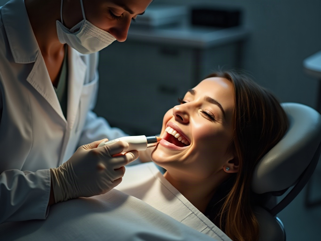
The reduction in weight is felt just as sharply by the person in the chair, and that is where the aesthetic and the clinical meet. A bulky wand held for a long capture forces the operator to lever against lips and cheeks, to rest the instrument’s weight where it should not rest, and to work slowly enough that the gag reflex has time to assert itself. A featherweight wand moves in smaller, gentler arcs, spends less time in the mouth because the scan finishes faster, and asks far less of the patient’s patience and jaw.
This matters most exactly where scanning is hardest to tolerate: children, anxious patients, strong gag reflexes, limited opening. A calmer capture is not merely kinder; it is more accurate, because a patient who is not flinching, swallowing, or repositioning holds the soft tissue still and lets the wand keep its rhythm. The comfort and the fidelity are the same thing viewed from two ends of the instrument. It is the same logic that makes an intraoral scan gentle enough to use for spotting early caries in children where a mouthful of hardware once made the exam a fight.
Everything else that had to shrink
A 106-gram wand is only the visible tip of a longer engineering story, because the weight could not fall until everything feeding it did. Older systems tethered the wand to a rolling cart and a separate power box, the cable itself adding drag and mass at the hand. The new generation collapses that: a single slim USB-C connection, the processing pushed into a laptop or a compact unit, no calibration ritual before each session, tips that heat quickly to keep the optics from fogging in the humid field.
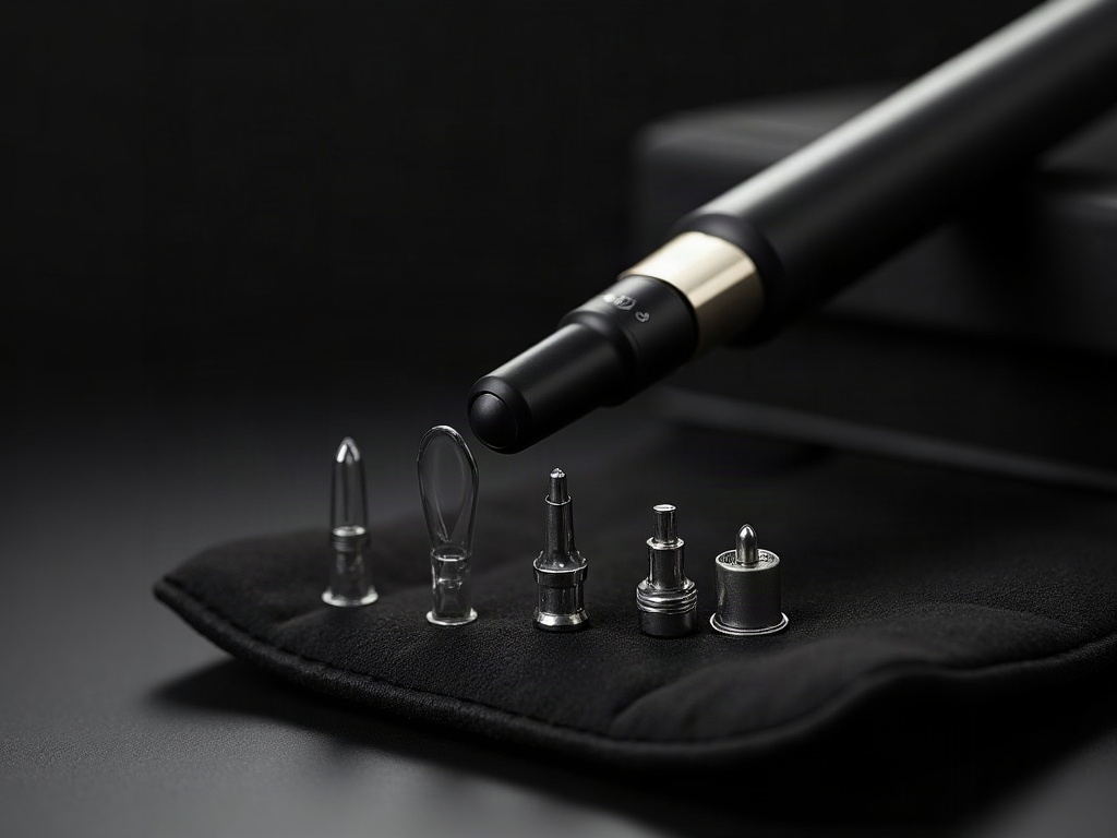
The tip strategy is part of the ergonomic argument too. A family of interchangeable tips — a smaller profile for pediatric or crowded mouths, a standard one for routine work — lets a single lightweight body serve every patient without compromise. And the optics have grown forgiving enough that the wand no longer has to hover at one exact distance; it tolerates the natural variation of a relaxed hand, which is the whole point of making the hand relaxed in the first place. Reduce the ceremony around the instrument and you reduce the ways a capture can go wrong.
The craft of a steady, unhurried pass
A lighter wand rewards technique rather than replacing it. The fundamentals of a clean digital impression are unchanged — they simply become easier to honor when the instrument cooperates:
- A consistent scanning path. Occlusal, then lingual, then buccal, in a deliberate unbroken sweep. A featherweight wand makes the return passes less tiring, so the path stays disciplined to the end of the arch instead of degrading as the arm tires.
- Constant distance and gentle overlap. The software stitches on shared geometry; keep the tip an even hover from the tissue and let each pass overlap the last. A balanced wand holds that distance almost on its own.
- A dry, retracted, motionless field. Saliva pools and moving tissue are still the enemy. Lightness buys speed, and speed buys a capture completed before the field floods again.
- Respect for the soft tissue. A light wand does not need to shove a cheek aside; retract deliberately with a mirror or retractor so the instrument records anatomy instead of deforming it.
Done well, the reward is a model clean enough to carry the whole downstream workflow — the kind of registration that lets a clinician fuse the surface scan with a radiograph into a single coherent picture, or feed a photogrammetric check of full-arch implant fit. A scan is only as trustworthy as the hand that carried the wand, and a wand you cannot feel is one the hand can carry perfectly.
Reading the finished surface
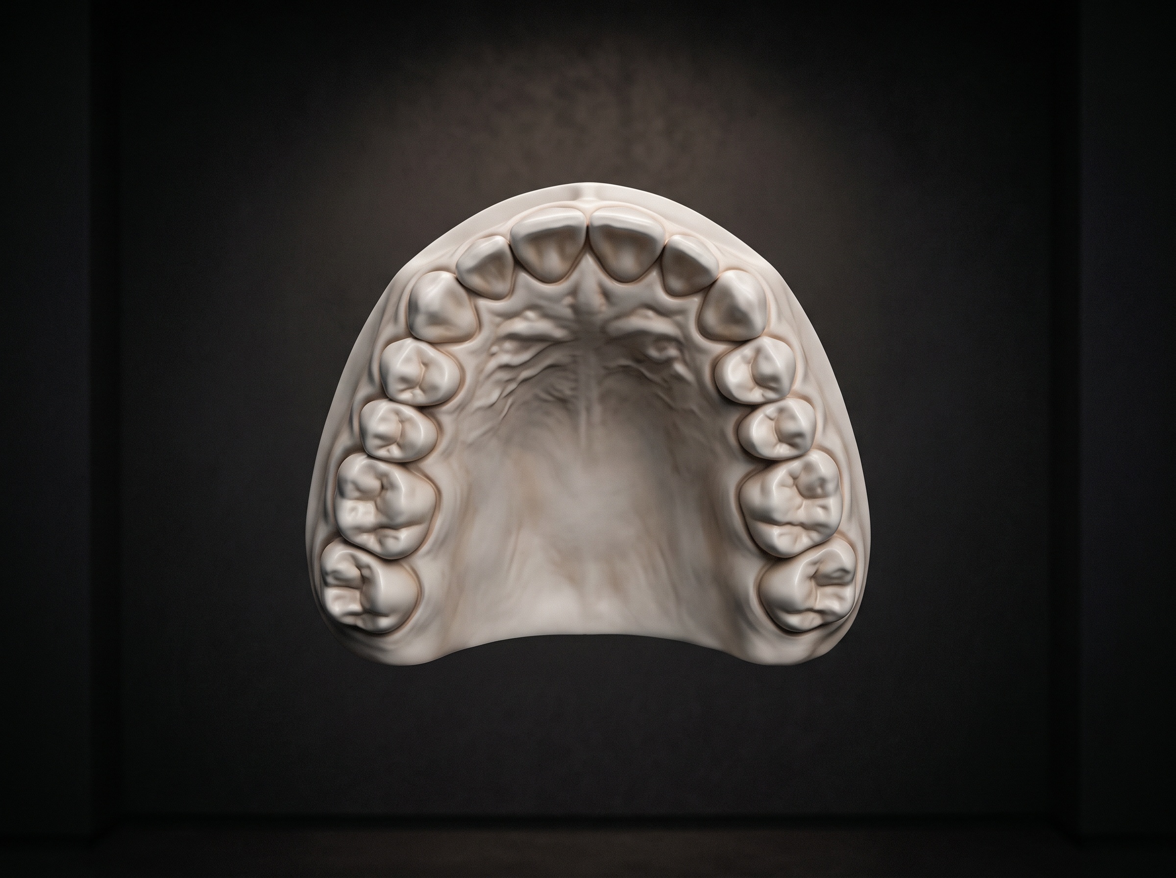
The proof of an ergonomic gain is in the model on the screen, and it rewards an honest look. A scan produced with a steady, unhurried pass shows continuous, watertight surfaces, crisp margins on a prepared tooth, and true morphology in the occlusal detail. A scan fought out with a heavy wand and a fatigued hand betrays itself the same way every time: holes where the path skipped, softened cusps where frames failed to register, a subtle warp across a long span where drift accumulated pass over pass.
None of that means the lighter instrument is doing the clinician’s thinking. It means the instrument has stopped adding its own errors to the reader’s. The wand’s job is to remove friction between intention and image; the judgment of what makes a margin acceptable, when a span needs rescanning, and whether the model is fit to send to the laboratory remains entirely human. A featherweight scanner simply makes the well-made scan the path of least resistance.
Future developments
The weight will keep falling, but the more interesting frontier is what the freed-up hand and processor now permit. On-board intelligence is already flagging under-scanned regions in real time, guiding a bite alignment, and cleaning the mesh as it forms; as that assistance matures, the wand becomes less a passive camera and more a collaborator that tells the clinician where the picture is still thin. Expect wireless bodies that shed even the last cable, tips that adapt their field of view to the anatomy in front of them, and capture that folds color, caries detection, and soft-tissue assessment into the same effortless sweep. The destination is a scan so light, so fast, and so forgiving that the instrument all but vanishes — and the entire craft collapses back to where it belongs: a steady hand, a still patient, and a true picture of the mouth. The lighter the instrument grows, the more the image becomes purely a matter of intention.
Sources & further reading:
Sorry, the comment form is closed at this time.