


September 6, 2026 Triangulating the Arch: How Photogrammetry Solves the Full-Mouth Implant Impression
There is a threshold in dentistry where the finest surface scan quietly stops being good enough. For a single crown, an intraoral scanner traces the prepared tooth and its neighbours in exquisite detail, and the restoration drops into place. For a full arch rebuilt on four, six, or more implants, the same instrument faces a harder geometry – and a more unforgiving master. A multi-implant framework is rigid. It cannot flex to accommodate a small error the way a natural tooth’s ligament forgives a tight contact. Every implant must be recorded not merely well, but in exact three-dimensional relation to every other, across the whole sweep of the jaw. Miss by a little at one end and the bridge that comes back from the laboratory will rock, strain the screws, or simply refuse to seat. The craft here is not drawing the mouth beautifully. It is measuring a handful of points in space with something close to perfection.
This is the problem photogrammetry was built to solve, and it solves it by refusing to do what every other imaging method in the operatory does. It does not capture a surface at all.
The Tyranny of the Passive Fit
A full-arch implant prosthesis lives or dies by a single property the profession calls passive fit: the finished framework should meet all of its implants simultaneously, seating fully onto each one with no pre-load, no tension, no gap closed only by tightening a screw. A framework that fits passively distributes chewing forces evenly and leaves the bone and the components in peace. One that does not is a slow source of trouble – screw loosening, component fatigue, marginal bone loss, and in the worst case fracture of the prosthesis or the implant itself. The tolerance is measured in tens of microns, and it applies across an arch that may span the better part of ten centimetres.
That span is exactly where surface scanning struggles. An intraoral scanner builds its model by capturing overlapping patches and stitching them together, each aligned to the last. As we described in Structured Light and the Stitched Mesh, this is a remarkable feat of real-time registration – but every join carries a tiny alignment error, and across a long edentulous arch with few landmarks and large smooth expanses of soft tissue, those errors accumulate. The mesh grows subtly less faithful the farther it travels from where the scan began. On a short span it is invisible. On a full arch it can drift past the tolerance the prosthesis demands.
Measuring Points, Not Surfaces
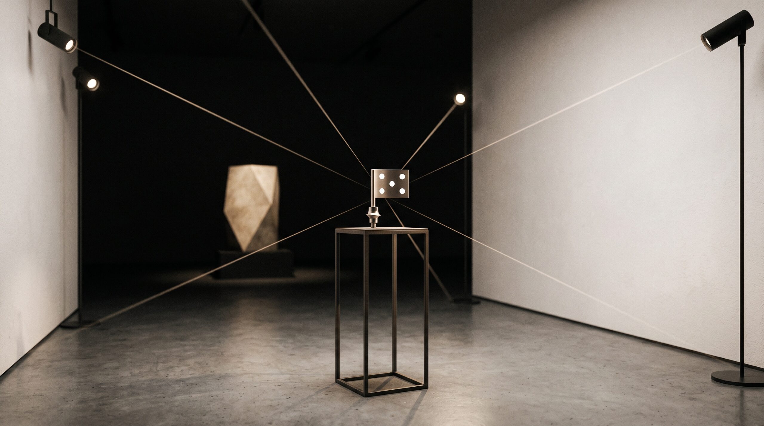
Photogrammetry sidesteps the accumulation problem by discarding almost everything a scanner works so hard to collect. Onto each implant the clinician threads a photogrammetric marker – often called a flag – a small plate carrying a pattern of coded reference dots. A specialized stereo camera then photographs these flags from many positions in quick succession. It ignores the gums, the tongue, the lips, the contours of the ridge. It cares only about the bright, unambiguous markers and where they sit.
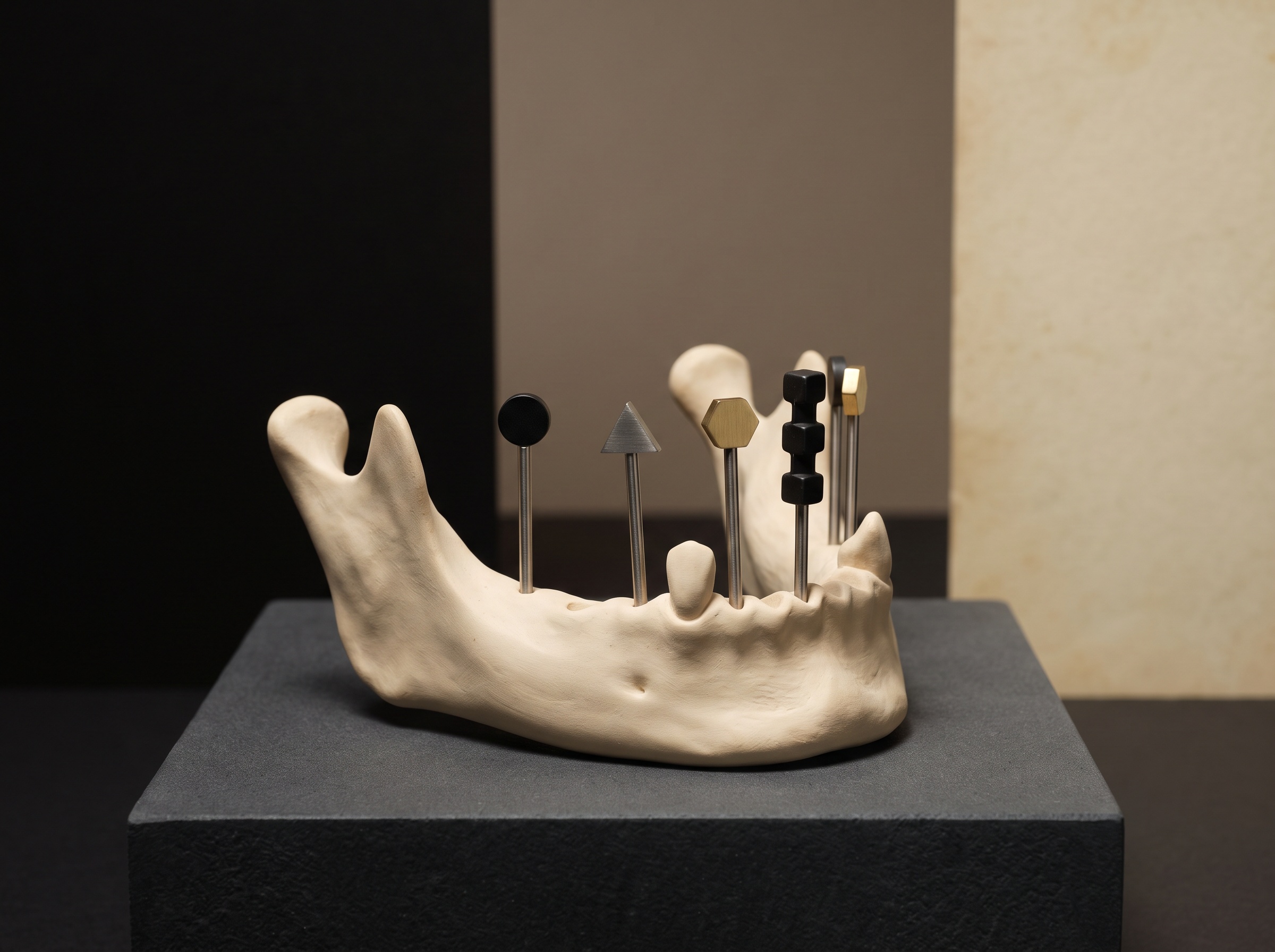
From dozens of images taken at different angles, software reconstructs the precise position and orientation of every marker – and therefore every implant – by triangulation. This is the same geometric principle that lets two eyes judge depth, that surveyors use to fix a distant peak, and that satellites use to place a receiver on the ground: a point seen from enough known viewpoints has exactly one location in space consistent with all of them. Because the markers are sparse, high-contrast, and designed to be found without ambiguity, the reconstruction is not weighed down by the smooth, feature-poor surfaces that trip up a surface scan. The reported numbers are striking. A recent clinical study in the prosthodontic literature places the linear precision of the method on the order of ten to fifteen microns and its angular precision at a small fraction of a degree – accuracy that holds across the full arch rather than decaying along it. It is measurement, not depiction.
What the Camera Deliberately Cannot See
The strength of photogrammetry is also, honestly, its limit. It returns implant coordinates and nothing else – no soft-tissue emergence profile, no shape of the ridge, no adjacent teeth, no bite. A prosthesis cannot be designed from a constellation of points alone; it needs the restorative context. So in practice the accurate skeleton captured by photogrammetry is combined with a conventional intraoral scan or a model that supplies the tissue and contour information, and the two data sets are merged – the implant positions taken as truth, the surface draped around them. The photogrammetric record becomes the rigid coordinate frame; the scan provides everything the human eye and the ceramist still need to see.
Those merged positions then have to travel. The coordinate file, the associated scan, and the design must move cleanly from operatory to laboratory to milling machine without loss or silent transformation – the same interoperability discipline we examined in The Image That Cannot Travel. Micron-accurate capture is wasted if the data degrades on the way to the mill. And upstream of all of it sits the plan itself: the implant positions were chosen on a three-dimensional radiographic study long before any flag was threaded, part of the same move toward reading the arch as a navigable volume that we explored in AI on CBCT volumes. Photogrammetry does not replace that planning imaging; it verifies, in the mouth, where the surgery actually placed what the plan proposed.
Three Ways to Record an Arch
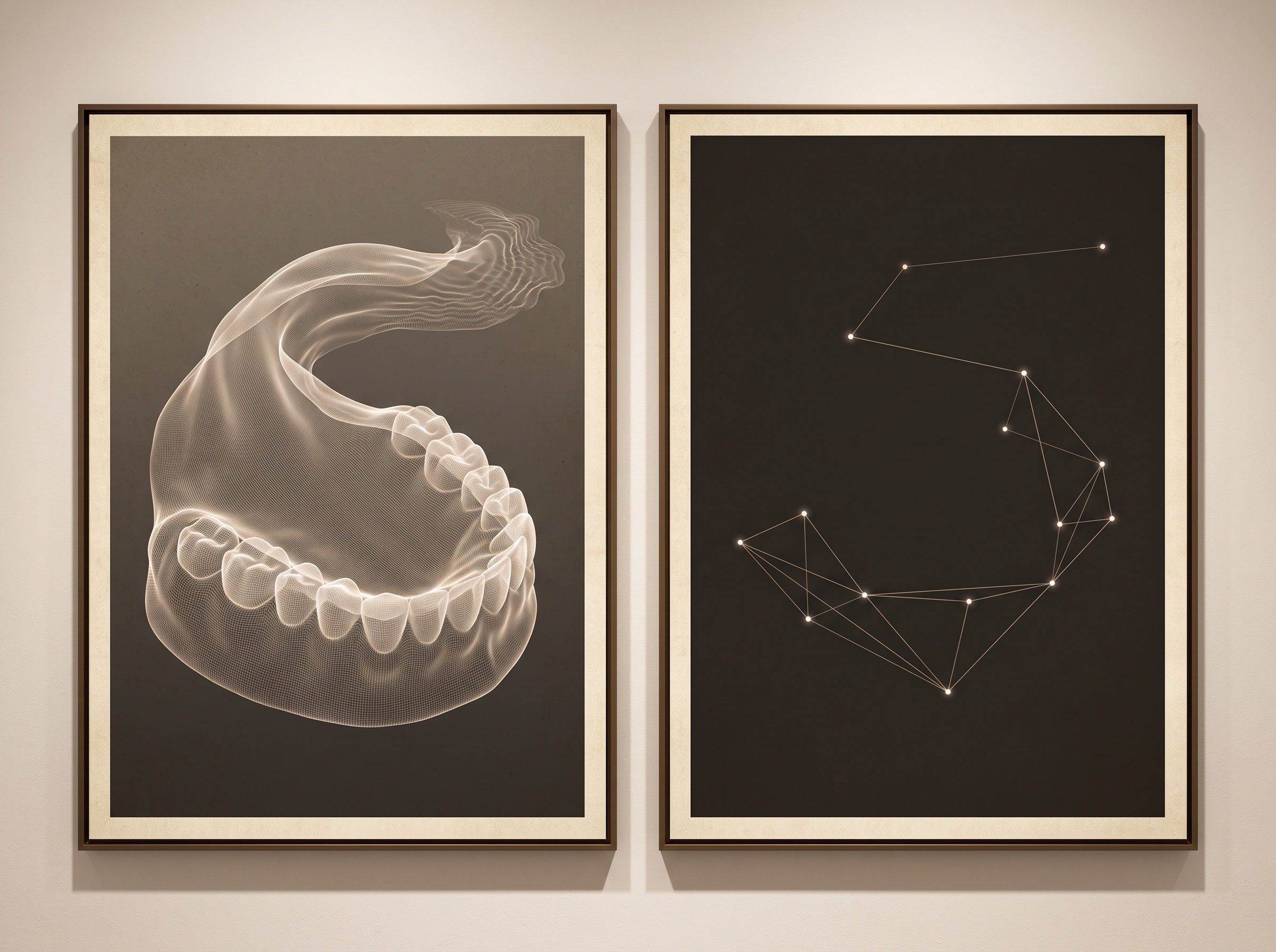
It helps to see the methods as complementary rather than rival. The traditional open-tray impression – splinting the implant copings in rigid material and pouring a physical model – remains a proven benchmark, but it is slow, materials-sensitive, uncomfortable, and analog to its core. Intraoral scanning is fast, patient-friendly, and rich in surface and soft-tissue detail, and for single units and short spans it is superb; its weakness is precisely the long, landmark-poor full arch. Photogrammetry is narrow and specialized – it does one thing – but that one thing is the hardest and most consequential part of the full-arch problem, and it does it with an accuracy that does not fade with distance. The mature workflow does not ask which method wins. It asks each to contribute what it captures best.
Future Developments
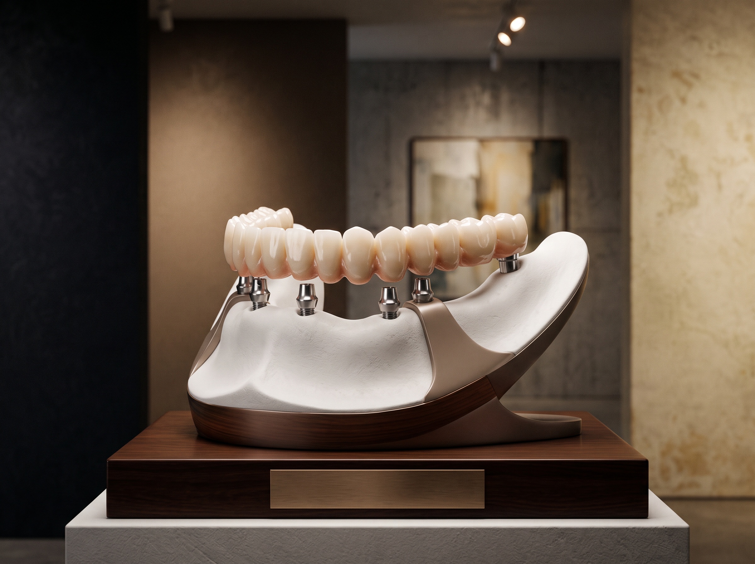
The trajectory is toward convergence. The dividing line today – photogrammetry for implant coordinates, surface scanning for tissue and contour, CBCT for the underlying anatomy – reflects the current limits of each sensor, not a law of nature. As cameras grow faster and reconstruction smarter, it is reasonable to expect a single capture episode that returns the accurate implant frame and the surrounding surface together, without the seam of merging two data sets by hand. The deeper lesson photogrammetry teaches is one worth carrying beyond implants: that accuracy and richness are different virtues, that the most faithful record of a thing is sometimes the one that captures the least, and that knowing exactly what to measure – and what to ignore – is itself a form of imaging artistry. For the patient, the reward is quiet and physical: an arch of teeth that seats the first time, meets the bone without strain, and is meant to last. The craft that delivers it began not with a beautiful picture of the mouth, but with a few bright points, measured to the micron, from every angle at once.
Sources & further reading:
Sorry, the comment form is closed at this time.