


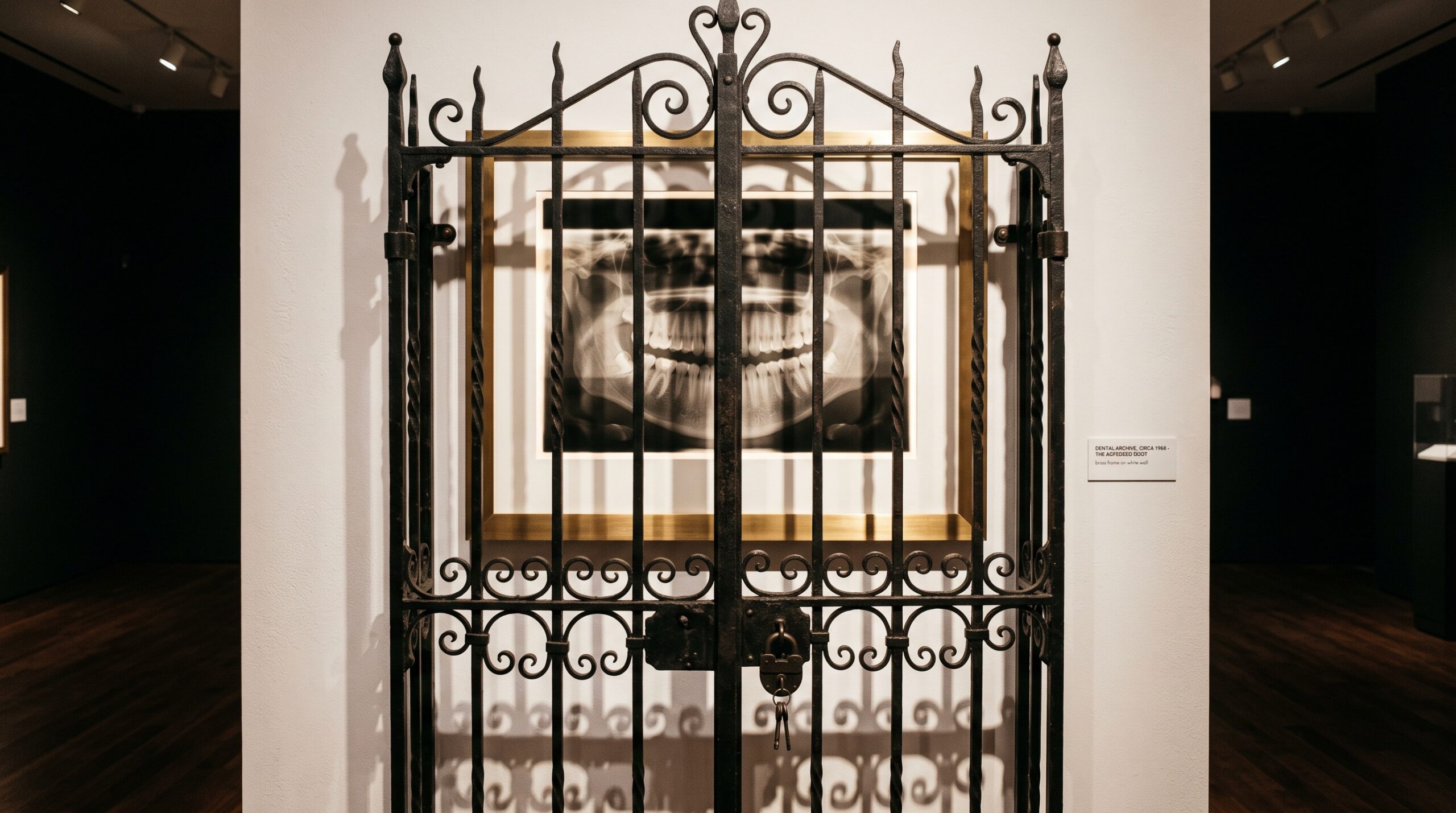
August 31, 2026 The Image That Cannot Travel: DICOM, Interoperability, and the Quiet Cost of a Radiograph That Will Not Move
Every dental image is made to be looked at twice. The first reading happens where it was captured – the operatory, the reviewing monitor a few steps away, the familiar software that knows exactly how to render it. The second reading is the harder one. It happens somewhere else: on the specialist’s screen across town, in the insurer’s review queue, on the laptop of a new dentist inheriting a patient’s history. And it is at that second reading that the beautiful, expensive, radiation-costed image so often fails – not because it was poorly made, but because it could not travel.
This is the quiet problem beneath the glossy surface of digital dentistry. We have spent two decades perfecting how a radiograph is captured and displayed. We have spent far less energy on the unglamorous question of what happens when it has to leave the building. In March 2026 the American Dental Association put the issue on the record, calling for improved interoperability standards and warning that the lack of them can degrade image quality, strip essential metadata, and lead to repeat imaging, added administrative burden, and unnecessary radiation exposure. That is a remarkable sentence. It says, in effect, that a data-handling failure becomes a dose of radiation delivered to a real patient.
The Radiograph That Reaches a Locked Door
Picture the ordinary journey. A general practice takes a clean periapical series, refers the patient to an endodontist, and sends the images along. In the best case they arrive as true diagnostic files the receiving software opens natively, windows correctly, and measures against. In the common case they arrive as a screenshot pasted into an email, a low-resolution JPEG, or a proprietary file that only the sending practice’s software can open. The endodontist, unable to trust a flattened copy, does the only defensible thing: retakes the image. The patient is exposed again, the schedule slips, and a perfectly good radiograph sits uselessly on a server twelve miles away.
The failure is rarely dramatic. No file is corrupted, no system crashes. The image simply arrives in a form that has quietly shed the very things that made it diagnostic – its full bit depth, its calibration, its scale. Interoperability is the discipline of making sure that does not happen: that an image made anywhere can be read, correctly and completely, everywhere.
What DICOM Was Meant to Do
The answer the profession settled on has a name: DICOM, the same Digital Imaging and Communications in Medicine standard that lets a hospital’s CT scanner, PACS archive, and reading workstation – often from three different manufacturers – speak one language. In 2000 the ADA resolved to pursue interoperability of digital dental images using DICOM as the backbone, and the standard has been extended to intraoral radiographs, panoramics, and cone-beam volumes alike.
DICOM’s ambition is larger than a file format. It specifies not just how the pixels are encoded but how they are labeled, transmitted, and requested from one system to another. Done fully, it means a radiograph carries its identity and its instructions with it, so that a receiving system does not have to guess how bright to make it or how many millimeters a pixel represents. The gap between that promise and daily reality is exactly the gap the ADA is now pressing to close.
The Quiet Damage of a Bad Export
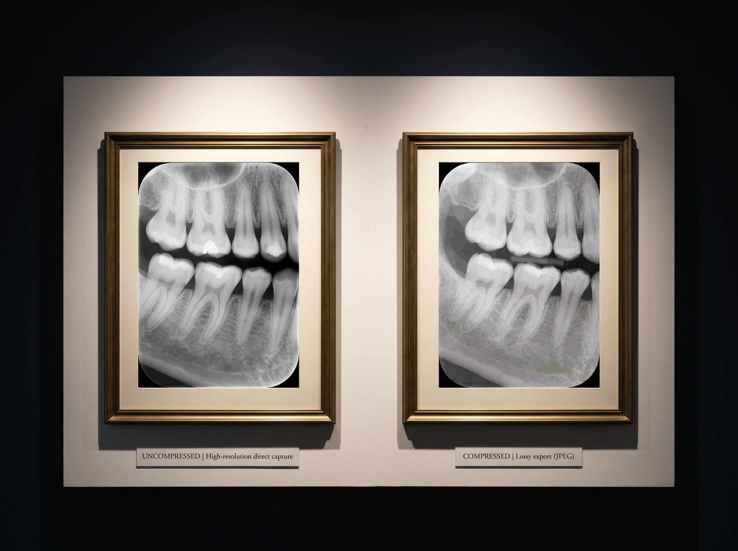
When an image is exported the wrong way, the loss is easy to miss because the picture still looks like a picture. A 14-bit sensor capture, flattened to an 8-bit JPEG for the convenience of email, has thrown away the deep reservoir of tonal information a clinician relies on to separate early decay from sound enamel. Re-compress it a second time and subtle banding creeps into the gradients. Take a screenshot instead of exporting the file, and the image inherits whatever contrast curve happened to be on the sending monitor – a cosmetic choice frozen into what is supposed to be objective data.
None of this announces itself. The receiving dentist sees a plausible radiograph and, if they are careful, distrusts it; if they are rushed, they read a degraded copy as though it were the original. This is the same fragility we have written about in the long survival of the archived image – only here the danger is not the slow erosion of years but the instant, invisible damage of a single careless transfer.
The Metadata Is the Meaning
The most under-appreciated casualty of poor interoperability is not the picture at all – it is everything attached to it. A proper diagnostic image is an object, not a snapshot: alongside the pixels it carries the acquisition parameters, the calibration that governs how it should be displayed, the scale that makes measurement honest, and the record of who acquired it, on what device, when. Strip that away and you are left with a pretty grayscale rectangle of uncertain provenance.
Calibration in particular does not survive a screenshot. The whole point of the DICOM grayscale standard that governs how a radiograph is displayed is that the same image looks the same on any conforming screen; an image torn from that framework is at the mercy of whatever display it lands on. And the metadata cuts the other way too – because some of what travels with an image is deeply personal. Understanding what a dental image really carries when it leaves the practice is the necessary companion to sharing it: interoperability and privacy are the same conversation viewed from two directions.
From Physical Media to Secure Exchange
For years the fallback for a stubbornly unmovable image was the burned disc – a CD handed across the counter, viewer software bundled on board, obsolete before the patient reached the parking lot. The ADA’s current guidance points firmly away from that world and toward secure electronic image exchange: images moving directly between systems, over networks built to preserve fidelity and metadata end to end. It is the difference between mailing someone a photograph of a painting and lending them the painting itself.
For a practice, the practical questions are unglamorous but decisive. Can your system export true DICOM, not just JPEG? Does it strip or preserve the header when it does? When you receive an outside study, does it open natively and measurably, or only as a flat image? These are the questions that decide whether a patient’s imaging history is a portable asset that follows them – or a hostage held by whichever software happened to capture it first.
Future Developments
The direction of travel is clear even where the pace is slow. Regulators and standards bodies are converging on the expectation that a medical image belongs, functionally, to the patient and their care – not to a vendor’s database schema. Expect interoperability to become a purchasing criterion rather than a footnote, expect cloud-native archives that speak DICOM as a first language, and expect the burned disc to finish its long-overdue retirement. The most beautiful radiograph is a private pleasure until it can be shared without loss; the next chapter of dental imaging is less about how sharply we can see and more about how faithfully we can pass that seeing on. An image that cannot travel is only half an image. Teaching it to move, intact, is the quiet work that turns a picture into care.
Sources & further reading:
- ADA calls for improved interoperability standards for dental imaging – American Dental Association
- Standards support capture, exchange of high-quality digital radiographs – American Dental Association
Sorry, the comment form is closed at this time.