


July 30, 2026 What the Gallery Keeps: Compression, DICOM, and the Long Survival of the Dental Image
Most of the attention an imaging system receives is spent on the instant of capture — the sensor, the exposure, the geometry, the light. But a dental image is rarely made for the appointment it was taken in. A bitewing recorded today may be pulled up in three years to judge whether a watched lesion has moved, or in ten to settle a question no one has thought to ask yet. In that long afterlife the picture must survive not only time but the very thing that makes storing it practical: compression. Making a file smaller is quiet, invisible work, and done carelessly it spends the image’s future fidelity to buy present convenience.
The Image That Has to Outlive the Appointment
A radiograph is a legal record, a diagnostic baseline, and, for a practice that treats its images seriously, an archive of a patient’s changing anatomy. None of those roles are served by the picture as it looks on the screen this afternoon; they are served by what remains retrievable years later. That shifts the question from how good an image looks to how much of it endures. Storage is not a passive shelf. The moment an image is written to disk it is usually encoded, and how it is encoded quietly decides whether the version recovered later is the one that was captured — or merely a convincing approximation of it.
What Compression Actually Trades
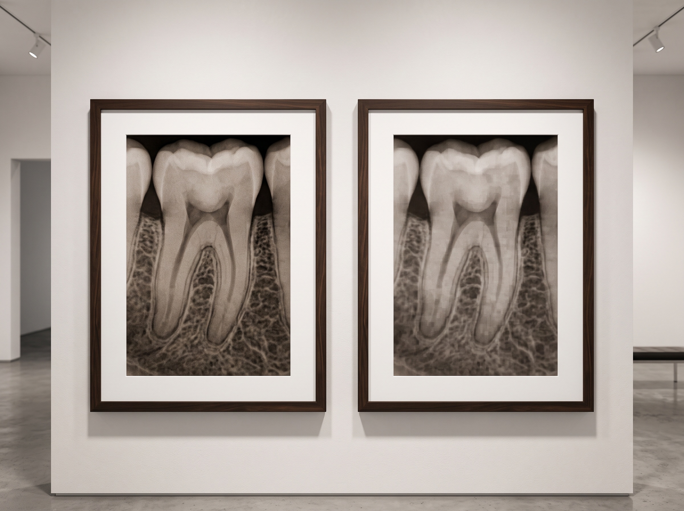
Compression comes in two fundamentally different kinds, and the distinction is the whole story. Lossless, or reversible, compression shrinks a file by encoding its data more efficiently — finding repetition and patterns and describing them compactly — while guaranteeing that the decompressed image is bit-for-bit identical to the original. Nothing is discarded. The cost is modesty: lossless methods typically manage only about 1.5:1 to 3:1, because a detailed radiograph is not very repetitive to begin with.
Lossy, or irreversible, compression plays a different game. It permanently removes information the algorithm judges least perceptible, and in exchange it reaches ratios of roughly 10:1 or far higher. The saving is dramatic and the picture can still look, to a casual glance, untouched. But the discarded data is gone for good — no future viewer, no better monitor, no clever software can bring it back. The word the field uses is exact and unforgiving: irreversible.
The Diagnostic Cost of a Smaller File
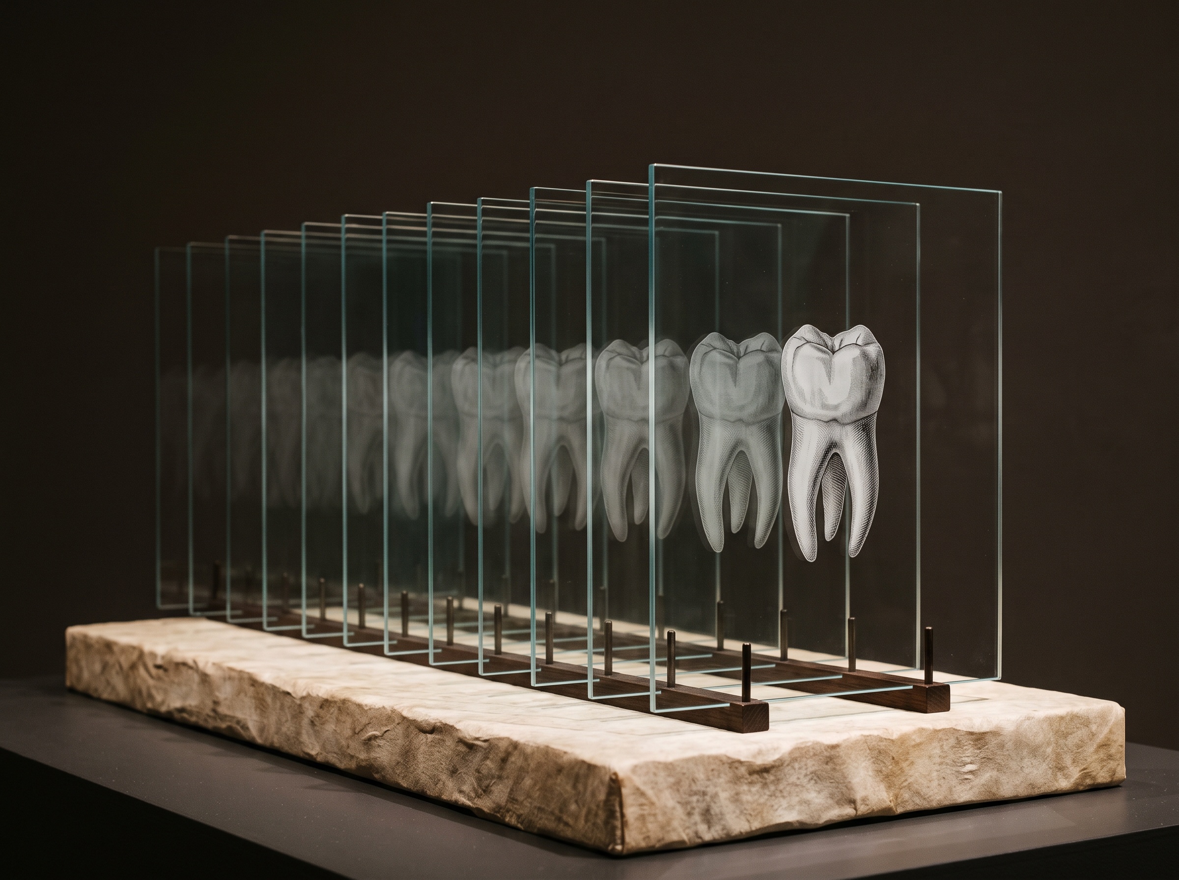
The danger is that lossy compression fails gracefully in the wrong direction. It preserves the bold, high-contrast structures the eye checks first — the crown, the obvious restoration, the gross outline of bone — while thinning exactly the low-contrast, fine-grained detail that carries the earliest diagnostic signal. An incipient interproximal lesion, the faint demineralization at an enamel margin, the subtle density shift at the floor of a restoration: these live in precisely the tones an aggressive algorithm considers expendable.
This is not hypothetical. Research on storage-phosphor dental images has examined how far lossy compression can go before caries detection begins to suffer, and the lesson is consistent across medical imaging: there is a threshold beyond which the smaller file is no longer telling the diagnostic truth. The industry even has a name for the careful middle ground — diagnostically acceptable irreversible compression — the level at which a study concludes that a given modality tolerates a given ratio without measurable loss of interpretive accuracy. The phrase is a warning as much as a permission: acceptability has to be earned per modality and per task, not assumed.
DICOM and the Rules of Preservation
Medical imaging did not leave this to chance. The DICOM standard, the common language of diagnostic images, specifies how images may be compressed and carries the scheme with the file so any compliant system knows exactly how to decode it. It embraces both philosophies deliberately. For reversible storage it supports lossless transfer syntaxes — including JPEG Lossless, the predictive scheme long used where every value must be preserved — and for controlled space savings it supports irreversible ones. JPEG 2000, in particular, is prized because a single wavelet-based codec spans both worlds: it can operate fully lossless, or scale smoothly into lossy at chosen ratios, from one well-understood pipeline. The point of standardization is not merely interoperability; it is that a preserved image announces, honestly and permanently, how it was made smaller.
The Whole Object, Not Just the Picture
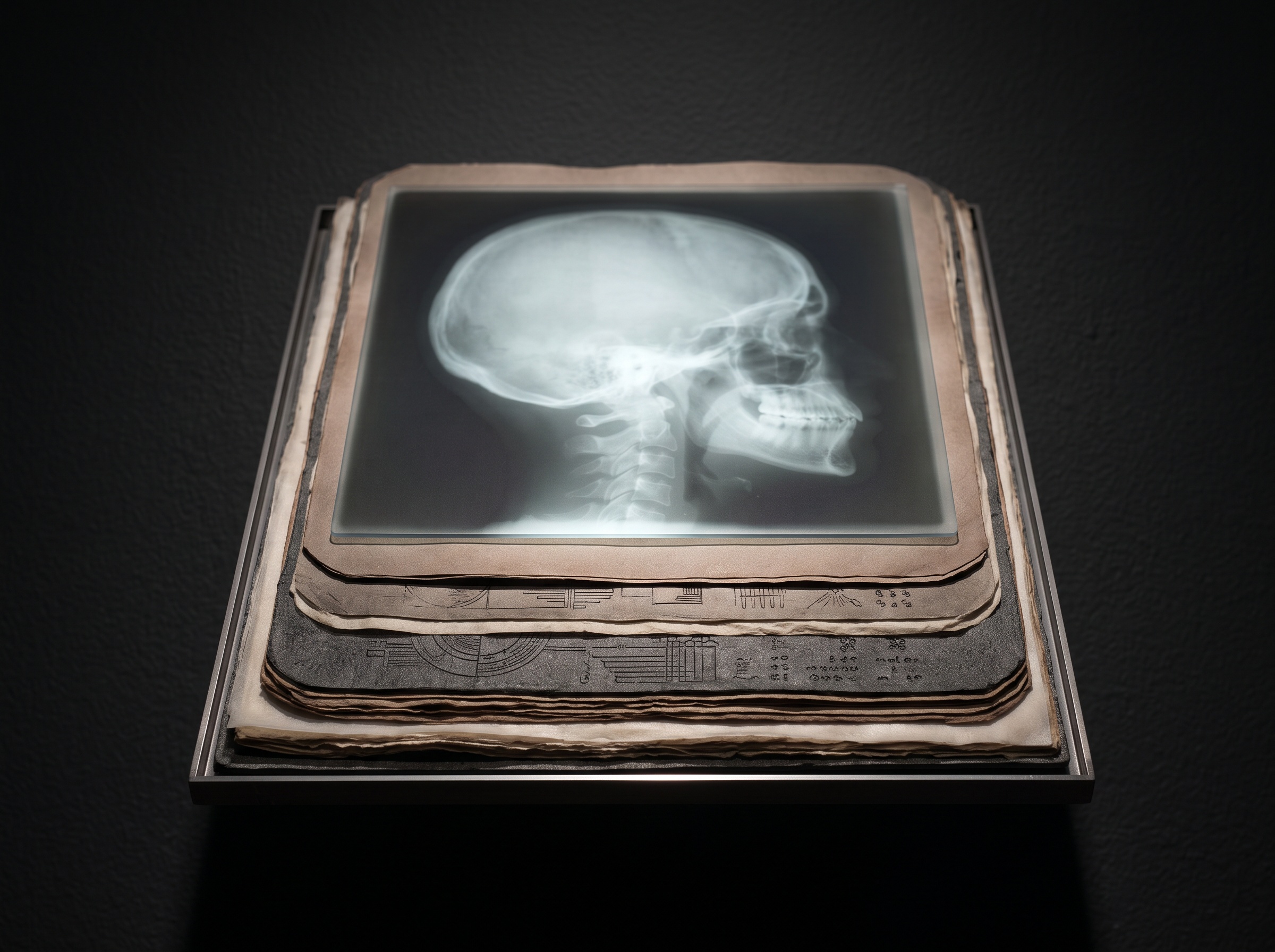
Preserving a dental image well means more than choosing a codec. The visible picture is only the surface of the artefact. Beneath it sits the full bit depth — a diagnostic radiograph often carries far more shades of gray than a screen shows at once, and that latent range is what lets a clinician window into the shadows later. Flatten a rich 12-bit or 16-bit capture into an 8-bit consumer image to save room, and the headroom for future re-examination quietly vanishes even if today’s view looks fine. Alongside the pixels lives the metadata: who, when, which device, which settings, which orientation. Strip or scramble it and the image becomes an anonymous, unmoored picture — technically intact, evidentially worthless. Conservation means keeping the object whole, not just the photograph legible.
The Slow Fragility of Format
Even a perfectly stored file faces a subtler threat: the format itself ages. Codecs fall out of support, proprietary viewers are discontinued, and media and systems are replaced on cycles far shorter than the decades an image may need to remain readable. An archive is therefore never finished at the moment of saving. It has to be tended — verified against corruption, held in open and widely supported encodings rather than a vendor’s private dialect, and migrated forward as the technology beneath it turns over. A dental image outlives the software that made it, and preservation is the discipline of making sure it also outlives the software’s absence.
Future Developments
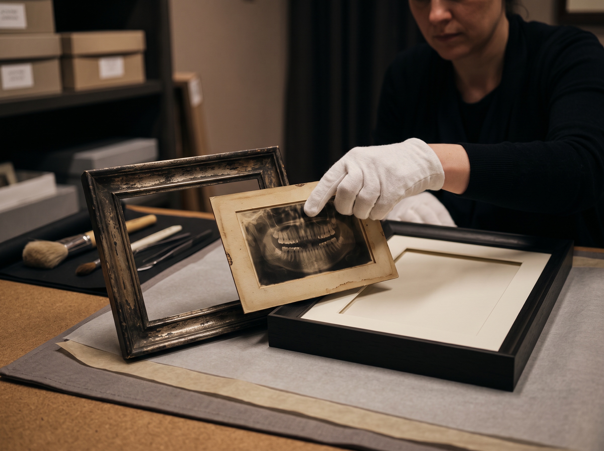
The direction of travel is toward archives that understand what they hold. Perceptually and anatomically aware compression is maturing — codecs guided by where diagnostic signal actually lives, so that saving is done around the lesion rather than through it. Self-verifying storage is spreading, with integrity checks and provenance records that let a system prove an image has not silently degraded since capture. And the long shift toward open, self-describing formats and routine migration is turning preservation from a one-time act of saving into a continuous act of care. The ambition is a simple and demanding one: that the image recovered years from now is not a flattering copy but the captured original, its faintest tones still intact — so that what the gallery keeps is not merely a picture of the patient, but the whole truth it was entrusted to hold.
Sources & further reading:
- Diagnostically acceptable irreversible compression — Wikipedia
- Impact of lossy image compression on accuracy of caries detection in digital images taken with a storage phosphor system — PubMed
Sorry, the comment form is closed at this time.