


July 26, 2026 The PHI Hidden in the Pixels: What a Dental Image Really Carries When It Leaves the Gallery
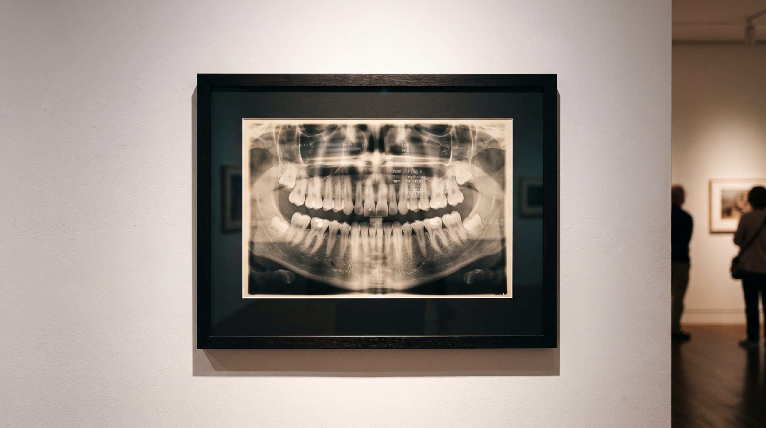
Hang a radiograph on a gallery wall and you see a single, quiet object: a lattice of enamel and bone rendered in silver and grey. But a dental image is never only the picture. Travelling with it, unseen, is a second image made entirely of words and numbers – the patient’s name, their date of birth, the referring clinician, the machine’s serial number, the exact minute of capture. When a case leaves your office for a specialist, a laboratory, a study, or a lecture slide, that invisible companion goes too. Understanding what it contains, and how to remove it cleanly, is one of the quieter disciplines of modern imaging – closer to the work of a conservator than a photographer.
The second image behind the picture
Most clinical images move through the world as DICOM files, and a DICOM file is a container with two compartments. One holds the pixel data – the visible radiograph. The other holds the metadata: a long, structured list of tagged fields describing who the picture is of, who made it, on what device, and when. This header is what lets imaging systems file, sort, and retrieve a study reliably. It is also, field for field, a dossier of protected health information.
The identifying fields are not incidental. They include the obvious – PatientName, PatientID, birth date, sex – and the less obvious: the accession number that ties the study to a record, the referring physician’s name, the institution’s name, the operator who ran the exposure, the station and its serial number. Even the unique identifiers that DICOM uses internally to distinguish one study from another can, left intact, act as a thread back to the individual. To share an image safely is to treat this header as the first thing to be attended to, not the last.
The gallery label names the work, not the sitter
A useful way to think about de-identification is the museum label. Beside every framed work hangs a small plate: title, medium, date. It tells you everything about the object and nothing private about the person who once sat for it. That is precisely the state a shared clinical image should reach – fully legible as a diagnostic object, silent about the patient behind it.
Emptying that plate is more than deleting a name. HIPAA’s Safe Harbor method enumerates eighteen categories of identifiers that must be removed before information is considered de-identified: names, all geographic subdivisions smaller than a state, every date more specific than a year that relates to the individual, phone and fax numbers, email addresses, record and account numbers, device identifiers and serial numbers, web and network addresses, and more. In DICOM terms this means scrubbing or generalizing the patient and encounter fields, stripping the institution and operator fields, cutting dates back to the year, and clearing private vendor tags and device serials. Unique identifiers are not simply deleted but regenerated under a deterministic, collision-safe scheme, so the study still holds together internally without pointing outward at a real person.
The text painted into the pixels
Here is the trap that catches careful people. You can perfect the header – blank every tag, regenerate every identifier – and still ship a file that betrays the patient, because some identifiers were never in the metadata at all. They were burned directly into the image.
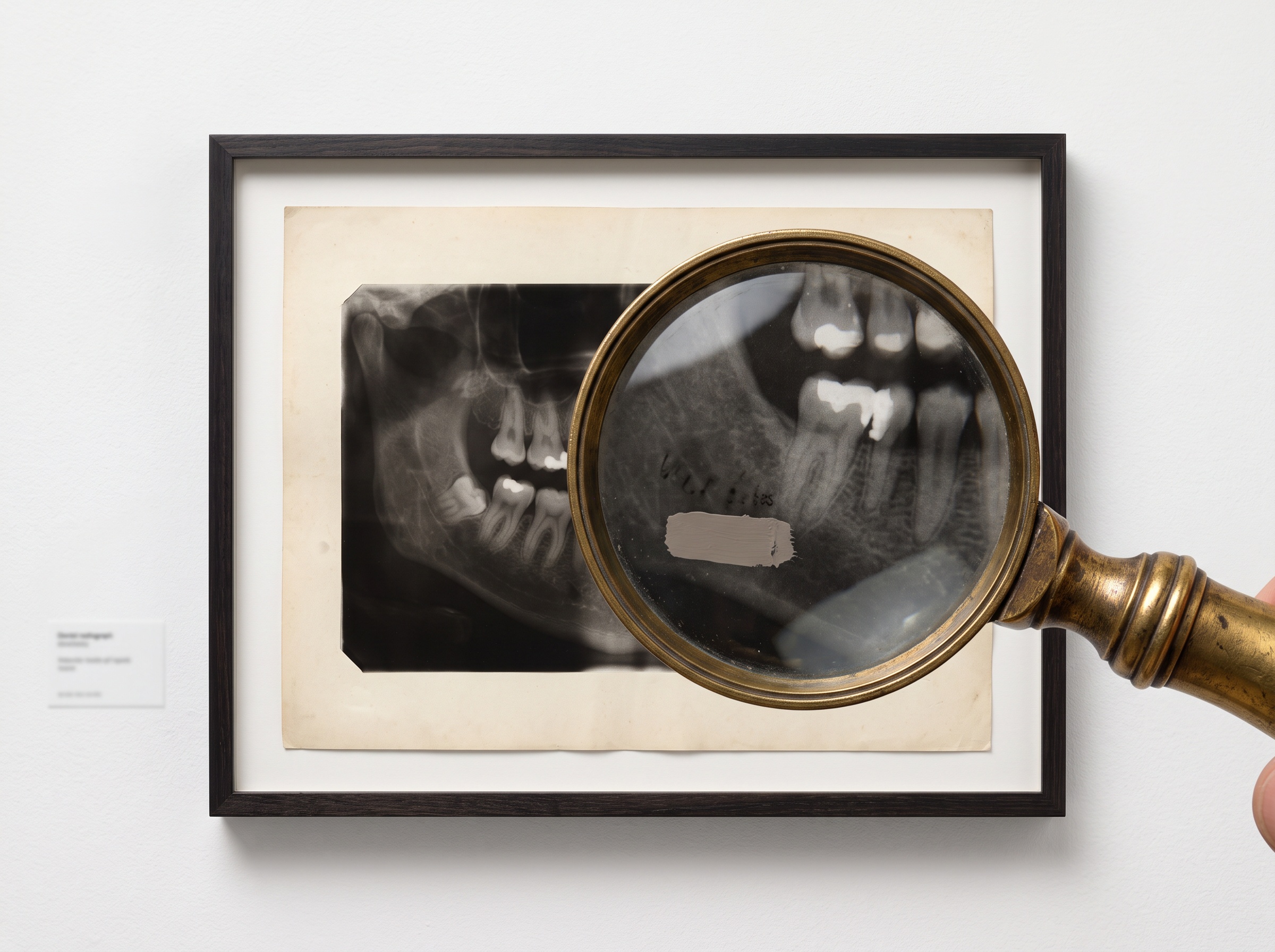
Secondary-capture images, screenshots from imaging software, scanned films, and certain acquisition modes routinely stamp text onto the picture itself: a name in the corner, a date along the margin, a clinic banner across the top. To the file format these are not data fields; they are pixels, indistinguishable from bone. Tag-level cleaning walks straight past them. Genuine de-identification therefore has to work on two fronts – clearing the header and inspecting the pixels – detecting and masking any burned-in annotation before the image is released. It is the imaging equivalent of checking not only a painting’s label but the artist’s signature hidden in the lower corner of the canvas.
Two doors to the same room
Regulation offers two routes to the same destination, and the choice between them is a matter of temperament as much as compliance. The first is Safe Harbor: the fixed eighteen-item checklist. Remove everything on the list and the data is deemed de-identified, no further argument required. It is a rule-straight door – narrow, prescriptive, and reassuringly unambiguous.

The second is Expert Determination. Here a qualified professional analyzes the specific dataset and its intended use, applies accepted statistical and scientific methods, and documents that the risk of re-identification is very small. This door is wider and more forgiving – it can permit useful detail that Safe Harbor would strip, such as finer dates for a longitudinal study – but it demands reasoned judgment and a written trail rather than a checklist. For most day-to-day sharing of dental images, Safe Harbor is the practical path; when the image needs to retain elements the checklist would forbid, Expert Determination is what makes that defensible.
Keeping the provenance, losing the person
The art of this work is subtraction without damage. A de-identified image is worthless if the diagnosis has been degraded, and dangerous if the identity has not been fully removed. The goal is a file that a receiving clinician can still trust completely – the geometry intact, the exposure faithful, the internal links between series and study preserved through regenerated identifiers – while every thread back to the individual has been cut.
That is why deleting a name in a viewer is not de-identification, and why the task rewards the same standardization that governs good capture. Established tooling exists precisely because the field is unforgiving of shortcuts: purpose-built de-identifiers let a practice configure which attributes to remove or replace and, critically, obscure burned-in text rather than trusting that none is present. Treating de-identification as a documented, repeatable procedure – not a one-off manual edit – is what turns a private clinical record into a shareable object without ever exposing the patient. The provenance survives; the person disappears.
Future Developments
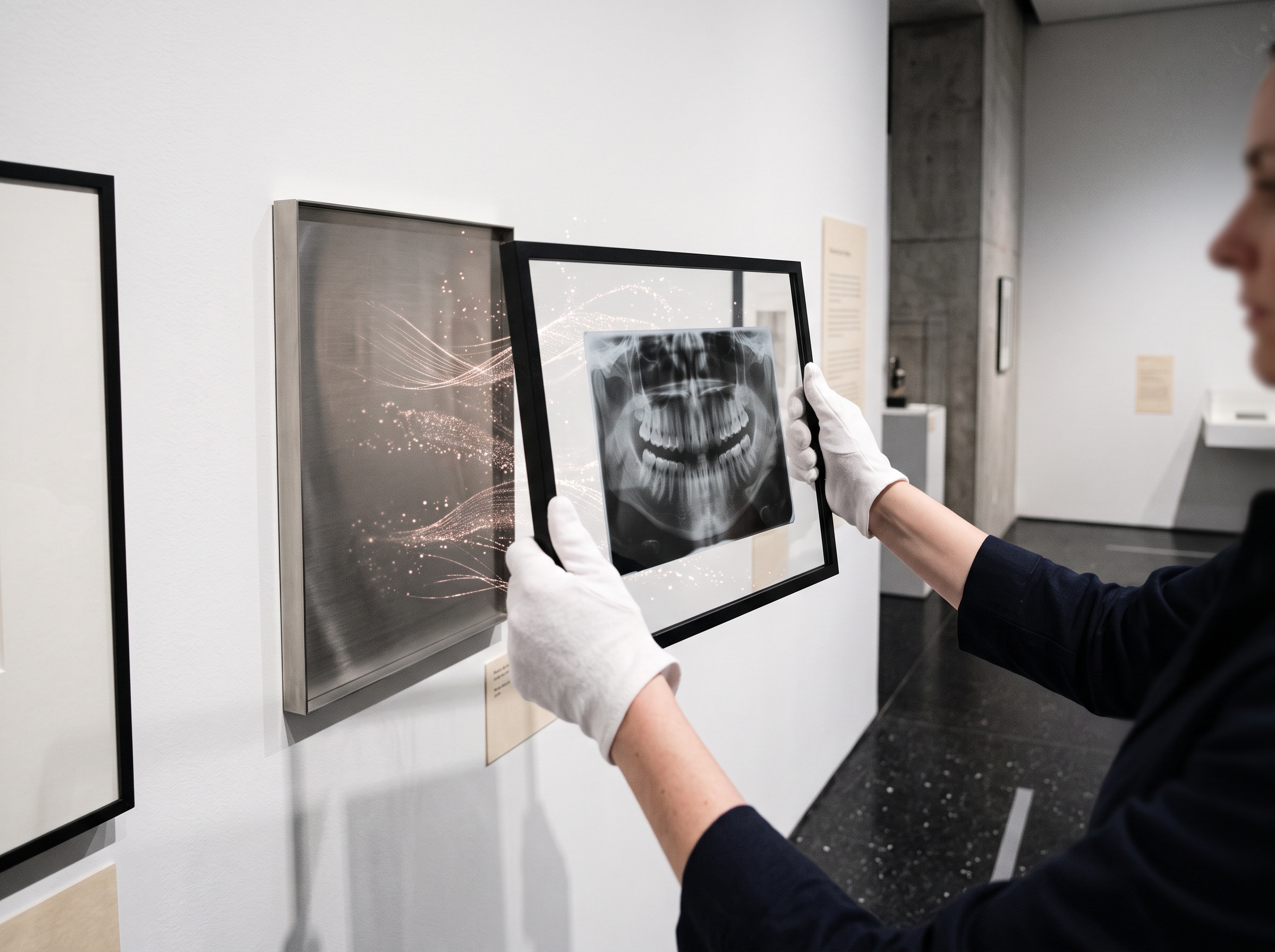
The next chapter belongs to automated conservation. Rule-based scrubbing of headers is mature, but the harder problem – reliably finding identifiers hidden in the pixels across the messy variety of real-world captures – is where hybrid systems now combine deterministic rules with machine vision that can read, locate, and redact burned-in text without human review. Deterministic identifier regeneration is making it possible to de-identify an entire longitudinal series while preserving the linkages that make follow-up studies meaningful. And as dental imaging feeds ever more into shared research sets and AI training, the standard is quietly rising from “remove the obvious” to “prove the risk is negligible.” The through-line does not change: an image is a work worth sharing, but the person who lent their body to it never consented to travel along. The craft is to send the picture and keep the patient home.
Sources & further reading:
- DICOM Files and HIPAA: How to Protect PHI and Stay Compliant
- Comprehensive Guide to HIPAA De-Identification Methods
Related Reading
- What the Gallery Keeps: Compression, DICOM, and the Long Survival of the Dental Image
- The Collection Behind the Machine: Why a Public, Pixel-Labeled X-Ray Archive Is the Raw Material of Dental Imaging AI
- The Image That Cannot Travel: DICOM, Interoperability, and the Quiet Cost of a Radiograph That Will Not Move
Sorry, the comment form is closed at this time.