


October 12, 2026 The Collection Behind the Machine: Why a Public, Pixel-Labeled X-Ray Archive Is the Raw Material of Dental Imaging AI
We have grown used to speaking of dental imaging artificial intelligence as though the intelligence were the achievement — the model that spots a lesion, segments a tooth, flags a shadow on a panoramic film. But a model is a mirror; it can only reflect what it has been shown. Before any algorithm reads its first radiograph in the clinic, someone has assembled the pictures it learned from, cleaned them of anything that might identify a patient, and then — image by image, tooth by tooth — drawn the boundaries that tell the machine what it is looking at. The intelligence is downstream. The raw material is a collection. And like any collection worth the name, its value lies not in its size alone but in the care with which it was gathered, catalogued, and labeled.
A dataset is a curated collection, not a pile of pictures
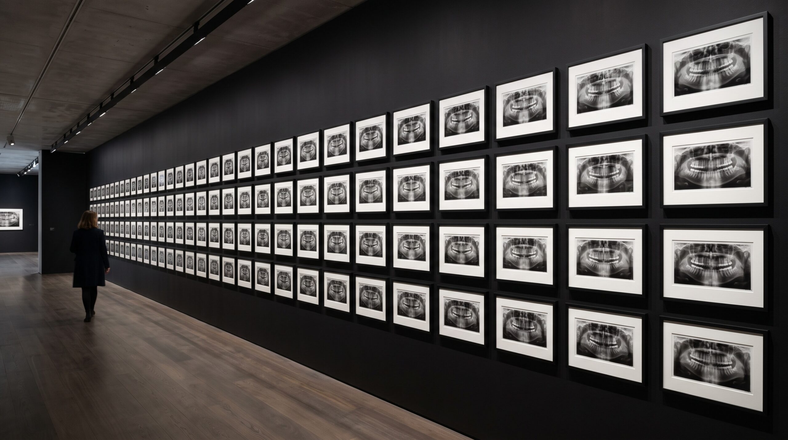
A newly published dataset in Scientific Data makes the point with unusual clarity. It gathers 8,655 de-identified oral panoramic X-ray images and pairs them with more than 30,000 pixel-level annotations — not loose captions or a single label per film, but boundaries drawn directly onto the pixels. The images were collected from a single hospital, stripped of identifying information, and then handed to a team of twenty dental imaging specialists who annotated them by hand, tracing polygonal contours around individual teeth and marking common oral conditions. The authors report testing the collection against several widely used deep-learning models and offering it as a foundation for tooth segmentation, lesion detection, and computer-aided diagnosis.
Read past the numbers and what emerges is a portrait of labor. Eight thousand images is a meaningful archive; thirty thousand hand-drawn regions is a mountain of patient, repetitive, expert work. This is the part of imaging AI that never photographs well and rarely gets celebrated, and yet it is the part on which everything else rests. The gallery metaphor is exact: a museum is not its building but its holdings, and its holdings are only as useful as the catalogue that describes them.
What a pixel-level label actually is
There are cheap ways to label an image and expensive ways, and the gap between them decides what a model can ultimately learn. The cheap way is a single tag for the whole picture — “caries present,” “implant present” — which teaches a model to answer yes or no but never to point. The next step up is a bounding box, a rough rectangle thrown around a region of interest. The pixel-level annotation in this dataset is the expensive end of that spectrum: a polygon traced along the true contour of each tooth and lesion, so that every pixel inside the boundary is claimed and every pixel outside it is not.
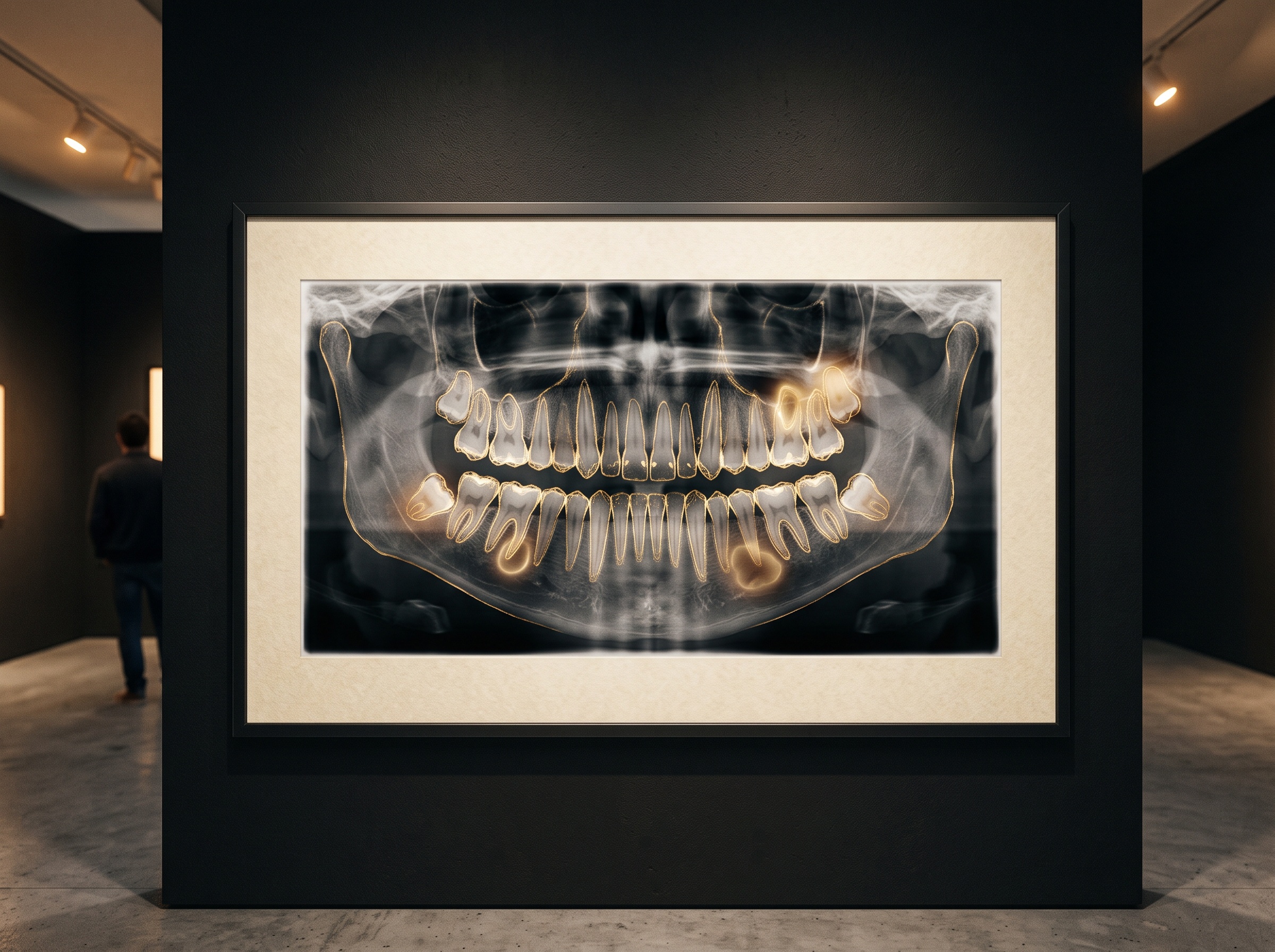
That precision is what makes semantic segmentation possible — the task of coloring in exactly which pixels belong to which structure. A model trained on boxes learns where a thing roughly is; a model trained on polygons learns a thing’s shape, its edges, the place where a crown ends and bone begins. This is the same conviction, carried into the training data itself, that animates the rest of modern dental imaging: that the image is not a vague impression but a precise field of measurable data, and that the more exactly we define its contents, the more exactly a machine can reason about them.
The quiet discipline of de-identification
A clinical radiograph is never only an anatomy; it arrives wrapped in a patient’s identity — a name in the corner, a date, an accession number, metadata threaded invisibly through the file. None of that can travel. Before an image can enter a public collection it must be stripped: identifying marks removed from the picture, identifying fields purged from the file, the radiograph reduced to the anatomy and nothing else. The dataset’s authors describe exactly this step, removing identifying information before the images ever reached the annotators.
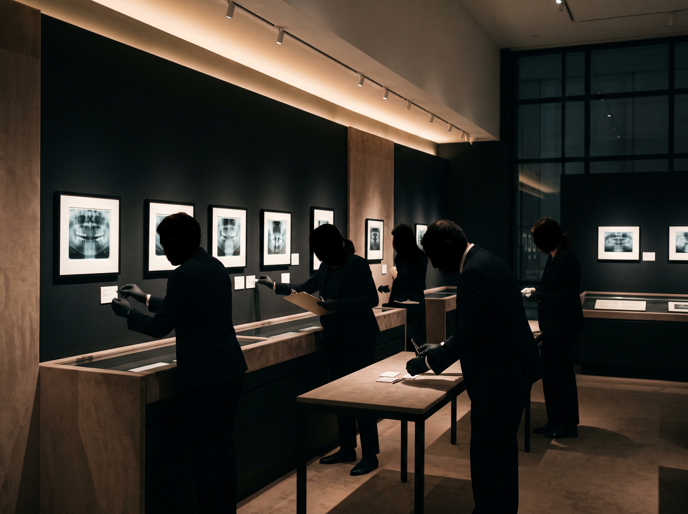
It is easy to treat de-identification as paperwork, but it is better understood as a curatorial act — the ethical condition that makes sharing possible at all. A picture that cannot be anonymized cannot be published, no matter how instructive it would be. Every public imaging dataset is therefore a negotiated object, assembled at the intersection of what is scientifically valuable and what can be ethically released, and the discipline that governs it is the same one that governs the operatory: the image serves the patient first, and the patient’s privacy is not a line item but the floor.
Why the archive sets the ceiling
Here is the harder truth that a published dataset forces into the open: a model cannot generalize beyond the world its training collection represents. The images in this archive came from a single institution, captured on its machines, reflecting the patients who walked through its doors and the conventions of the specialists who labeled them. That is a strength — consistency, shared standards, controlled quality — and it is also a boundary. A system trained on one hospital’s panoramics may stumble on films from a different unit, a different population, a different exposure habit, because the texture, geometry, and framing it learned to expect have quietly shifted beneath it.
This is the problem of domain shift, and it is why the same AI that performs brilliantly in validation can disappoint in a practice across the country. It is also why the raw image underneath still rules everything: feed a model films unlike those it trained on — noisier, differently processed, differently positioned — and its confidence will outrun its competence. The oldest lesson in imaging holds at the scale of the dataset too, that what the data omits, the algorithm will invent or ignore. A collection does not merely teach a model; it defines the edges of what that model can honestly claim to know.
The collection as shared infrastructure
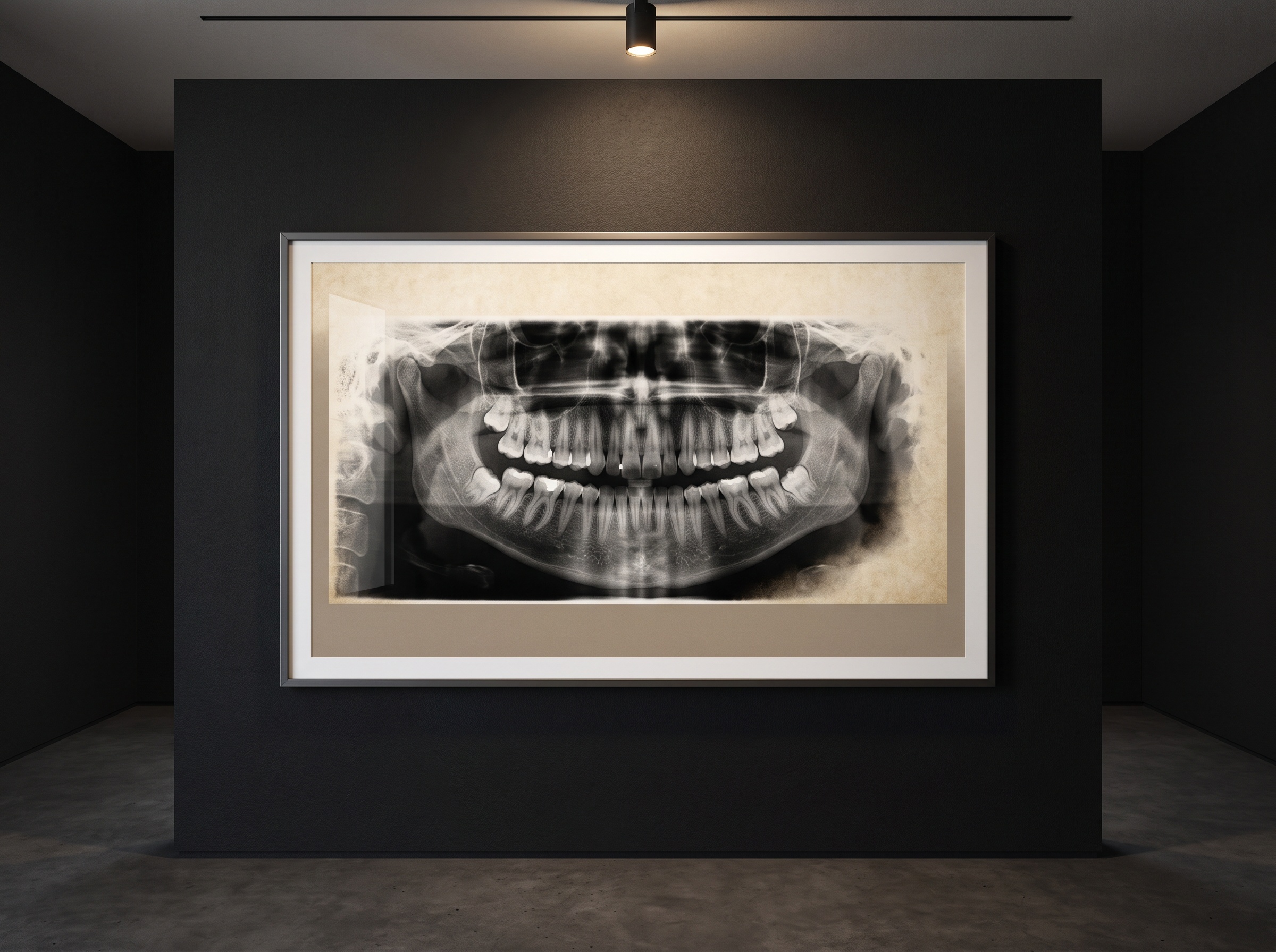
What makes a published dataset different from a private one is that it becomes common ground. When a collection is released with a clear provenance and open annotations, every research group can train against the same images, compare results on the same benchmark, and build on one another’s work instead of each quietly assembling an archive no one else can inspect. This is how a field moves from scattered, unrepeatable claims toward something that can be checked. A shared collection is to imaging AI what a reference specimen is to a museum: the fixed object against which every later interpretation is measured.
It also redistributes who gets to build. Assembling and annotating thousands of radiographs is expensive enough to exclude all but the largest institutions; a public, well-labeled dataset lowers that wall, letting a smaller team test an idea without first spending a year labeling. The same democratizing instinct runs through the tools that let a practice harness AI at all — the reconstruction methods that make low-dose deep-learning imaging practical on ordinary hardware — and it begins, upstream of all of them, with someone choosing to publish the collection rather than keep it.
Reading a dataset critically
If a published collection is raw material, then the clinician and the researcher alike owe it the same scrutiny a curator gives a new acquisition. Where did the images come from, and from how many sources? Who drew the labels, by what definitions, and was any second reader used to check them? How was the collection de-identified, and under what license may it be used? A dataset answers a model’s first question — what does the world look like? — and so its blind spots become the model’s blind spots. The right posture toward an imaging AI is therefore to ask not only how accurate it is, but what it was raised on, because a system is, in the end, an argument made entirely out of its training images.
Future Developments
The near future of dental imaging AI will be written as much in its datasets as in its architectures. Expect collections to grow past the single institution toward multi-center archives that span machines, populations, and labeling conventions, so that a model’s confidence is earned across difference rather than inside a comfortable monoculture. Expect annotation itself to become a hybrid craft, with algorithms proposing the first boundary and human specialists refining it — faster than the fully manual tracing behind today’s archives, but still anchored to an expert eye. And expect provenance to become a published field in its own right, a kind of wall label for the data, so that anyone deploying a model can read where its vision came from. The glamour will always gather around the machine that reads the radiograph. But the lasting work, the work that decides whether that machine can be trusted, happens earlier and more quietly — in the gathering, the stripping, and the patient labeling of the collection behind it. The art of imaging has always been an art of looking carefully. Teaching a machine to look begins by curating, with equal care, everything we ask it to see.
Sources & further reading:
No Comments