


November 1, 2026 Diagnocat vs Pearl: Two AI Readers of the Dental Radiograph, and When Each One Earns Its Place
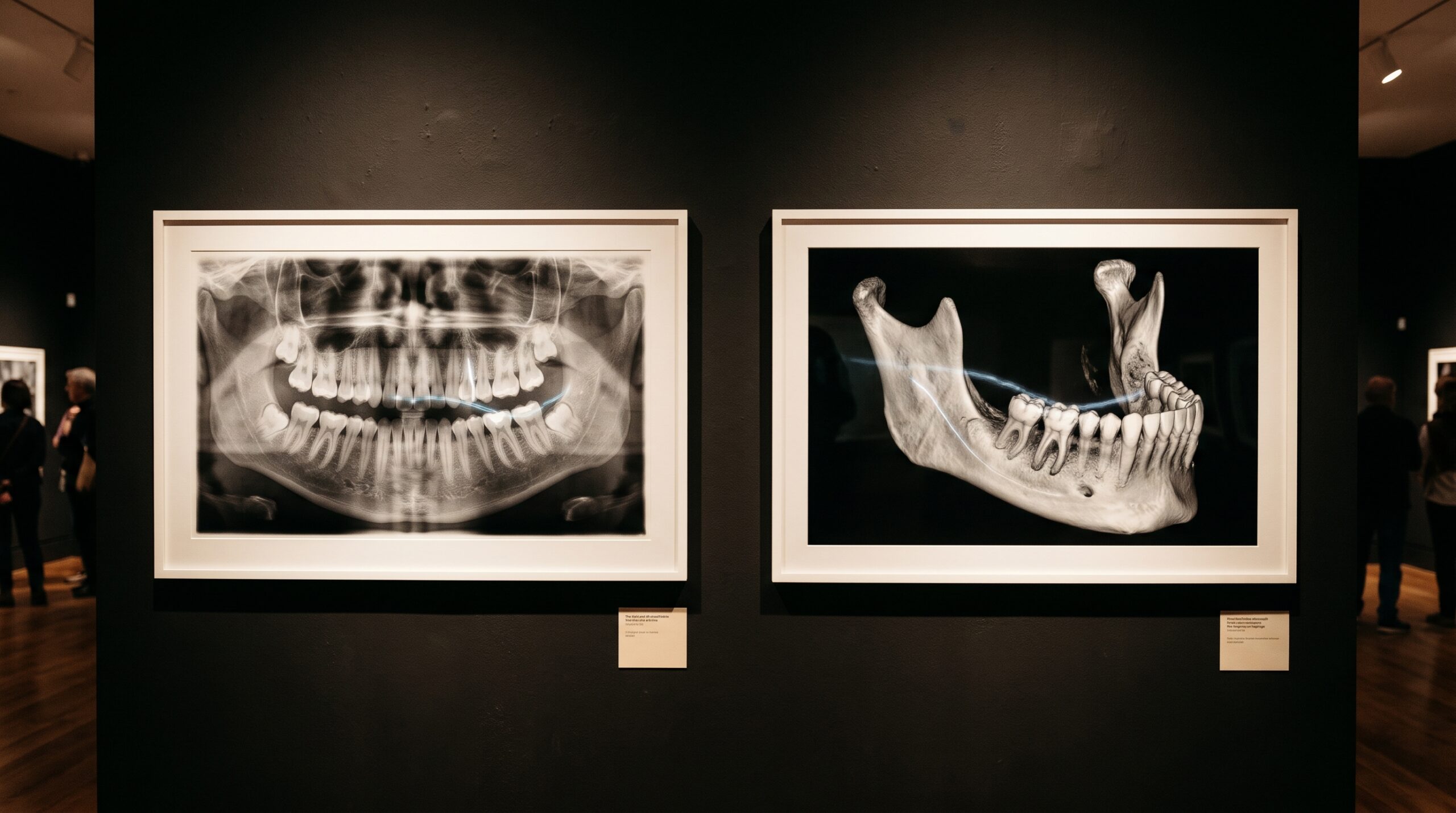
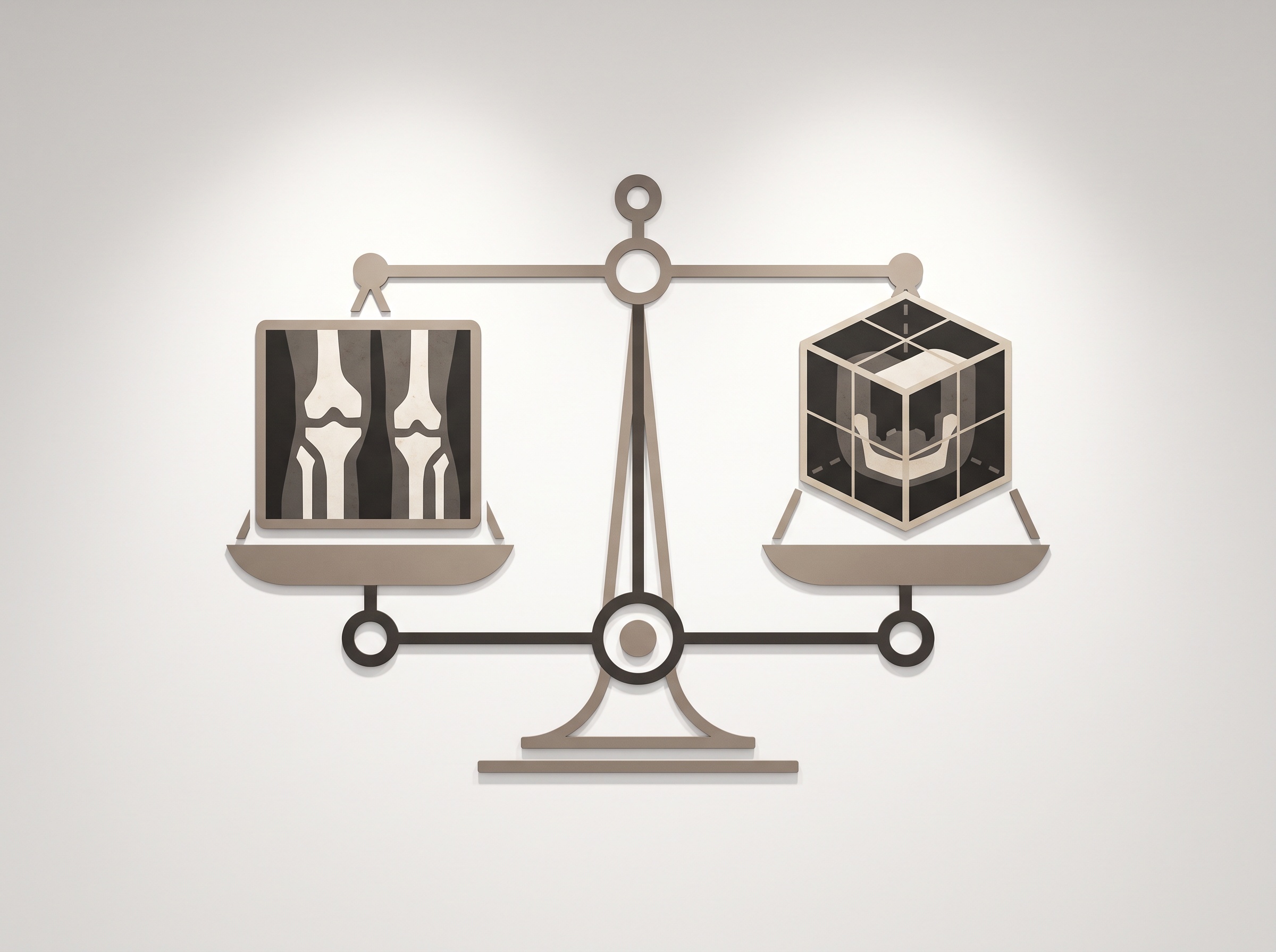
Put two AI systems in front of the same dental radiograph and you might assume they are competing to answer the same question. Diagnocat and Pearl are both independent, unaffiliated companies whose software is cleared by the United States Food and Drug Administration to help clinicians read dental images, and both are frequently named in the same breath. But they are not interchangeable, and the most useful thing to understand about them is that each was built to look most carefully at a different kind of picture. Pearl earned its reputation reading the flat, two-dimensional radiographs that fill an ordinary chart; Diagnocat has pressed hardest into the volumetric world of the cone-beam scan. Choosing between them is less a contest of accuracy than a question of which image a practice most needs a second pair of eyes on.
What follows is an even-handed comparison drawn from public, verifiable facts: regulatory clearances, stated intended use, and the capabilities each vendor describes. It is not an endorsement, and it deliberately avoids the unsourced accuracy claims that litter affiliate roundups. Where a number matters, it comes from a cleared study or the FDA record.
The Short Answer
Pearl is the broad two-dimensional reader. Its software analyzes bitewing, periapical, and panoramic radiographs for findings such as caries, calculus, periapical radiolucencies, and bone loss, and it is widely chosen for its patient-facing annotations that overlay findings directly on the image during a consult. Diagnocat is the practice that has gone deepest into three-dimensional analysis; its most recent United States clearance is specifically for flagging possible periapical radiolucencies on cone-beam CT volumes as a second-read aid, and it is known for automated CBCT analysis and 3D segmentation that the 2D-first tools do not attempt. If your diagnostic weight falls on everyday intraoral and panoramic films, Pearl is the natural starting point. If it falls on the cone-beam volume, Diagnocat is built for that room.
At a Glance
| Criterion | Pearl | Diagnocat |
|---|---|---|
| Primary image type | 2D: bitewing, periapical, panoramic (plus a cleared 3D/CBCT product) | 3D: cone-beam CT, with 3D segmentation; broader 2D analysis offered outside the US |
| US regulatory status | Multiple FDA 510(k) clearances, beginning with 2D analysis and extending to Second Opinion 3D for CBCT (cleared May 23, 2025) | FDA 510(k) clearance (K252934) granted January 15, 2026 for periapical radiolucency detection on previously acquired CBCT scans |
| Stated intended use | Aid to clinicians interpreting dental radiographs; findings presented for review, not autonomous diagnosis | Aid for a second reading of CBCT scans of permanent teeth in patients aged 22 or older; does not replace clinical judgment |
| Who may use the US-cleared tool | Dental professionals; panoramic clearance stated from age 16 | US clearance limited to clinicians with at least two years of experience reading and assessing CBCT scans |
| Signature strength | Everyday 2D workflow coverage and patient-facing annotation | Automated CBCT analysis and 3D segmentation |
One caveat on the table: a product’s marketed features and its FDA-cleared claims are not the same thing, and sources sometimes blur them. A cleared indication is narrow and specific; a feature list is not. Before relying on any claim, the honest move is to check the clearance itself in the FDA 510(k) database rather than a vendor page or a comparison blog.
Reading the One Number Worth Quoting
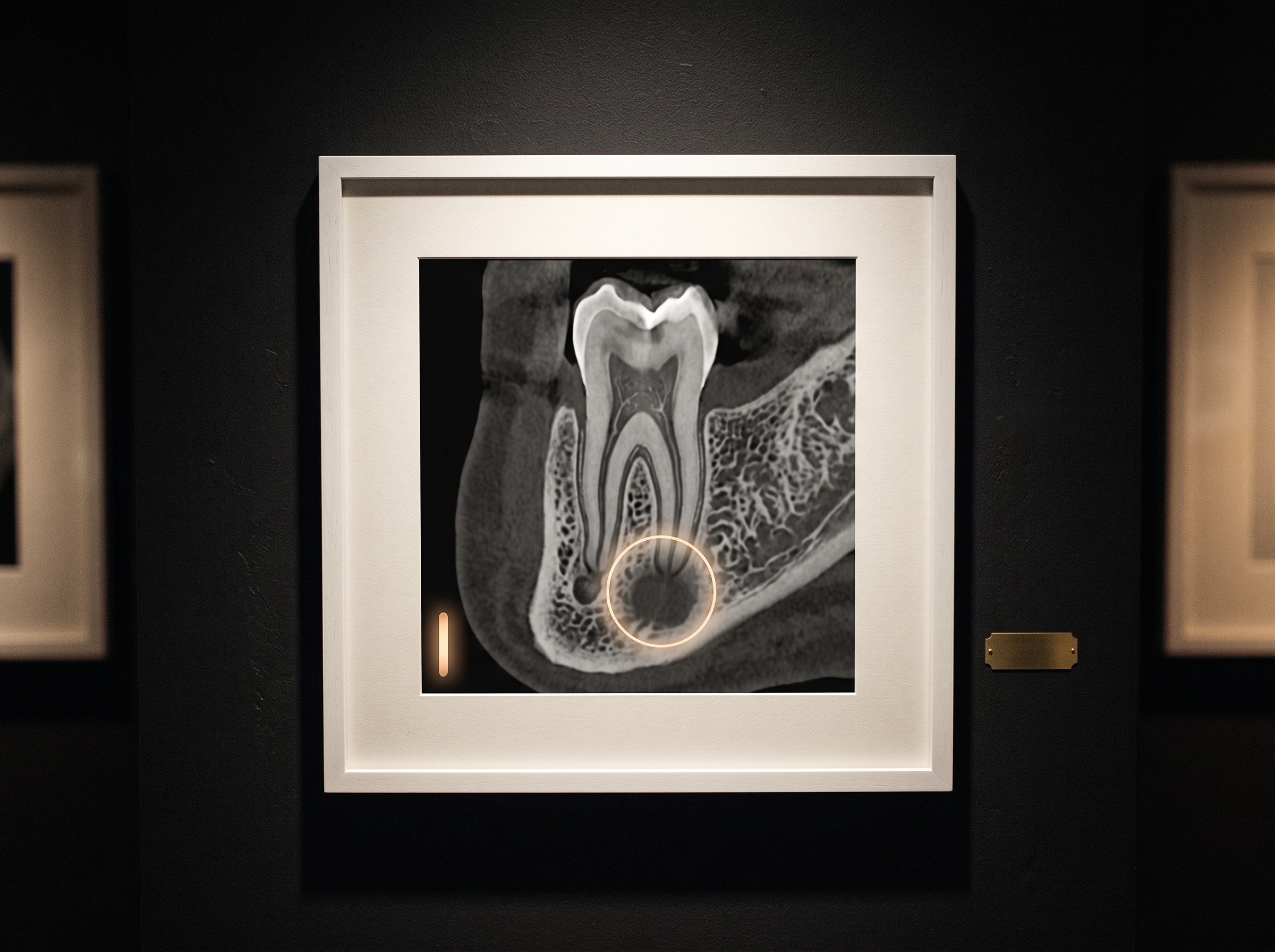
Accuracy claims in this field are often loud and rarely cited. The exception worth repeating is the study behind Diagnocat’s CBCT clearance, because it was submitted to a regulator and reports its figures plainly. In that reader study, clinicians interpreting cone-beam scans with the aid achieved an area under the curve of 0.921, compared with 0.894 unaided; the software’s standalone performance was reported at a sensitivity of 0.854 and a specificity of 0.991 for the periapical-radiolucency task. The shape of those numbers is as instructive as the magnitudes. A very high specificity with a more modest sensitivity describes a tool that rarely cries wolf but can still miss a subtle lesion, which is precisely why its cleared role is a second read beside an experienced clinician, not a replacement for one. That is the same honest boundary we traced in our look at how AI moves from merely flagging a finding to drafting a reading a clinician still owns.
When to Choose Diagnocat
Diagnocat makes the most sense where the cone-beam volume is central to the practice’s diagnostic life: in endodontics, oral surgery, implantology, and any setting where previously acquired CBCT scans are reviewed often and the cost of overlooking a periapical radiolucency is high. Its 3D segmentation, laying the anatomy out as separable structures rather than a single dense block, answers questions a flat film cannot, and its cleared second-read role fits a reader who already has the CBCT experience the United States clearance requires. If the recurring frustration in your operatory is the time and uncertainty of combing a volumetric scan, this is the tool built for that frustration. It presumes, rightly, that you have already decided the case genuinely warranted a cone-beam volume rather than a full-mouth series.
When Pearl Might Fit Better
For a general practice, the honest truth is that most diagnostic days are spent on 2D images, and that is Pearl’s home ground. A tool that reads bitewings, periapicals, and panoramics covers the volume of work most offices actually generate, and Pearl’s patient-facing annotations have a second, non-clinical virtue: they help a patient see on the screen what the dentist is describing, which supports understanding and case acceptance in a way a radiologist’s private read does not. If your practice rarely acquires CBCT, a CBCT-centric tool will sit idle; the broad 2D reader will be working every hour. An even-handed comparison has to concede this plainly, because the best tool is the one that matches the images you actually take, not the one with the newest clearance.
Frequently Asked Questions
Are Diagnocat and Pearl the same company or affiliated? No. They are separate, unaffiliated companies offering distinct dental-imaging AI products. They are often compared because both are FDA-cleared to help clinicians read dental radiographs.
What is the core difference between them? Pearl is best known as a broad analyzer of two-dimensional radiographs (bitewing, periapical, panoramic), while Diagnocat has gone deepest into three-dimensional cone-beam CT analysis and 3D segmentation. Both have expanded beyond their origins, but their centers of gravity differ.
Can either tool diagnose on its own? No. Both are cleared as aids to a clinician. Diagnocat’s United States CBCT clearance is explicitly a second-read aid for permanent teeth in patients 22 and older and states it does not replace clinical judgment. A general-purpose chatbot is not a substitute for cleared, task-specific software either.
How do I compare their integration with my practice software? Vendor integration models vary, from open interfaces to partner-gated ones, which shapes how easily a tool slots into your imaging pipeline. We examined that dimension in our piece on open versus partner-gated dental AI vendors.
Future Developments
The line between these two tools is already blurring, and it will blur further. Pearl has reached into the cone-beam world with its 3D product, and Diagnocat has long offered broader two-dimensional analysis in markets outside the United States; the regulatory record simply lags the engineering. It is reasonable to expect that within a few years the meaningful question will no longer be “Diagnocat or Pearl?” but “which of this vendor’s cleared indications actually covers the finding in front of me, and on which image?” That is a more demanding question, and a healthier one, because it keeps the clinician, not the brand, at the center of the reading. The radiograph remains what it has always been at this gallery: an image to be read with discipline and care. The newest instruments are extraordinary at drawing the eye to a shadow worth a second look, but the diagnosis, and the responsibility for it, still belongs to the person standing at the viewing screen.
Sources & further reading:
- Timeline: Five FDA clearances for 3D dental AI as Pearl launches CBCT software for general dentists – Oral Health Group
- FDA 510(k) Premarket Notification Database (search device K252934)
No Comments