


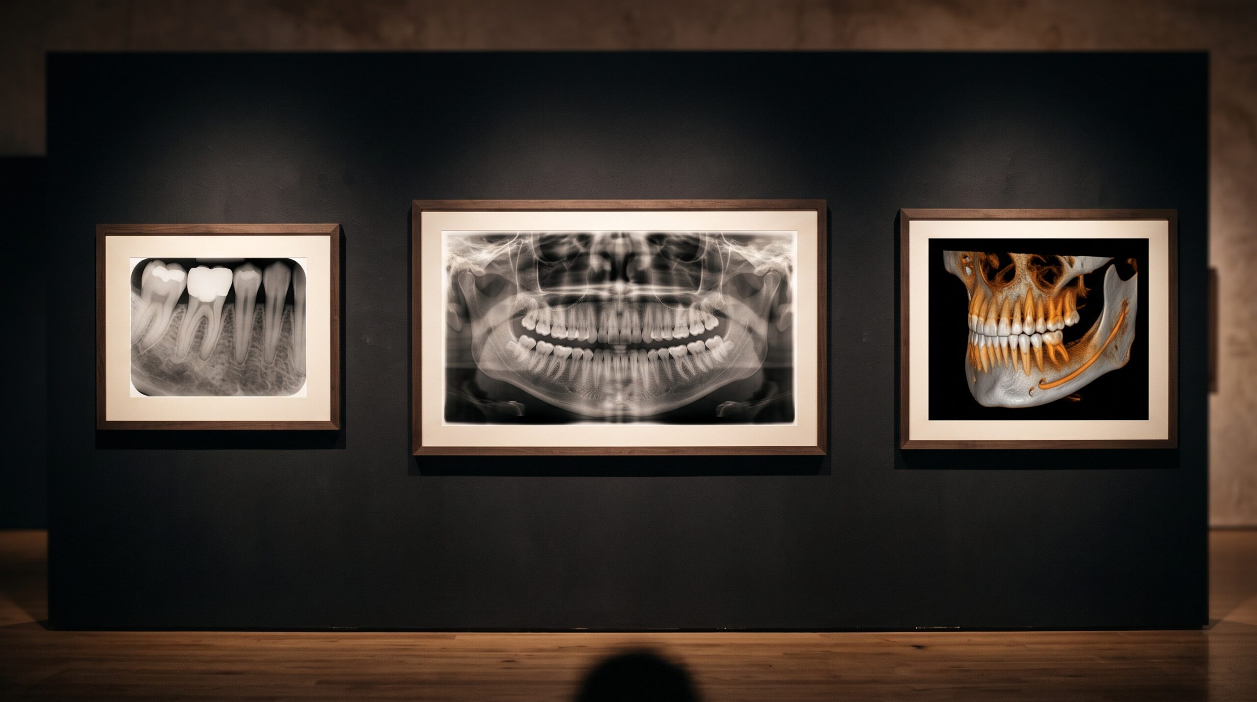
November 9, 2026 One Algorithm, Three Media: Why AI Reads the Intraoral X-Ray, Panoramic and CBCT So Differently
Hang an intraoral radiograph, a panoramic film and a cone-beam reconstruction side by side, and the eye immediately understands that these are three different kinds of picture. One is a jewel of local detail, one is a sweeping survey, one is a volume you can turn in your hand. It is tempting to assume that an artificial-intelligence reader, unburdened by the human habit of seeing, glides across all three with equal ease. A growing body of evidence, including a 2026 pilot study that put a single commercial AI platform to work across all three modalities, says otherwise. The algorithm’s accuracy shifts with the medium, and understanding why is less a question of software than of physics, geometry and the nature of the image itself.
The Modality Is the Medium, Not the Pipe
There is a quiet assumption in much of the marketing around dental AI: that the image is simply a container, and the intelligence lives entirely in the model. In practice the opposite is closer to the truth. A detection model can only find what the image encodes, and each modality encodes the mouth in a radically different way. An intraoral radiograph is a high-resolution two-dimensional projection of a small region. A panoramic image is a curved tomographic layer that flattens a three-dimensional jaw onto a single plane. A cone-beam volume is a reconstructed stack of hundreds of slices. These are not three qualities of the same photograph; they are three different media, each with its own grammar of signal and noise. When the same engine is asked to read all three, it is not performing one task three times. It is performing three related but genuinely distinct tasks, and its scorecard reflects that.
Where the Intraoral Image Lets AI Shine
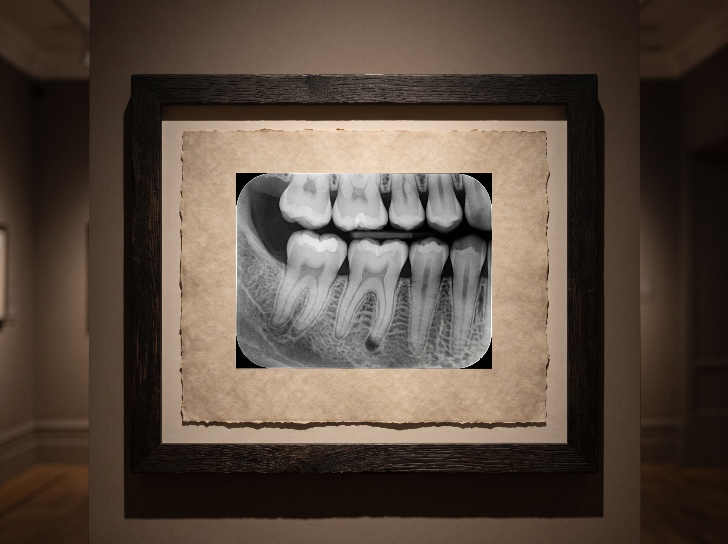
The intraoral radiograph is, in many respects, the friendliest input an algorithm can receive. The field is small, the resolution is high, and the geometry is relatively standardised. Caries at the contact point, a widened periodontal ligament, a periapical radiolucency, calculus on a root surface: these present as local contrast changes against a consistent background, and they are exactly the patterns that convolutional models learn efficiently. This is why the earliest and most numerous regulatory clearances in dental AI clustered around bitewing and periapical interpretation, and why real-world accuracy on these tasks is often reported as strong. The limiting factor is rarely the algorithm’s capacity and more often the humble question of technique: a foreshortened image, a cone-cut, or overlapping contacts degrade the input before the model ever sees it. The lesson is old and familiar to anyone who treats the radiograph as a crafted object rather than a mechanical output, a discipline that rewards the practice far more than any single piece of software. When two engines disagree on the same clean image, the difference tends to reflect how each was trained, a theme explored in our comparison of two AI readers of the dental radiograph and when each earns its place.
The Panoramic Film and the Problem of Overlap
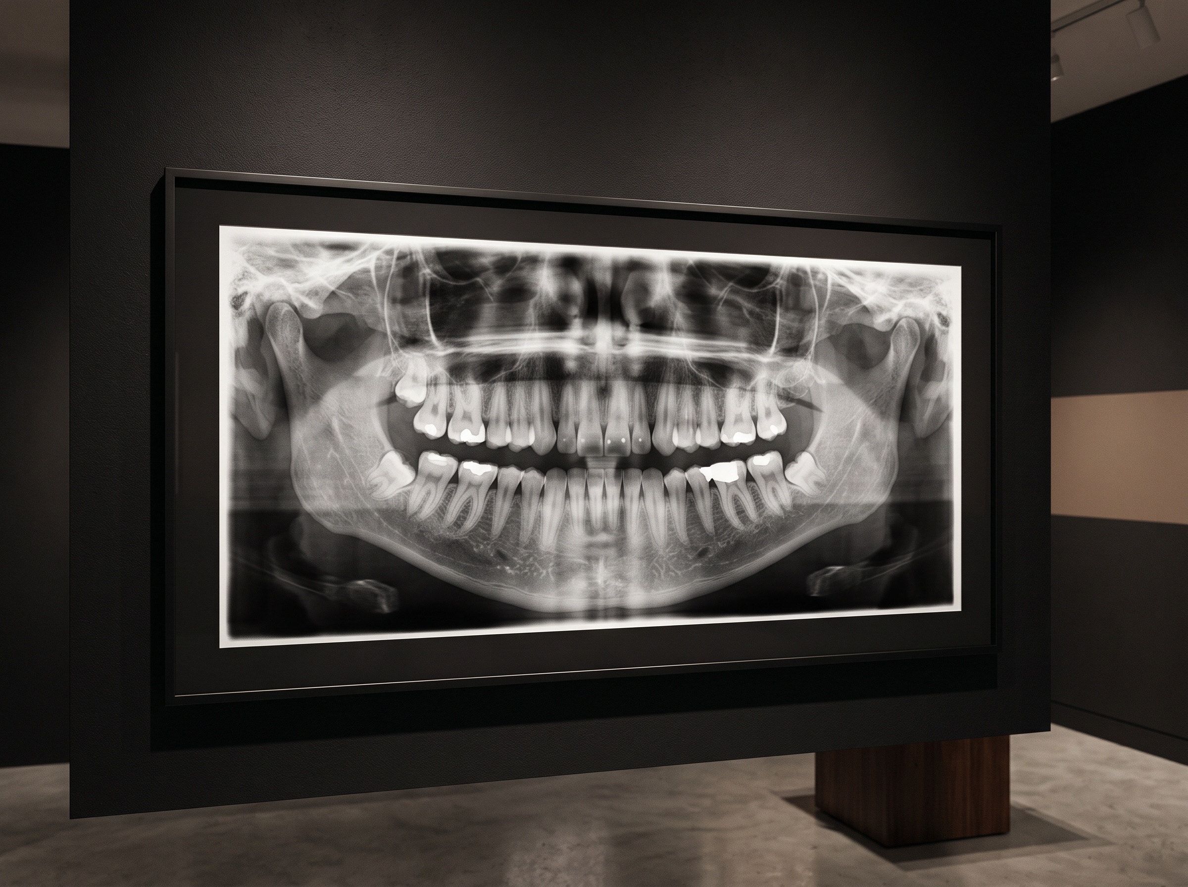
The panoramic image presents a harder problem, and for a revealing reason. Its great virtue, a single survey of both arches, the sinuses, the condyles and the surrounding bone, is bought with real optical compromises. Structures are magnified unevenly, anatomy superimposes, and the curved focal trough means that anything outside its narrow band blurs. Then there are ghost images: the phantom shadows cast when a dense structure on one side of the head is projected, reversed and magnified, onto the opposite side. A human reader learns to recognise and dismiss these as artifacts of the machine’s geometry. An algorithm trained primarily to find real pathology can mistake them, or be thrown by the overlapping anatomy that makes a subtle interproximal lesion ambiguous even to an expert. Accuracy on panoramic tasks therefore tends to sit between the crisp confidence of intraoral reading and the three-dimensional complexity of the scan, precisely because the modality asks the reader to separate true signal from the medium’s own distortions.
CBCT: More Data, More Ways to Be Fooled
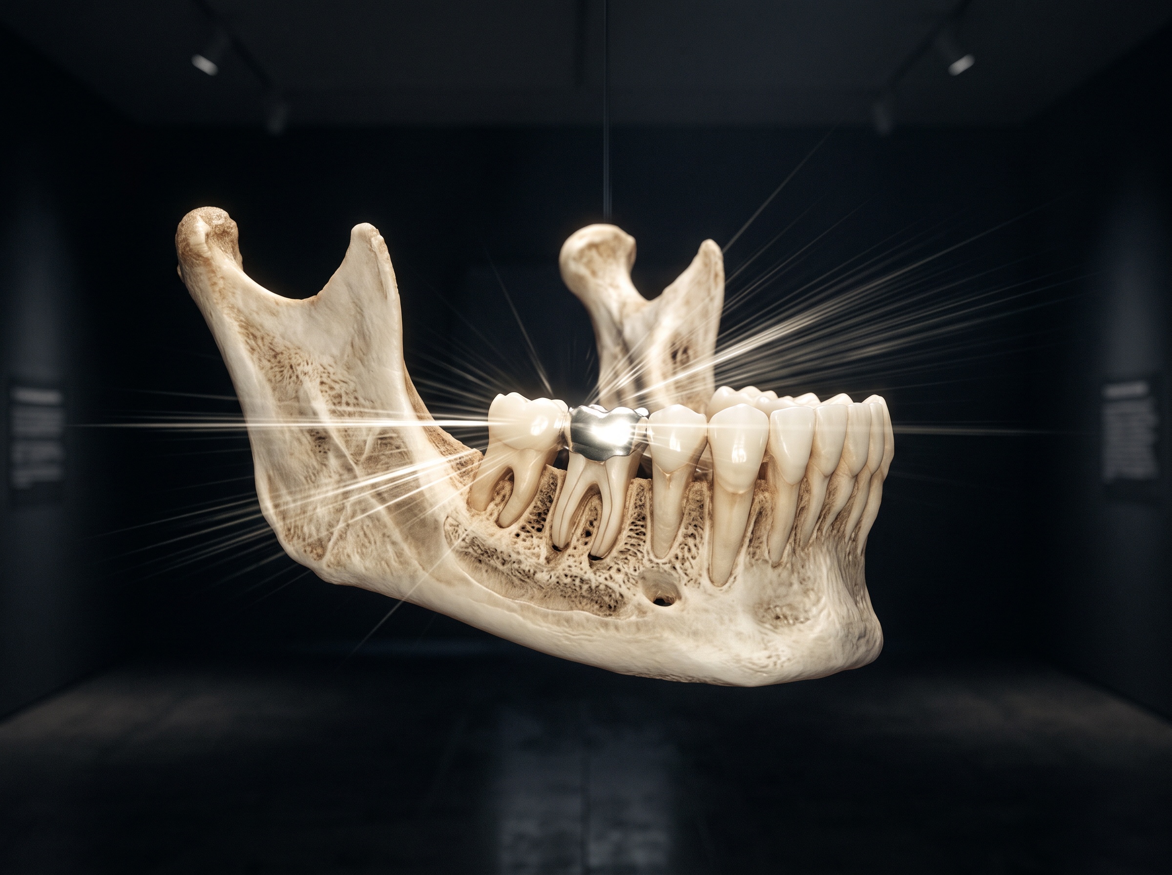
Cone-beam CT would seem, on paper, to be the richest input of all. It provides true depth, letting a model segment individual teeth, trace a canal, measure a bony defect or map an airway in three dimensions. Automated tooth segmentation on CBCT has become genuinely impressive, and volumetric analysis is one of the most compelling arguments for AI assistance. Yet the same abundance of data carries new hazards. The first is noise: cone-beam reconstructions are generally noisier than medical CT, and low-dose protocols trade image clarity for patient safety. The second, and the stubborn one, is the metal artifact. Dense restorations and implants scatter and harden the beam, throwing bright and dark streaks across the reconstruction that obscure exactly the regions a clinician most wants to examine. Because these streaks are reconstruction errors rather than anatomy, a model trained mainly on cleaner volumes can be led astray by them. The result is a modality that unlocks questions no two-dimensional image can answer, while quietly imposing its own ceiling on reliability. When the scan is read well, it reveals things the flat image never could, as in reading a CBCT for the breathing you cannot see.
Reading the Accuracy Numbers Honestly
None of this means one modality is better than another, and it certainly does not mean an AI result should be dismissed. It means that a single headline accuracy figure for a dental AI product is nearly meaningless unless it specifies the modality, the task and the reference standard it was measured against. A model may be excellent at flagging caries on a bitewing and far less dependable at calling a fine periapical lesion on a noisy panoramic, and both facts can be true of the same software on the same afternoon. The reference standard matters too: a pilot study measured against a single expert reader is a feasibility signal, not a guarantee, and small study sizes are common in this young field. The practical posture is to treat every AI finding as a prompt for attention rather than a conclusion, to know the specific blind spots of the modality in front of you, and to confirm on the original image before the result touches a treatment plan. This is also why the better tools now try to show their reasoning rather than merely assert it, a shift we examined in the visual grammar of explainable AI on the dental radiograph.
Future Developments
The trajectory is clear, and it points toward models that understand which medium they are reading. Regulatory clearances are already extending from two-dimensional into three-dimensional imaging, and the most interesting research is in modality-aware systems: artifact-reduction networks that clean a metal-streaked volume before interpretation, architectures that fuse the local certainty of the intraoral image with the spatial context of the scan, and training sets large and varied enough that a ghost image is recognised for the phantom it is. The craft of imaging will not be automated away by any of this. If anything, it becomes more important, because an algorithm inherits every virtue and every flaw of the picture it is handed. The practice that produces a clean, well-posed, well-exposed image, and that chooses the right modality for the question it is asking, gives the machine its best chance to be right. In the gallery of the dental record, the frame has always shaped the art. Artificial intelligence does not change that truth; it simply makes it impossible to ignore.
Sources & further reading:
No Comments