


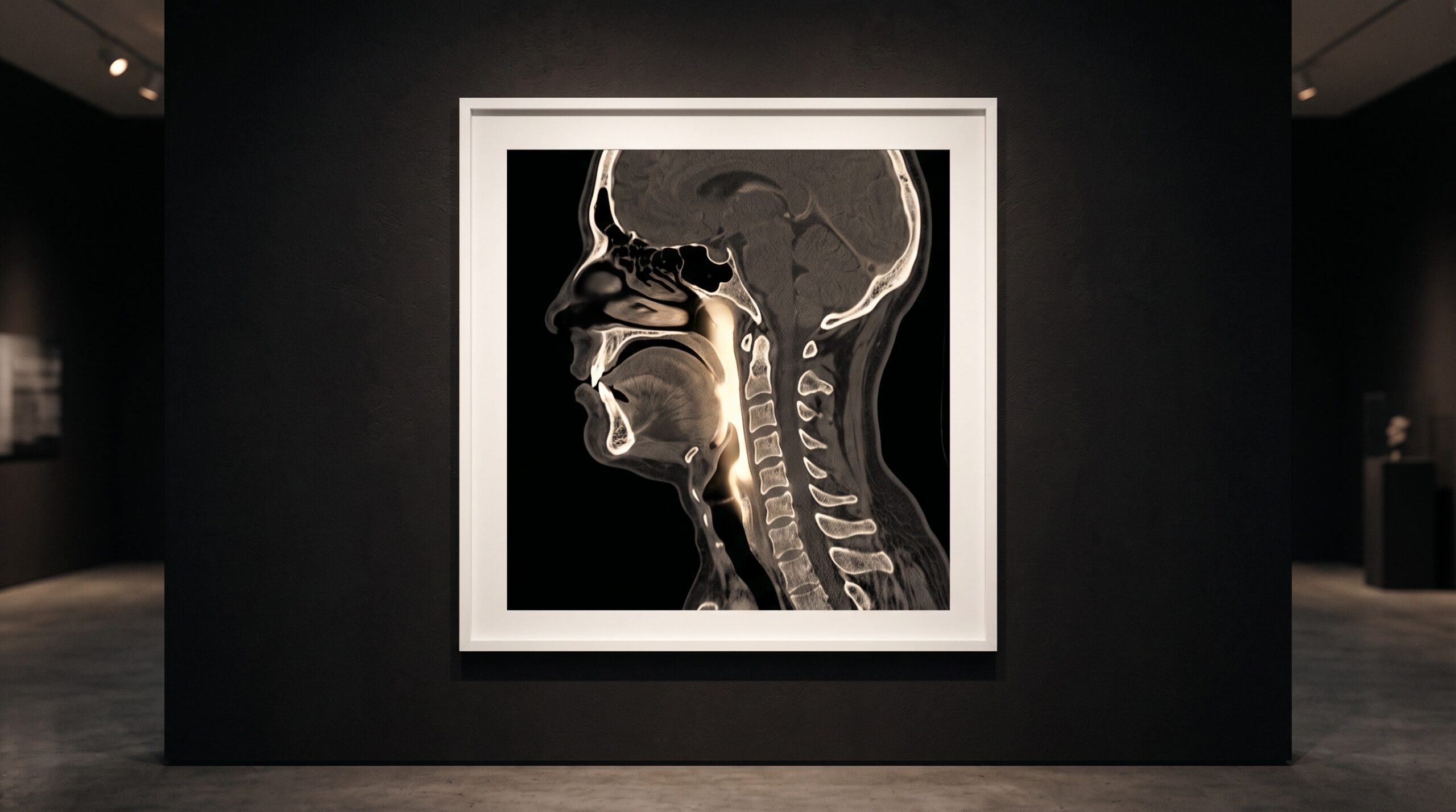
October 29, 2026 The Airway in the Scan: How Dentists Read a CBCT for the Breathing You Cannot See
Most cone-beam scans are ordered for something solid. An implant site, an impacted third molar, a root fracture, an orthodontic workup — the clinician is looking for bone and tooth, the hard architecture a flat film cannot fully show. Yet inside that same volume, running behind the tongue and soft palate and down toward the throat, sits something the scan records whether or not anyone asked for it: a column of air. That dark channel is the upper airway, and learning to read it — with discipline, and with a clear sense of what the reading can and cannot say — has become one of the quieter crafts in dental imaging.
The gallery has always held that an image deserves to be read in full, not merely for the finding that prompted it. The airway is the clearest example of why. It is the part of the scan most often ignored and, for some patients, the part that matters most.
The Passage the Scan Already Holds
A cone-beam volume with a field of view large enough to include the pharynx captures the full course of the upper airway: the nasopharynx behind the nose, the velopharynx behind the soft palate, the oropharynx behind the tongue, and the hypopharynx below. Because air is radiolucent and the surrounding soft tissue is not, the passage appears as a dark, continuous channel threading through the gray — a shape that can be isolated from everything around it. Whether that channel deserves attention depends partly on why the scan was taken in the first place, which is the same disciplined question of justification that governs choosing cone-beam CT over a simpler radiograph at all. An airway worth reading is a bonus of a scan properly indicated for another reason — not a reason to irradiate on its own.
From a Dark Channel to a Measurable Volume
Reading the airway seriously means turning the passage into something that can be measured, and that is the work of segmentation. The software thresholds the volume by density, separating the low-attenuation air from the tissue walls, and reconstructs the passage as a three-dimensional isosurface — a smooth sculptural object that can be rotated, volumized, and sliced. Two numbers dominate the reading. The first is total airway volume, the sheer amount of space in the column. The second, and the more clinically telling, is the minimum cross-sectional area: the single narrowest slice along the channel, the pinch point where airflow is most likely to fail. A generous total volume can still hide a dangerous constriction at one level, which is why the narrowest cross-section, not the bulk volume, tends to carry the weight. This is the same shift from picture to measurement that drives so much of modern practice, the move toward reading the image as data that also animates machine cephalometric analysis of the growing face.
Why a Dentist Looks at All
The dentist is not in the business of diagnosing sleep disorders, and should never pretend to be. But the dental chair is an unusually good vantage point onto the structures that govern breathing. Orthodontists planning to advance or restrict a jaw are altering the very walls of the airway, and a narrow pharynx changes the stakes of that plan. Clinicians fabricating a mandibular advancement appliance want to understand the space they are trying to open. And a strikingly narrow oropharynx in a patient who snores, wakes unrefreshed, and carries the daytime fog of poor sleep is a signal worth not ignoring. Seen this way, the airway is simply another instance of the principle that a scan shows more than the tooth — the same reason the full volume is read for everything it legitimately reveals, no more and no less.
The Limits Written Into the Image
Here the craft turns on honesty, because the airway measurement carries limits so fundamental they are built into the picture itself. A cone-beam scan is almost always taken upright, in a single held breath, during wakefulness. Obstructive sleep apnea, by definition, happens lying down, over hundreds of breaths, during sleep, when muscle tone collapses. The scan therefore measures a posture and a moment that are precisely the opposite of the condition it is tempting to read into it. Head position, tongue placement, and even the phase of respiration at the instant of capture can swing the numbers substantially. A narrow airway on an upright daytime CBCT raises a question; it does not answer one. Treating a static volume as a diagnosis of apnea is a category error — the imaging equivalent of mistaking a confident description for a careful reading, a trap this gallery has examined before in the context of tools that produce fluent output without true sight.
Reading Responsibly, Then Referring
What, then, is the airway reading actually for? It is a screening observation, not a verdict. The disciplined posture is to segment and measure when the field of view and the clinical question warrant it, to note a markedly constricted passage as a finding, to place it beside the patient’s history and symptoms rather than above them, and — when the picture and the story align — to refer for the one test that can actually diagnose the disorder: a sleep study. The dentist who sees the narrowing owes the patient not a diagnosis but a door. Documented plainly, measured consistently, and handed to the right specialist, an incidental airway finding on a scan ordered for other reasons can be the moment a serious, under-recognized condition finally gets named.
Future Developments
The near horizon is about closing the gap between the snapshot and the night. Research is pushing toward airway segmentation that runs automatically and reproducibly, stripping out the operator-to-operator variance that makes two clinicians trace the same passage differently. Computational fluid dynamics is being layered onto the static model to estimate how air actually moves through a given geometry, turning a shape into a simulation of flow and resistance. And efforts to pair upright imaging with supine or even sleep-state data aim squarely at the posture problem that limits today’s reading. None of it changes the discipline the image already demands: measure what is truly there, say only what the measurement supports, and know the difference between a channel of air captured in a single waking breath and the living, collapsing airway of sleep. The gallery has always insisted that an image be read with both ambition and restraint. The airway, quietly present in scans we took for other reasons, is where that balance matters most.
Sources & further reading:
No Comments