


October 25, 2026 The Ethic of the Exposure: New ADA Guidance and the Discipline of Imaging in Moderation
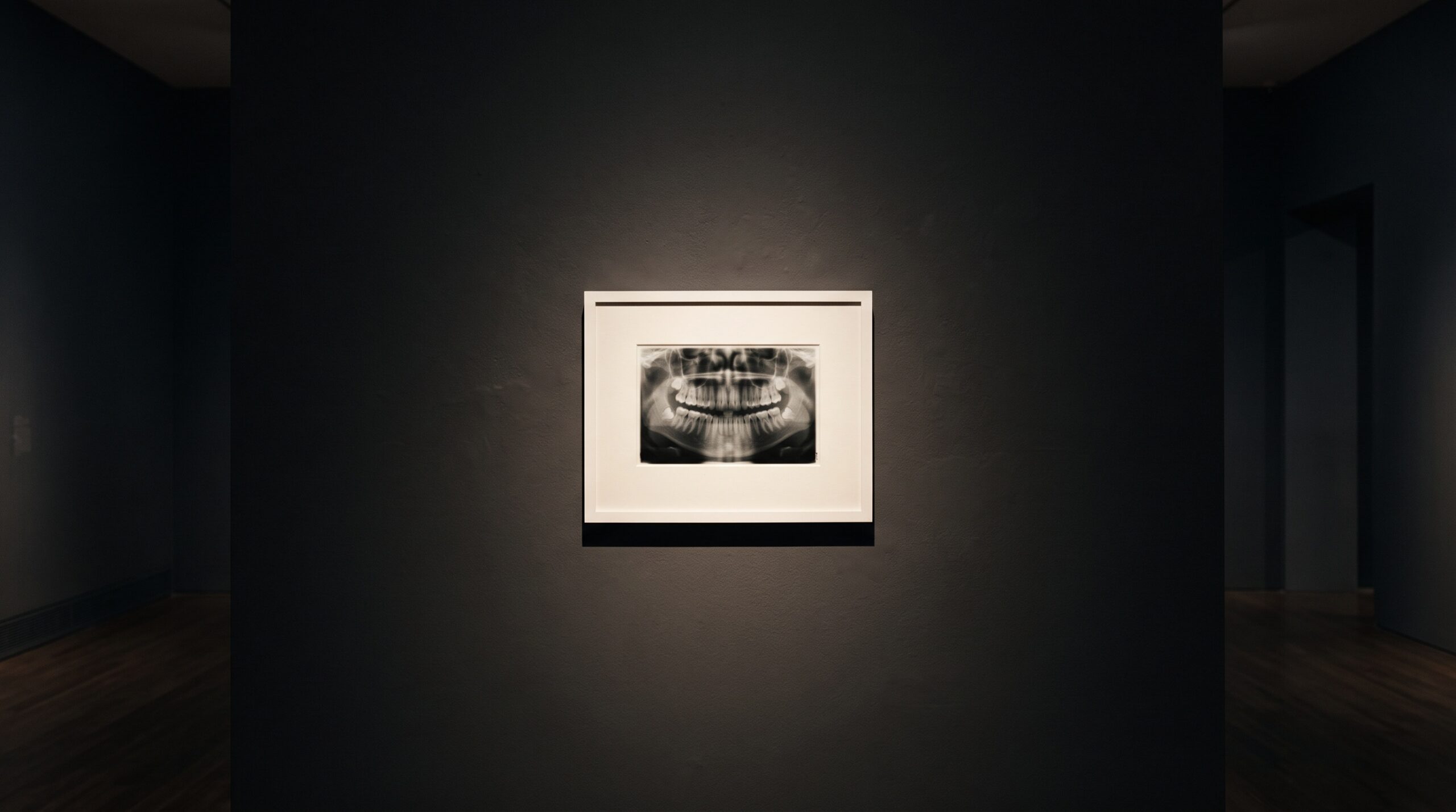
Every gallery is defined as much by what hangs on its walls as by what it leaves in storage. A curator’s real discipline is restraint: the willingness to let a vast dark wall hold a single, deliberately chosen image rather than crowd it with everything the archive could supply. For most of radiography’s history, dental imaging has leaned the other way. The exposure became a reflex — a standing order attached to the recall visit, a full-mouth series taken because the calendar said so rather than because a question demanded it. In January 2026 the American Dental Association published its first patient-selection guidance for dental X-rays in more than a decade, and its quiet thesis is one any gallerist would recognise: the image is most powerful when it is chosen, not when it is automatic.
The recommendations, developed by an expert panel of the ADA Council on Scientific Affairs and endorsed by the American Academy of Oral and Maxillofacial Radiology, confirm that dental X-rays are most effectively used in moderation — ordered only when clinically necessary to minimise radiation exposure for patients and clinicians alike. It is the first guidance of its kind to address both two-dimensional planar radiography and three-dimensional cone-beam computed tomography together, organised around specific clinical scenarios rather than a blanket schedule. The shift is subtle but profound. It moves imaging from the realm of routine to the realm of decision.
Look First, Then Decide
At the centre of the new guidance is an order of operations that reads almost like a studio discipline. The clinical examination comes first. Before an image is made, the clinician is asked to examine the patient, review the medical and dental history, and consult prior radiographs — and only then to determine whether a new exposure will actually support diagnosis, treatment planning, or clinical management. Radiographic exams, in this framing, should not be routine or automatic. The image is the answer to a question the examination has already posed; it is not a substitute for asking the question in the first place.
This is the opposite of imaging by default, and it reframes the radiograph as an instrument of inquiry rather than a box to tick. A gallery that photographs every object the same way, at the same interval, regardless of what the object is or what has changed, is not curating — it is cataloguing on autopilot. The craft lives in the judgement that precedes the shutter: this patient, this history, this clinical finding, therefore this image, at this resolution, now. And just as often, the judgement runs the other way — the examination resolves the question, the prior record already holds the answer, and the most skilful act is the exposure not taken.
CBCT and the Weight of the Decision
Nowhere does this discipline matter more than with cone-beam CT. A volumetric scan is a magnificent thing — a jaw rendered in three dimensions, sliceable and rotatable, a gift to surgical planning and complex diagnosis. But its dose is not that of a periapical film, and its radiosensitive targets — thyroid, salivary glands, the oral mucosa — deserve respect. The literature is consistent that CBCT effective doses run meaningfully higher than their two-dimensional counterparts, and that the youngest patients, with longer lifetimes ahead and more vulnerable tissue, carry the greatest share of any risk. The new guidance’s decision to treat 2-D and 3-D imaging within one selection framework is an acknowledgement that the question is never simply which image, but whether this image, at this dose, for this benefit.
Justification is the word radiation physicists use, and it is best pictured as a balance: on one pan, the diagnostic clarity an exposure promises; on the other, the dose it costs. CBCT belongs on that scale more squarely than any other modality in the operatory. The modern answer to the dose question is not to abandon powerful imaging but to pair restraint at the point of decision with efficiency at the point of capture — the kind of dose economy that photon-counting detectors and optimised low-dose protocols are beginning to deliver. Choosing to image less often and to image more efficiently are two halves of the same ethic.
The Archive as a Radiation-Protection Tool
There is a line in the guidance that imaging-minded clinicians should frame and hang on the wall: review the prior radiographs before making a new one. It sounds administrative. It is, in fact, one of the most elegant dose-reduction strategies available, and it costs nothing but attention. An image already in the gallery — properly captured, faithfully stored, and above all retrievable — frequently answers the clinical question without a fresh exposure. This is where the unglamorous disciplines of image quality and interoperability become acts of radiation protection in disguise. A radiograph that was captured to standard and can move freely between systems is a radiograph that need not be retaken. Every retake avoided is dose avoided. The gallery that keeps its collection well does more for the patient than it ever appears to.
The Discipline Behind the Picture
It is worth remembering that this guidance arrives on a continuing trajectory of measured confidence rather than fear. The same scientific current that, in 2024, retired the routine thyroid collar and lead apron — on the evidence that optimised technique and low modern doses had made them largely unnecessary — now asks us to be deliberate about the exposure itself. These are not contradictory impulses. They are the same impulse: let the evidence, not habit, govern the image. Shed the ritual protections that no longer earn their place, and decline the ritual exposures that never did.
For a practice that treats its imaging as a craft, none of this is a constraint. It is a refinement. The most beautiful radiograph is not merely the sharpest or the most elegantly composed; it is the one that needed to be made. Restraint, in imaging as in art, is not the absence of ambition but its highest form — the confidence to leave the wall dark until the right image, for the right reason, asks to hang there.
Future Developments
The direction of travel is clear, and it points toward an imaging practice that is at once more powerful and more sparing. Expect selection criteria to grow more granular and more personal — risk-stratified recall intervals tuned to the individual rather than the schedule, and perhaps, in time, decision support that weighs a patient’s history and findings to suggest when an image will genuinely change management. Dose-efficient hardware will keep lowering the cost side of the justification scale, and better-kept, freely-exchangeable archives will keep answering questions without new exposures. The gallery of the near future will not be the one that captures the most. It will be the one that has learned, with the quiet authority of evidence, precisely when to look — and when the most skilful act is to set the instrument down.
Sources & further reading:
- New Recommendations Confirm Dental X-Rays Most Effectively Used in Moderation (ADA)
- New ADA recommendations confirm dental imaging most effectively used in moderation (ADA News)
- Using thyroid collars during radiographic exams no longer recommended by ADA (ADA News, 2024)
No Comments