


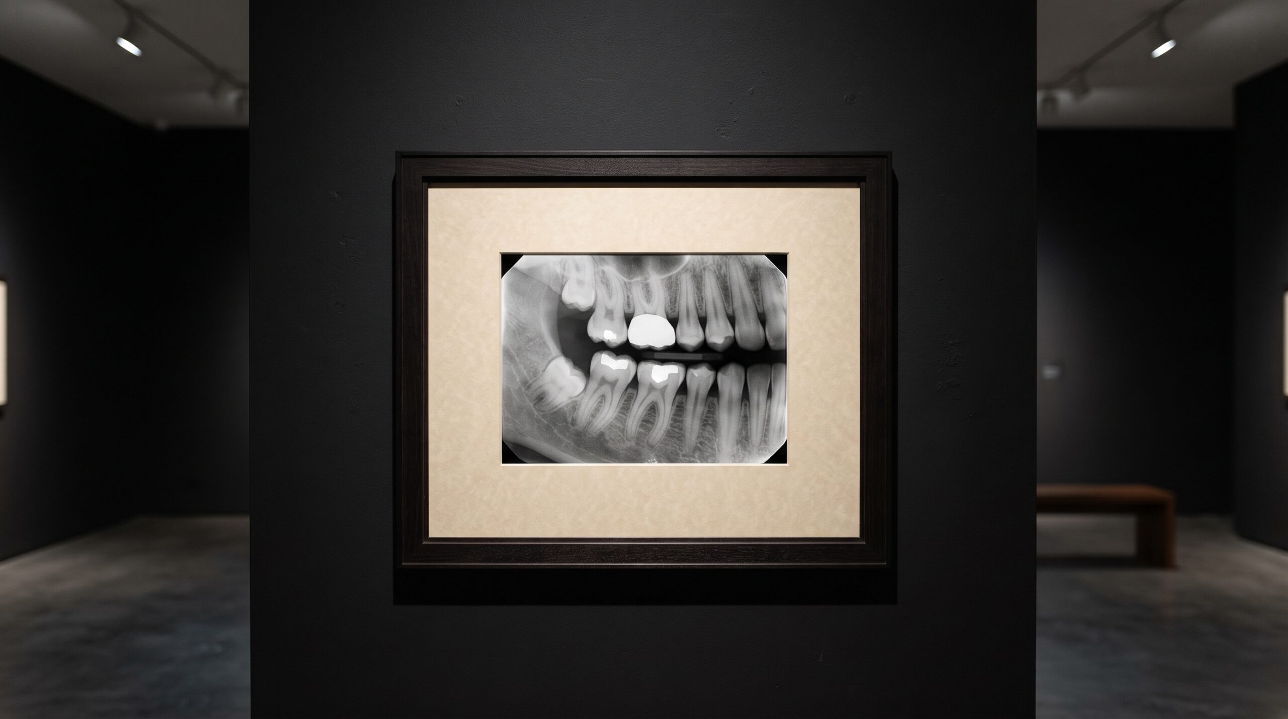
October 23, 2026 The Two Standards Behind a Trustworthy Radiograph: Quality at Capture and the Freedom to Exchange
In a field intoxicated by the new, it is easy to overlook that a radiograph has two separate ways of failing that have nothing to do with the cleverness of the machine that made it. The first is that the image is simply not good enough at the instant it is captured — underexposed, poorly positioned, soft where it needed to be sharp — a flaw no later brilliance can honestly repair. The second is quieter and crueler: the image is excellent, and then it cannot move. It will not open on the specialist’s workstation, it loses its calibration in transfer, it arrives stripped of the detail that made it diagnostic. Two unglamorous documents from the American Dental Association exist precisely to close these two failure modes, and in 2026 the Association is pressing to strengthen both. They are not detectors or algorithms. They are standards — the frame and the hanging-wire that let the picture survive.
Why a picture needs a rulebook
We tend to think of a standard as bureaucracy, something that happens to an image after the interesting work is done. It is closer to the opposite. A diagnostic image is a claim — this is what the bone, the margin, the canal truly looks like — and a claim is only as good as the discipline behind how it was produced and preserved. The ADA’s Standards Committee on Dental Informatics, through its working group on digital imaging, writes the documents that give that discipline a shape: what counts as an adequate radiograph, and how one practice’s image can be trusted by another’s system. For a discipline that treats the image as the art, this is the conservation science beneath the gallery — the unseen protocols that decide whether what hangs on the wall is authentic and whether it will last.
The first standard: quality at the moment of capture
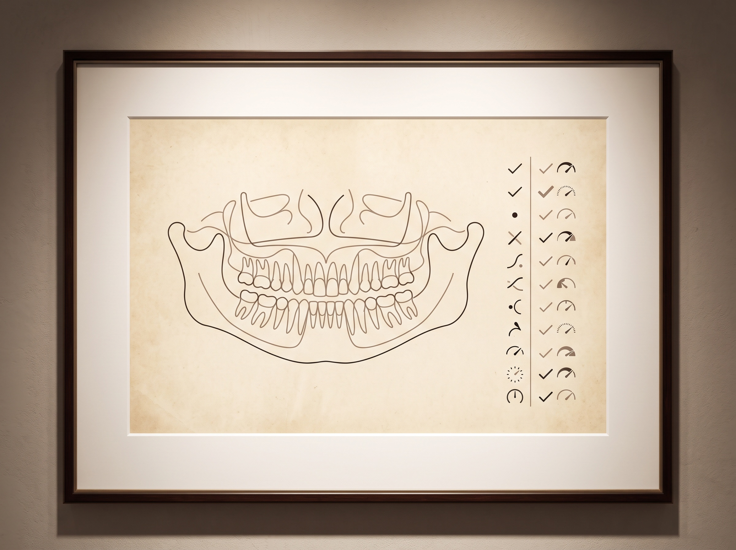
The quieter of the two concerns is quality assurance, and it answers a deceptively hard question: what, measurably, makes a digital panoramic or intraoral radiograph good enough to diagnose from? ANSI/ADA Standard No. 1099 addresses exactly this for digital panoramic and intraoral radiography — turning the intuitions a seasoned clinician carries about sharpness, contrast, density, positioning and the absence of artifact into defined, checkable requirements. This matters because the digital sensor quietly removed the old honest feedback of film: a poorly exposed digital image can be dragged toward looking acceptable on screen, masking a capture that was never diagnostic to begin with. A quality-assurance standard restores an objective floor. It is the same instinct that drives the work we described in how deep learning now grades the radiograph itself — the recognition that image quality is not a matter of taste but something that can, and should, be held to a specification.
The second standard: the freedom to be exchanged
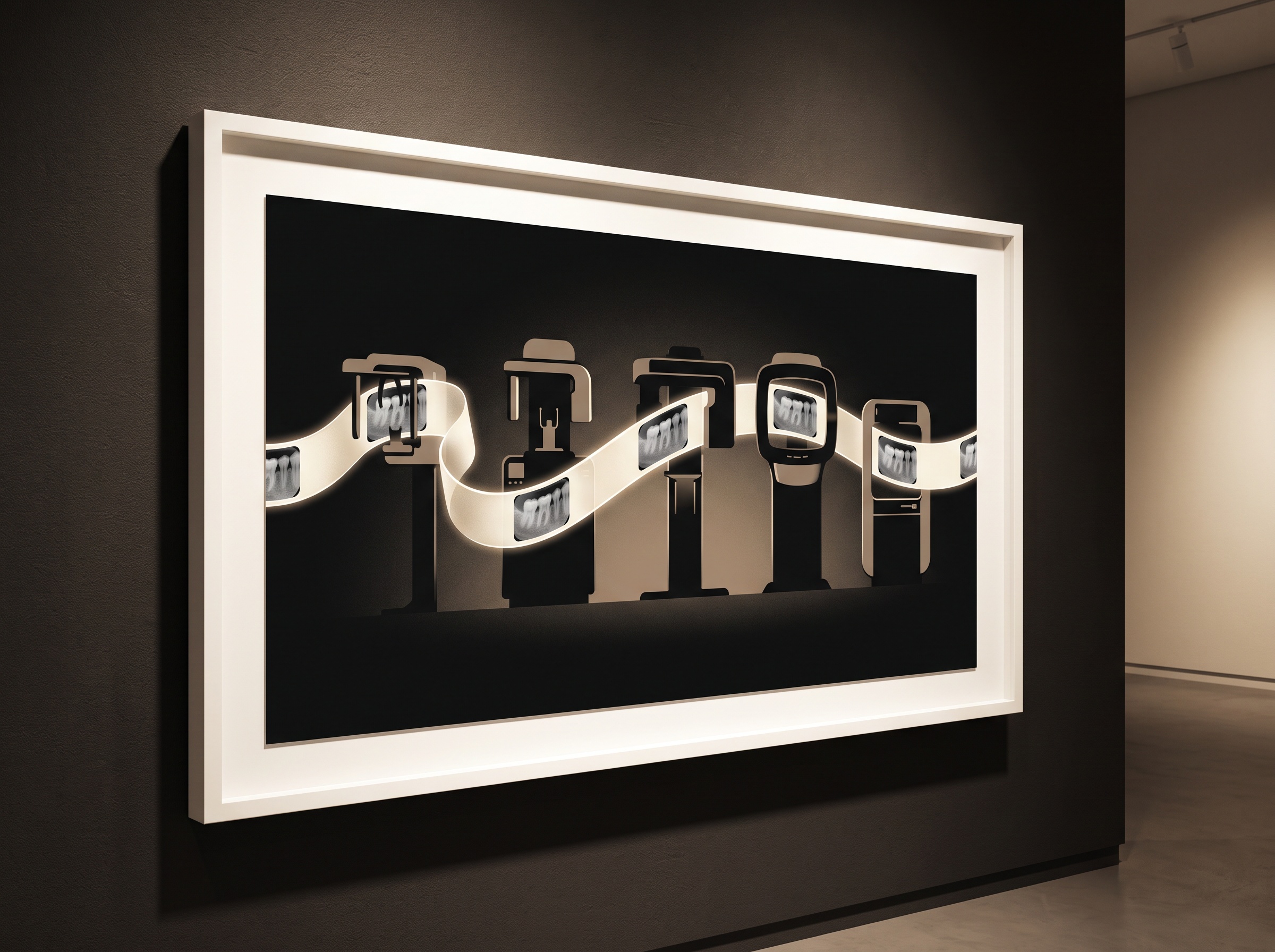
The louder, more visible problem is movement. A radiograph that cannot travel is, for every purpose beyond the room it was made in, only half an image — a difficulty we explored at length in the quiet cost of a radiograph that will not move between systems. Here the governing instrument is DICOM, the international Digital Imaging and Communications in Medicine standard, which the ADA supports as the basis for formatting, storing and exchanging digital radiography files so they remain interoperable across imaging systems, practice-management software and the offices of referring colleagues. ANSI/ADA Standard No. 1114 goes further, specifying the requirements for using DICOM effectively within dentistry — because supporting a standard in name and conforming to it in practice are not the same thing, as anyone who has watched a CBCT volume arrive subtly broken on a different vendor’s viewer can attest.
What a conformant image carries that a picture does not
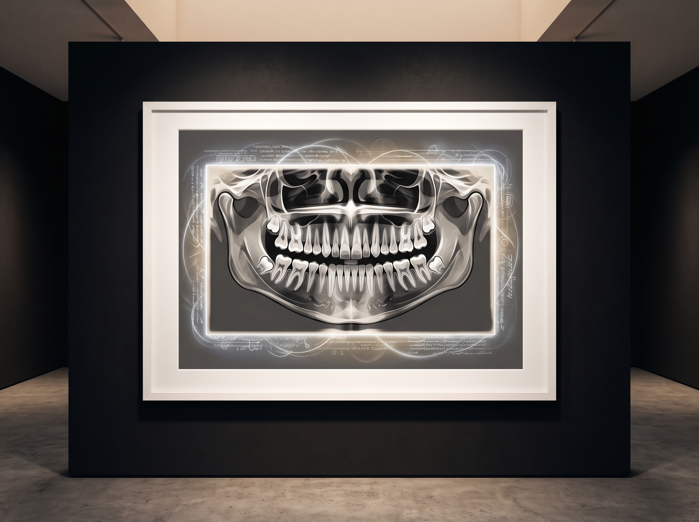
The deepest value of the exchange standard is something a casual viewer never sees. A properly formatted DICOM file is not merely a picture in an envelope; it carries a structured body of information with the image — the acquisition parameters, the geometry, the identifiers and provenance that let a later reader understand not just what the image shows but how it came to be. That embedded ledger is what makes an image trustworthy once it leaves its origin, and it is also, handled carelessly, what makes it a liability — the same hidden payload that governs long-term compression and archival integrity, and that must be respected when images cross between practices. A standard for exchange is therefore quietly also a standard for honesty: it insists the image travel with the context that keeps it diagnostic rather than arriving as a bare, uncertain shadow of itself.
The 2026 push, and why it is not finished
In March 2026 the ADA renewed its call for improved interoperability standards in dental imaging, pointing to the suite of informatics standards — 1114 for DICOM in dentistry among them — as the lever by which images, especially three-dimensional cone-beam volumes, can be exchanged between systems without degradation. It is worth being candid that this remains an aspiration pursued against friction. Conformance is uneven; manufacturers implement DICOM to differing depths; three-dimensional data strains formats designed in a two-dimensional age; and a standard is only as strong as the market’s willingness to adopt it over the soft convenience of proprietary lock-in. That the Association is still calling for improvement two decades into digital dentistry is not a sign the standards have failed — it is a sign the work of standardization is never a single act but a continuous negotiation between what is technically possible and what is commercially comfortable.
Future Developments
The horizon these two standards are converging toward is an image that is born trustworthy and remains so for its whole life. Picture a capture that validates its own quality against a defined specification at the moment it is taken, flags itself when it falls short, and is written from the first instant into a fully conformant, richly documented format — so that the question of whether it can be read elsewhere, now or in fifteen years, is simply never in doubt. As cone-beam and the newer three-dimensional modalities become routine, the pressure on the exchange standard will only grow, and the quality standard will have to learn to speak about volumes as fluently as it speaks about flat films. For a gallery that cares about the image as both science and art, there is something clarifying in giving these humble documents their due. We lavish our attention on the brilliance of capture and forget that brilliance undocumented and unexchangeable is brilliance that dies in the room it was born in. The detector makes the picture; the standard makes it endure. The most advanced radiograph in the world is worth little if it cannot be trusted the day it is made and still be read, faithfully, long after the machine that made it is gone.
Sources & further reading:
- ADA calls for improved interoperability standards for dental imaging (ADA News, March 2026)
- Standards support capture, exchange of high-quality digital radiographs (ADA News)
- X-Rays / Radiographs — ADA Oral Health Topics
No Comments