


February 9, 2026 When the Machine Becomes the Critic: Deep Learning Now Grades the Radiograph Itself
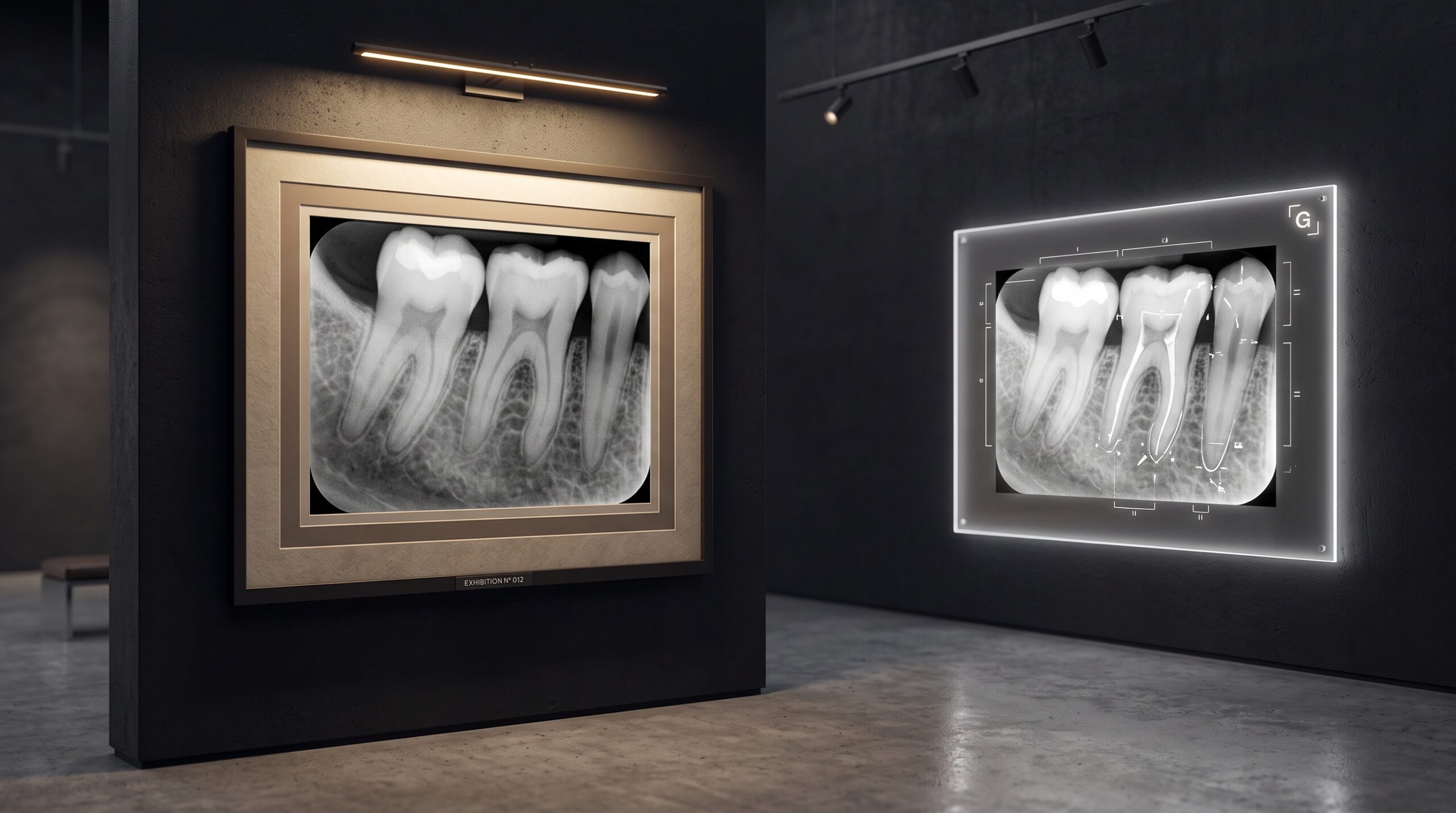
For as long as we have made radiographs, the final judgment of an image has belonged to a human being. Someone leans toward the screen, reads the apex, checks the framing, weighs the density against the diagnosis it must support, and decides — quietly, in a second or two — whether the picture is good enough or whether the patient must be exposed again. That judgment has always been a form of connoisseurship, the trained eye of a curator deciding whether a work belongs on the wall. It is now being taught to a machine. A deep-learning model, presented with a periapical radiograph, can appraise its diagnostic quality in the instant it is captured, and the implications reach far beyond convenience.
The Retake Nobody Frames
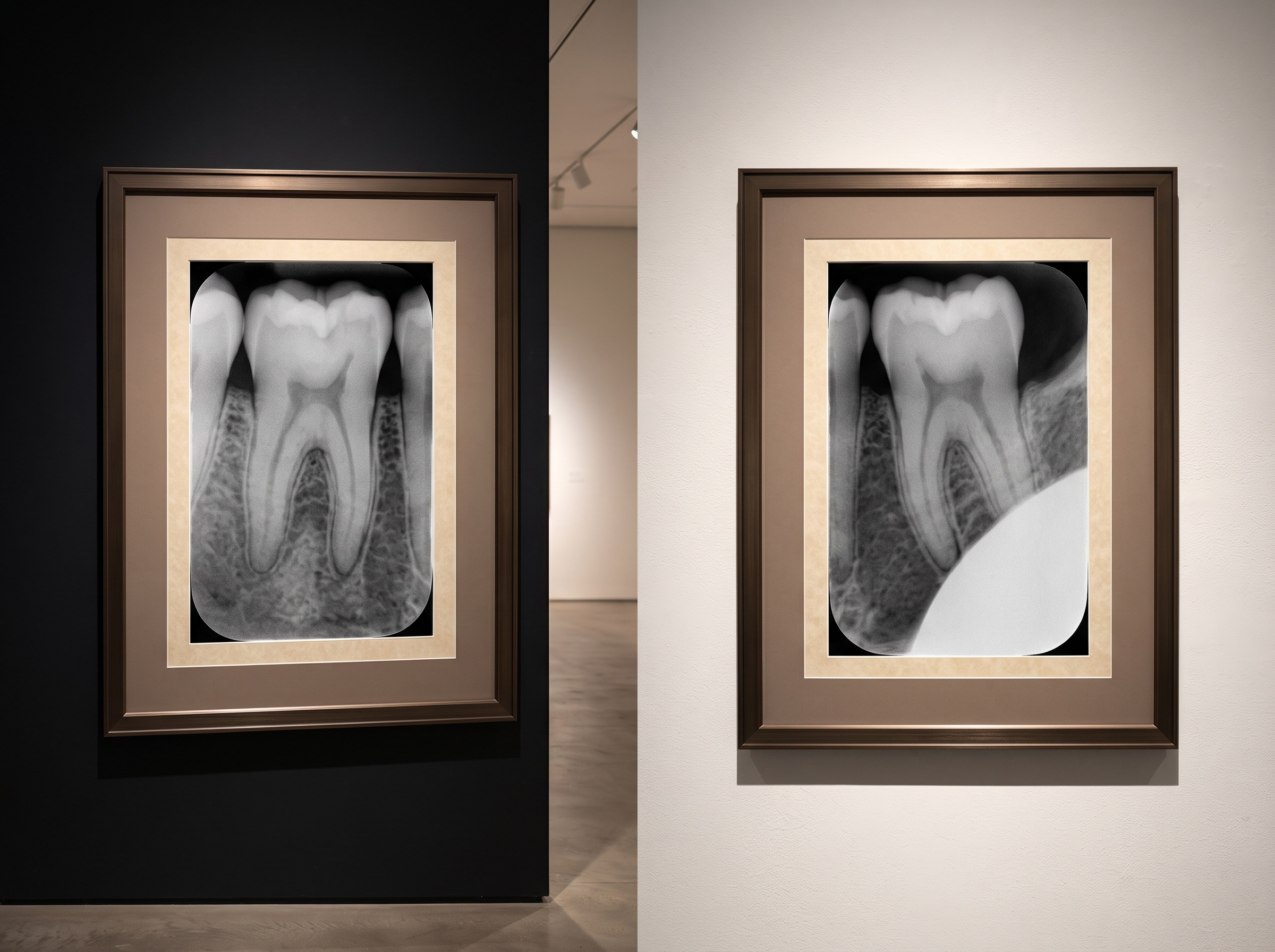
Every clinic keeps a private, unspoken tally of images that never make it to the wall. A cone cut across a corner, a root apex lost off the edge, a molar stretched by foreshortening until the crestal bone becomes a guess — each one is a small failure of geometry, and each one carries a real cost. The retake means another exposure, another increment of dose to a patient who was promised the least necessary, and a fracture in the workflow while the sensor is repositioned and the shot repeated. Studies of periapical technique have long shown that a meaningful fraction of images fall short on positioning or exposure. The retake is the photograph we quietly discard, and until now the only safeguard against it was the vigilance of a busy operator between patients.
What Makes a Periapical Worthy of the Wall
Before a machine can grade an image, we have to be honest about what “good” means, and in radiography it is refreshingly concrete. A diagnostic periapical shows the entire tooth including several millimeters beyond the apex, without the vertical distortion of elongation or foreshortening that a poorly angled beam introduces. It is free of the pale, dose-wasting arc of a cone cut, where the collimated beam missed the receptor. Its proximal contacts are open rather than overlapped, so early interproximal caries has somewhere to hide no longer. And its density and contrast sit in the narrow band where enamel, dentin, bone, and the delicate periodontal ligament space all remain legible at once. These are not matters of taste. They are measurable properties, which is precisely why they can be learned.
Teaching a Network to See Quality
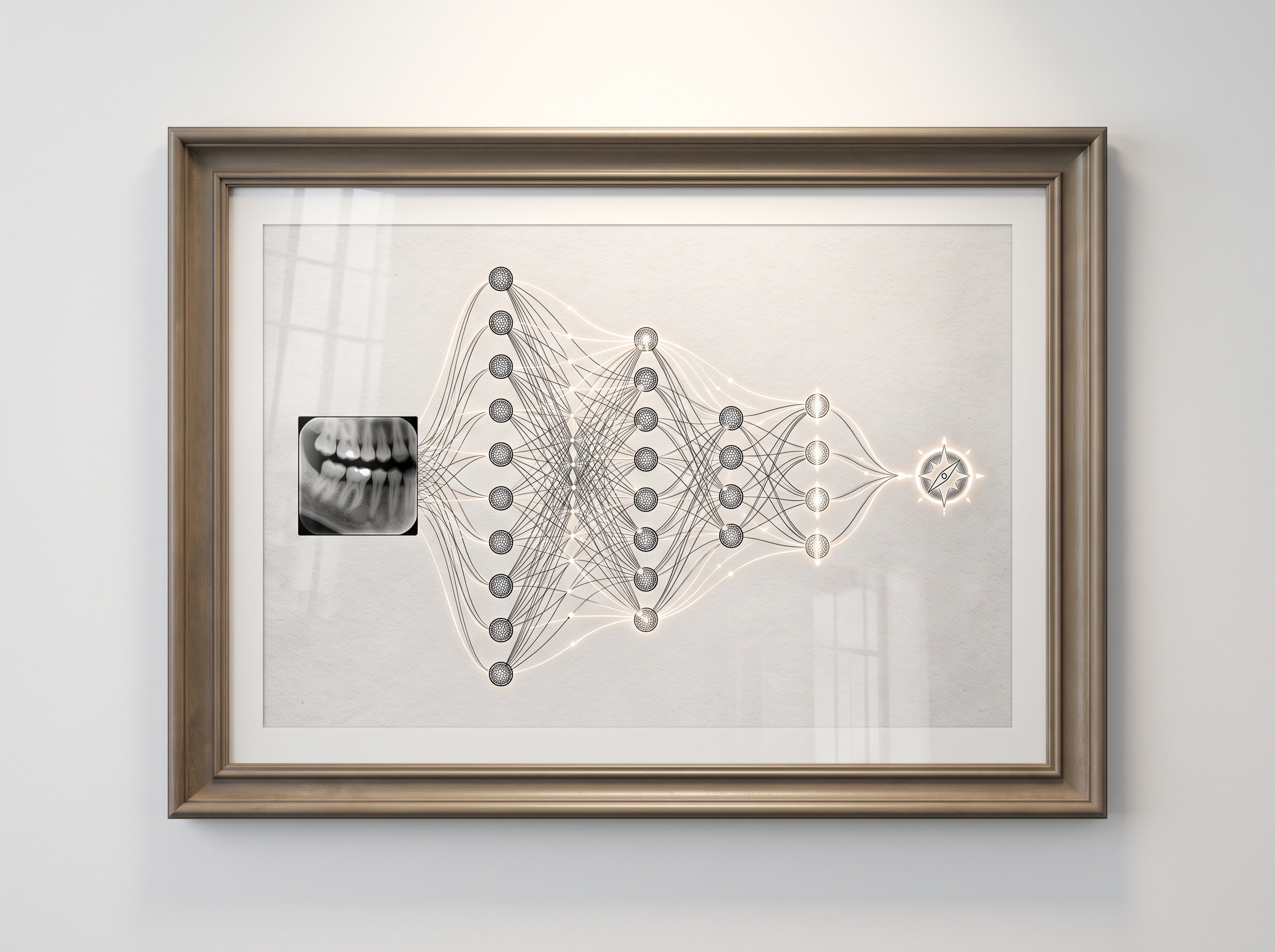
The approach at the center of recent work is elegant in its restraint. Rather than asking artificial intelligence to make the diagnosis — to find the caries or read the lesion — it asks the far humbler question a supervising radiographer asks: is this image good enough to diagnose from at all? Researchers reporting in Scientific Reports trained a convolutional neural network on large sets of periapical radiographs that human experts had graded for quality, letting the model learn the visual signatures of adequacy and inadequacy across thousands of examples. The result is a system that classifies image quality with striking consistency, mirroring expert assessment without an expert present. Its authors are candid that broader, multi-center validation is still required before clinical deployment, but the proof of concept is unambiguous: quality itself is a pattern a machine can recognize.
Objective Feedback at the Moment of Capture
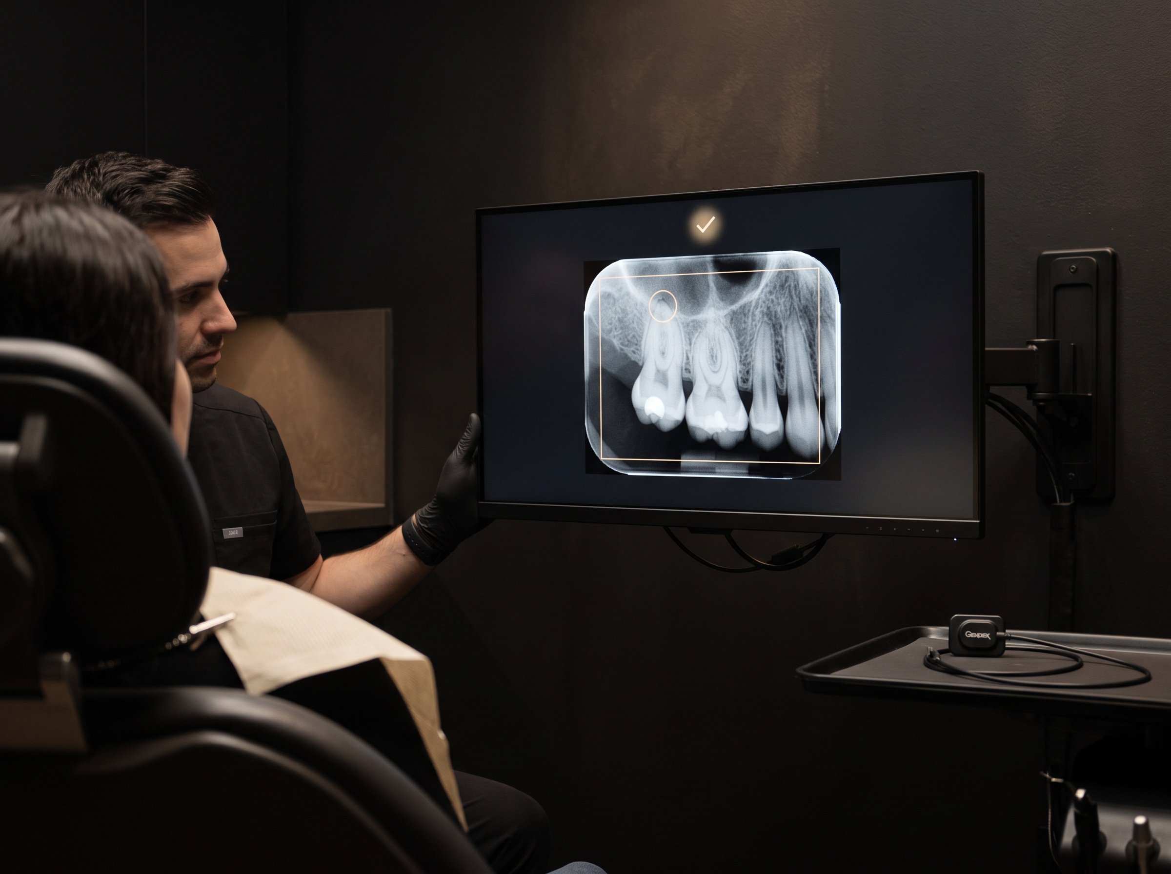
The power of this idea is not in grading old images but in grading new ones the instant they appear. Commercial dental AI platforms have begun to fold quality checks into the capture workflow, flagging a substandard radiograph on the monitor before the sensor has even left the mouth. The value proposition is deceptively simple and genuinely profound: real-time, objective feedback closes the loop while the patient is still in the chair. A retake becomes a second click rather than a second appointment. The gain is threefold — sharper diagnostic images, fewer wasted exposures and therefore less cumulative dose, and a smoother clinic that no longer discovers its bad radiographs after the patient has gone home. Objectivity is the quiet virtue here; the algorithm does not tire toward the end of a long day, and it holds the same standard for the first image of the morning and the last of the evening.
Consistency as a Form of Craft
There is an aesthetic argument buried inside this engineering one, and it is the argument this gallery cares about most. A single beautiful radiograph is a fine thing, but a consistent body of work is a greater one. When every image in a patient’s record is captured to the same standard of framing, density, and geometry, the archive becomes legible as a whole — one radiograph comparable to the next across years, so that the slow drift of a bone level or the subtle progress of a lesion reveals itself against an unwavering baseline. An algorithmic critic, applied uniformly, is how a practice earns that consistency at scale. It transforms image quality from a matter of who happened to be operating the sensor into a property of the system itself, a shared standard hung on every wall.
Where the Human Eye Still Presides
None of this dissolves the clinician’s role; it sharpens where that role truly lies. A machine that grades technical quality is answering a narrow question — is the image well made — not the deeper one of whether it was the right image to make, aimed at the right tooth, prescribed for the right clinical reason. It can certify that an apex is captured and a contact is open, but the meaning of what appears there, and the decision that follows, remain irreducibly human. There is a subtler caution as well: a quality score is a convenience, not an alibi. An operator who defers entirely to a green indicator risks unlearning the very connoisseurship that made the tool possible. The healthiest arrangement is the oldest one in craft — a skilled hand, now with a tireless second opinion.
Future Developments
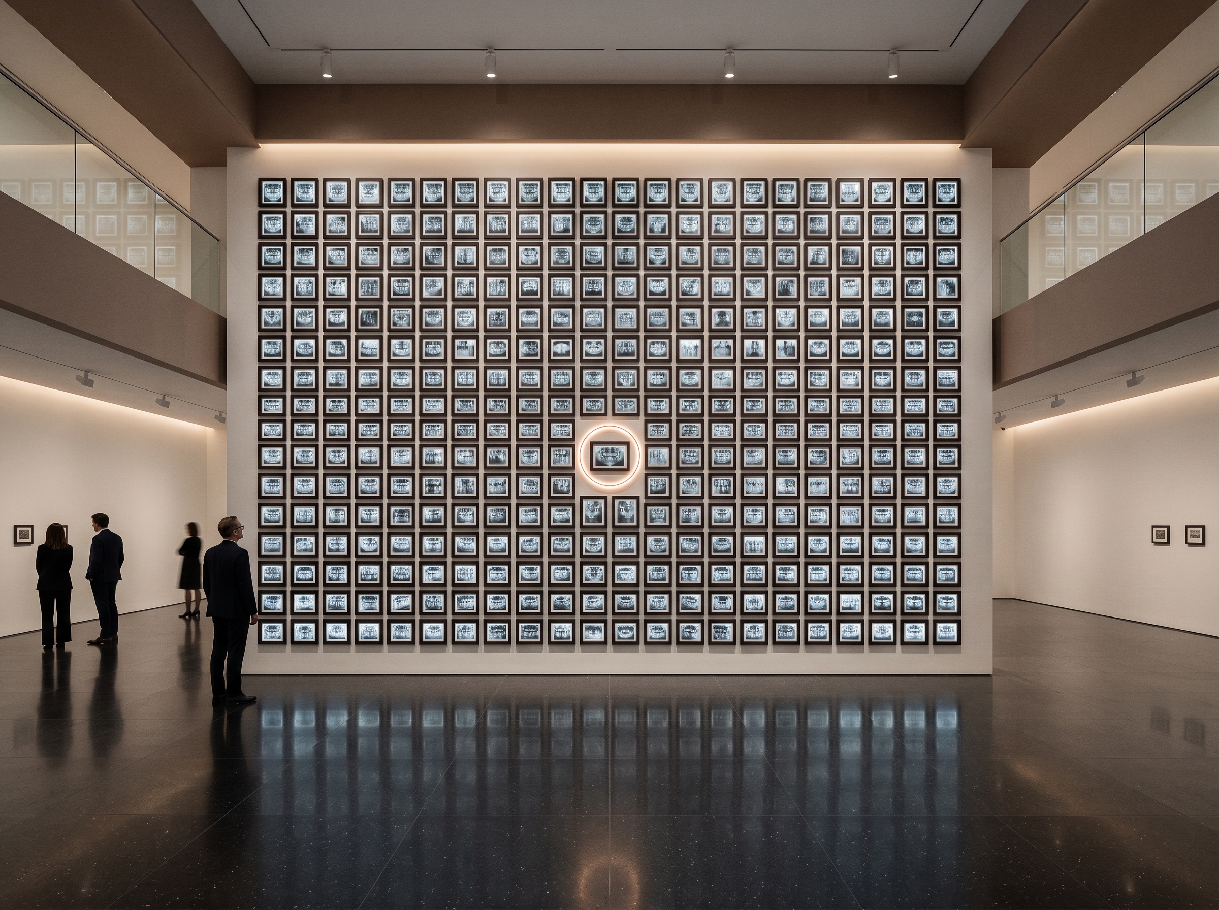
The near horizon is a quiet integration, the kind that becomes invisible once it works: quality assessment folded directly into every sensor and software suite, so that objective feedback at capture is simply how radiographs are made, the way autofocus became simply how photographs are taken. From there the ambitions deepen. A model that recognizes why an image failed — too much vertical angulation, a receptor placed too shallow — could coach technique rather than merely flag the result, teaching positioning in the moment. Federated learning across many practices could refine these judgments without patient images ever leaving the building, answering the validation and privacy questions in a single stroke. What endures beneath all of it is the shift we are living through now: the radiograph has acquired a critic of its own. To make images of the mouth as both science and art has always meant holding oneself to an exacting standard. For the first time, that standard can look back at us, image by image, and quietly insist we meet it.
Sources & further reading:
- Deep learning-based assessment of periapical radiographic image quality (Scientific Reports)
- AI dental radiograph analysis and image interpretation (Pearl)
Sorry, the comment form is closed at this time.