


September 8, 2026 Landmarks by Machine: How AI Traces a Cephalometric X-Ray for Orthodontic Analysis
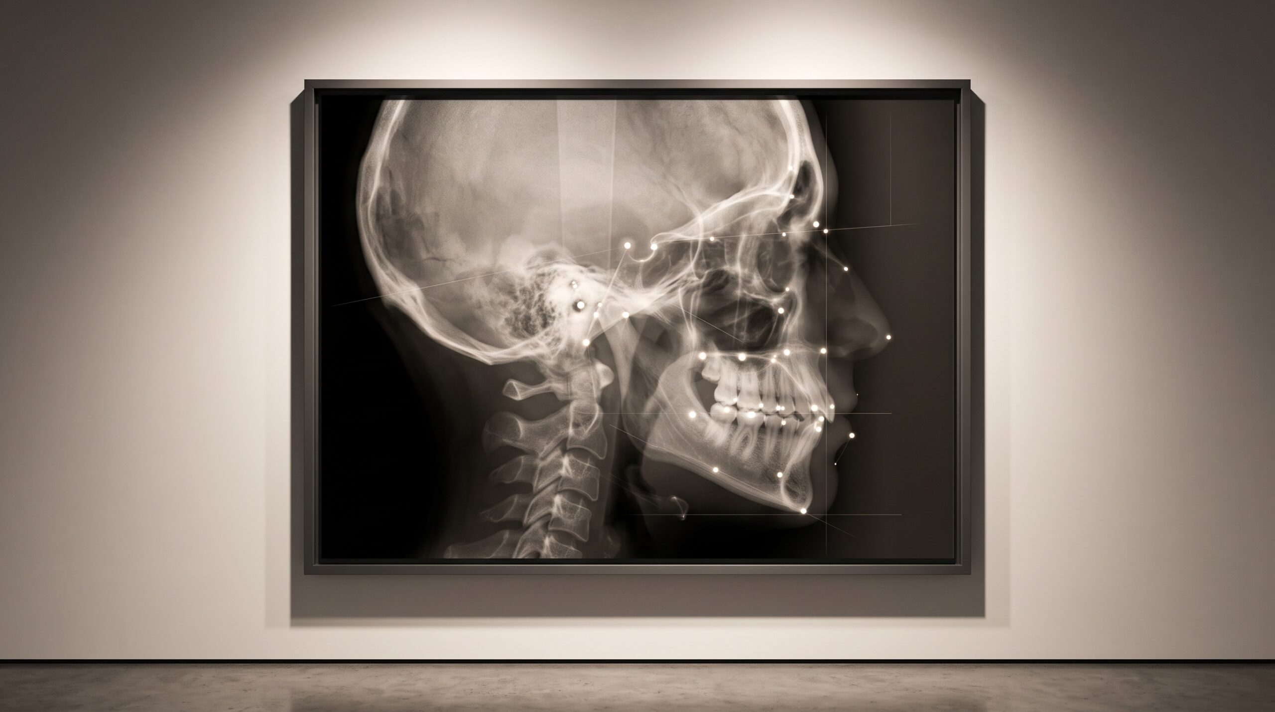
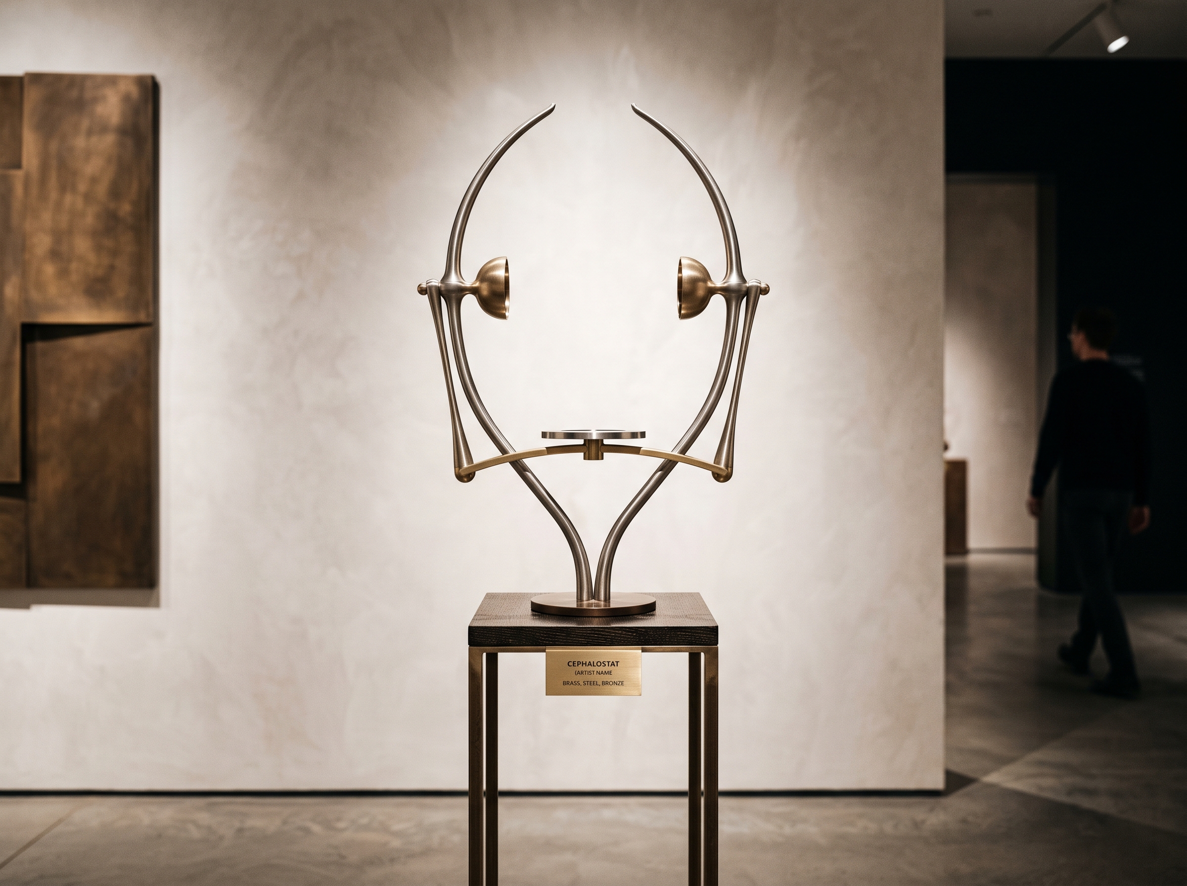
Among all the images dentistry makes, the lateral cephalogram is the strict one. A bitewing or a periapical is captured in the geometry of the moment – wherever the sensor sat, whatever angle the beam took. The cephalogram refuses that freedom. It is made with the head clamped in a cephalostat, two ear rods seating the skull in exactly the same orientation every time, at a fixed and deliberately long source-to-object distance so that magnification is a known constant rather than an accident. The point of all that rigour is reproducibility: a radiograph so standardized that the same anatomical landmarks can be found on it this year and again in three years, and the difference between the two traces can be trusted to mean growth rather than a shift in technique. It is, in the most literal sense, imaging built to be measured. And for the better part of a century, the measuring was done by hand.
That handwork is the craft this article is really about, because it is the part a machine has now quietly taken over. To analyse a cephalogram, a clinician first has to find its landmarks – a set of named anatomical points scattered across the skull profile. Sella, the centre of the pituitary fossa. Nasion, where the frontal and nasal bones meet. A point, the deepest concavity on the front of the upper jaw; B point, its counterpart on the lower. Pogonion and Menton on the chin, Gonion at the angle of the jaw, Orbitale and Porion defining the horizontal reference plane. None of them announces itself. Each is a judgement about where, precisely, a soft curve of bone reaches its extreme.
Why the Points Matter
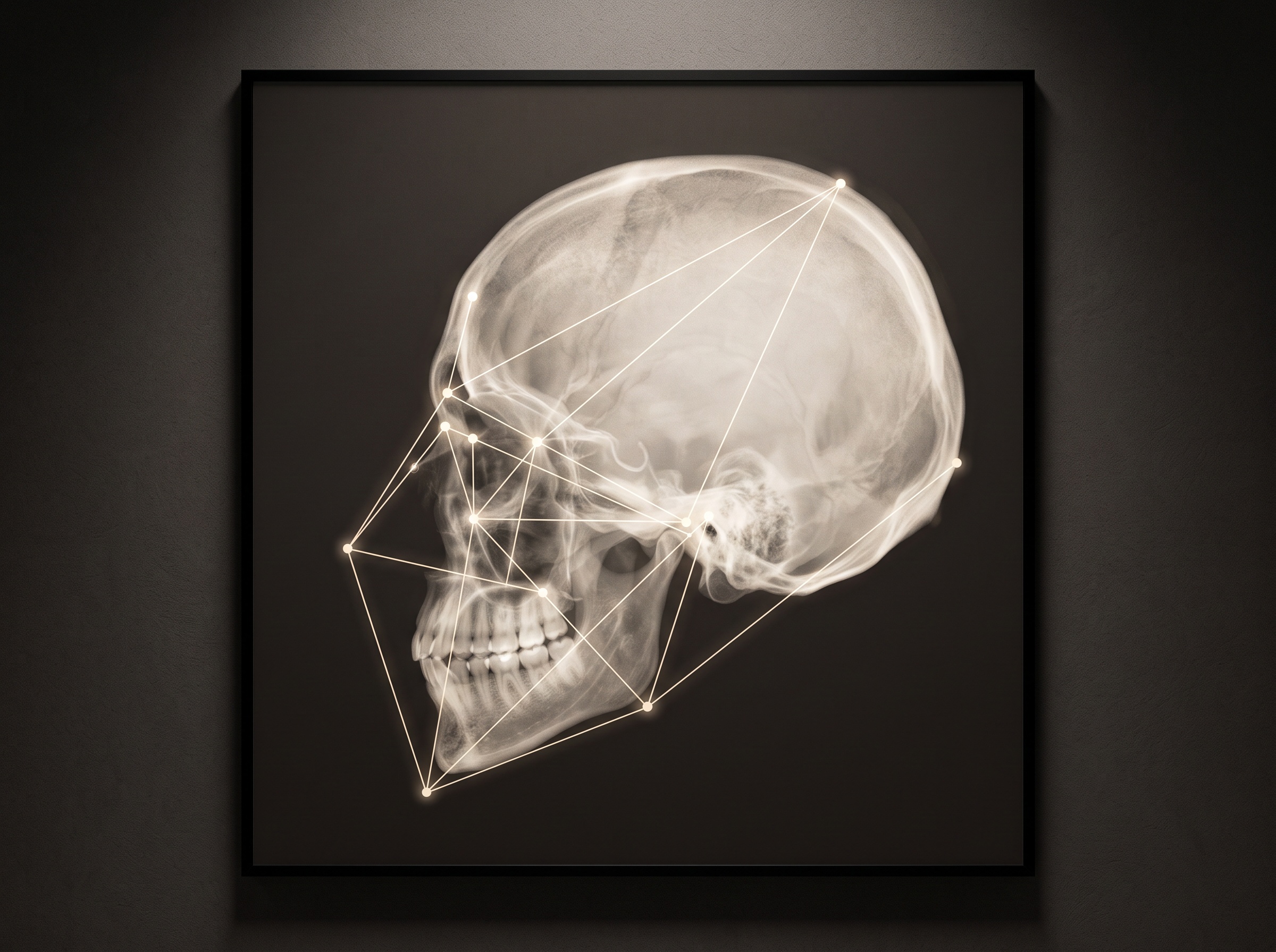
Those points are not the destination; they are the coordinates from which meaning is computed. Connect Sella to Nasion to A point and you have the SNA angle, a measure of how far forward the upper jaw sits beneath the skull base. Swap A for B and you have SNB, the same question asked of the lower jaw. The difference between them – the ANB angle – is the number that classifies a patient’s skeletal relationship into the familiar orthodontic categories and, with a handful of companion measurements from the Steiner, Ricketts or Wits analyses, drives the treatment plan itself. This is the same movement from raw image to clinical statement that we traced in From Detection to Diagnosis: the picture is only the substrate, and the diagnosis lives in what is extracted from it. On a cephalogram, everything downstream depends on where those first points land.
The Drift in the Human Hand
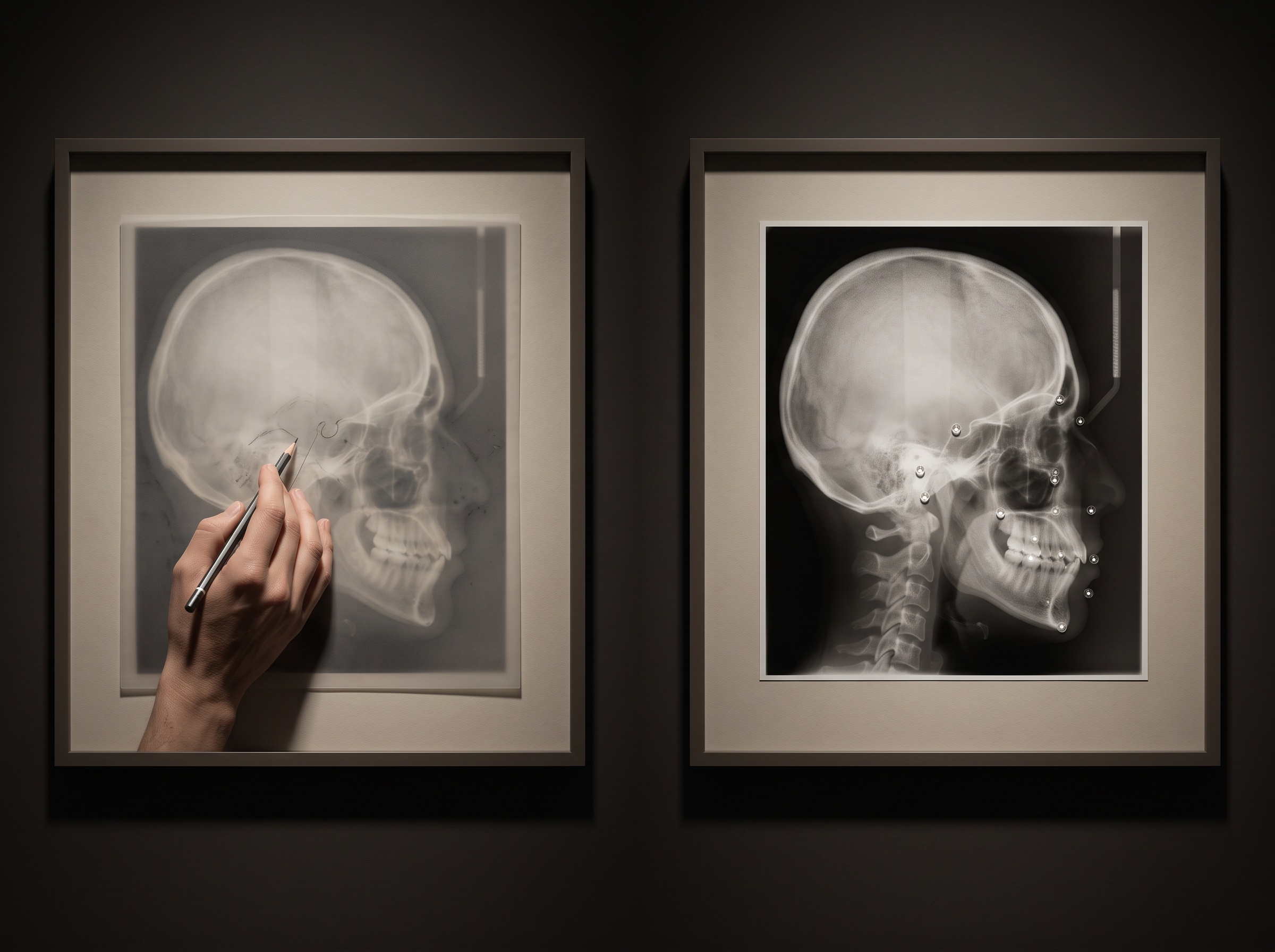
And this is where manual tracing shows its weakness. It is slow – fifteen or twenty patient minutes with a fine pencil over an acetate overlay, or the digital equivalent of clicking point after point. Worse than slow, it drifts. Ask two experienced orthodontists to trace the same film and their landmarks will not coincide exactly; ask the same clinician to trace it twice and the second attempt will differ from the first. The variability is not uniform, either. A high-contrast point like Sella, cradled in an obvious bony fossa, is placed consistently. But a landmark like Gonion, sitting on a smooth curved jaw angle with no crisp feature to anchor it, or Porion, buried under the superimposed shadows of both sides of the skull, wanders by a millimetre or more between observers. That wander does not stay put; it propagates into every angle built on the point, so a soft placement of B point quietly falsifies the ANB that a treatment decision rests on. The measurement inherits the uncertainty of the human who found the points.
How the Machine Learns to Look
The modern solution is a convolutional neural network trained on thousands of cephalograms that skilled clinicians have already traced by hand. Rather than reasoning about anatomy the way a person does, most systems learn to produce a heatmap for each landmark – a probability field across the image that glows brightest where the point most likely sits – and then read the coordinate off the peak. Because the network has seen so many hand-traced examples, it internalises the same visual cues an expert uses: the shape of the sella turcica, the concavity that defines A point, the curvature that marks Gonion. What it does not do is tire, rush, or place the point differently on a Friday afternoon than a Monday morning. Given the same image, it returns the same coordinates – the very consistency the human process cannot guarantee. In seconds it lays down the full constellation of points and computes every downstream angle.
Measuring the Machine Honestly
A field that grew up obsessing over reproducibility was never going to accept a black box on faith, and the metrics reflect that. Automated landmarking is judged by the mean radial error – the straight-line distance, in millimetres, between where the machine placed a point and where a reference examiner did – and by the success detection rate, the fraction of landmarks that fall within a clinical tolerance, conventionally two millimetres. Two millimetres is the threshold because errors smaller than that rarely change the diagnostic interpretation of an angle. The best contemporary systems now clear that bar for the large majority of standard landmarks, placing them within a range that approaches the disagreement between two human experts – which is the honest ceiling, since the ground truth is itself only a consensus of fallible people. The claim is not that the machine is perfect. It is that the machine is no less reliable than the humans it learned from, and far more consistent.
Where It Still Struggles
The failure modes are worth naming plainly, because they follow directly from the physics. A lateral cephalogram is a two-dimensional projection of a three-dimensional skull, so bilateral structures superimpose – the left and right jaw angles overlap into a single ambiguous shadow, and a network can be pulled toward the wrong edge just as a human is. Unusual anatomy and pathology, underrepresented in any training set, degrade accuracy exactly where careful measurement matters most. And the whole enterprise rests on input quality: a cephalogram with poor contrast, motion blur, or incorrect positioning gives the network the same impoverished evidence it gives a person, and no amount of learned skill invents detail that the exposure never captured. This is the cephalometric case of a rule that holds across all of imaging AI, one we made in Garbage In, Garbage Out and again in How AI Reads a Bitewing: the algorithm is only ever as good as the radiograph handed to it. The standardized image was always the foundation; the machine simply makes the cost of a bad one more visible.
Future Developments
The most consequential shift ahead is dimensional. As cone-beam volumes become routine, cephalometric analysis is migrating off the flat projection and into three dimensions, where the superimposition problem that plagues the lateral film simply dissolves – the left jaw angle and the right are finally separate objects, and a landmark can be placed in true space rather than on a shadow. AI is what makes 3D cephalometrics tractable at all, since hand-placing points through a volume is prohibitively slow. Alongside that, the more mature systems are learning to express uncertainty – to return not just a coordinate but a confidence, flagging the landmark it found ambiguous so a clinician’s eye goes straight to it. That is the mature form of this technology, and the one most in keeping with how PatientGallery reads the whole field: the machine does the patient, repetitive, drift-free work of measurement, and hands the human back a cleaner canvas on which to exercise judgement. The craft was never in the clicking of the points. It was always in knowing what the angles mean – and that remains, gratifyingly, the clinician’s art.
Sorry, the comment form is closed at this time.