


July 25, 2026 Who Teaches the Machine to See? The Overlooked Craft of Annotating Panoramic Radiographs
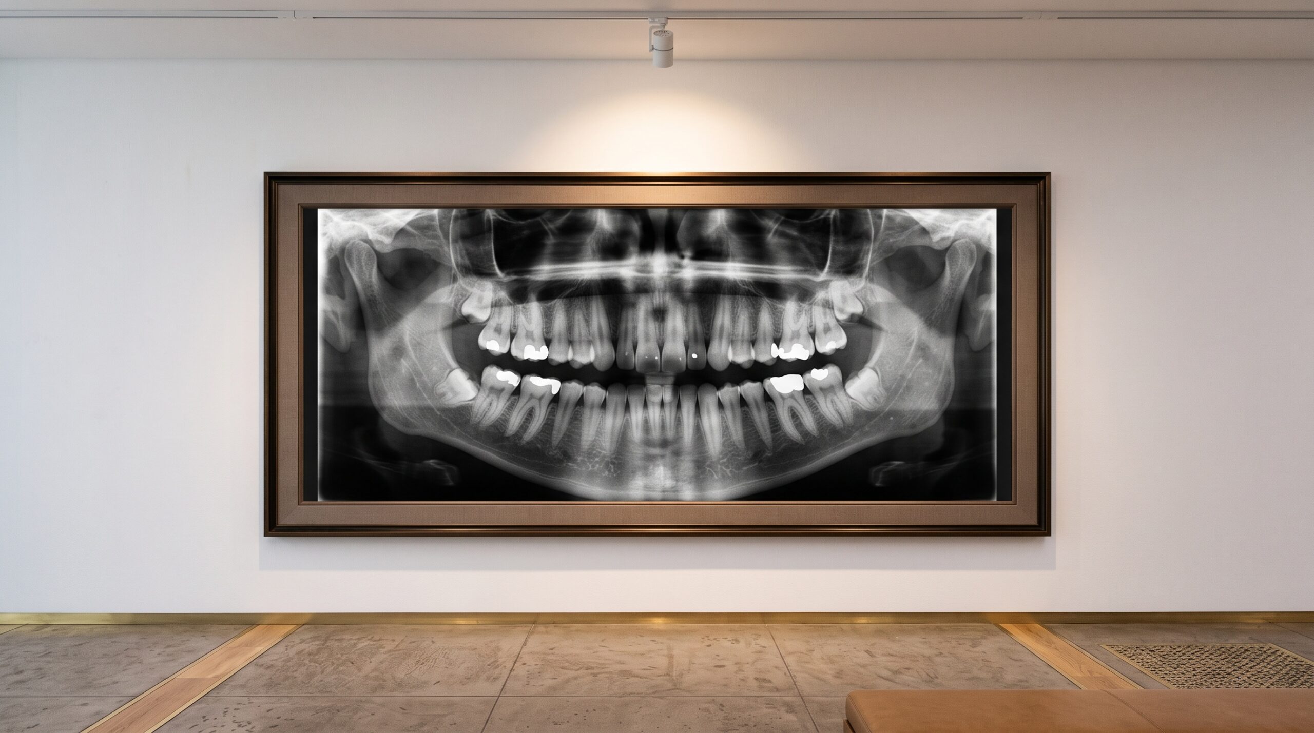
Hang a panoramic radiograph on a wall and it behaves like any good portrait: the eye travels the sweep of the arch, lingers on a bright restoration, reads the quiet geography of bone. It looks complete, self-evident, finished. Yet the moment we ask software to see the same image, that illusion of completeness falls away. A machine sees only a field of grey values until someone, pixel by pixel, tells it what each region means. Behind every algorithm that flags a lesion or counts a tooth stands a quieter and largely uncredited craft, and a newly published open dataset of multi-focus panoramic images, annotated at the level of the individual pixel, is a fine occasion to look at it directly.
The Portrait and Its Hidden Draftsmen
We tend to speak of dental artificial intelligence as if it learned to read radiographs the way a resident does, by absorbing thousands of films over years of practice. The truth is more deliberate and more human. Before a network can recognize a molar, a person must first mark, on real images, exactly where every molar is. That marking is the training data, and its quality places a hard ceiling on everything the model will ever do. A brilliant architecture trained on careless labels produces confident nonsense.
The dataset that prompts this reflection does something unusually rigorous: it labels panoramic films not with loose rectangles but at pixel-level fidelity, tracing the true contour of each structure. It is painstaking work, closer to manuscript illumination than to data entry, and it is performed by clinicians whose names will never appear in the products their labor makes possible.
What Pixel-Level Really Means
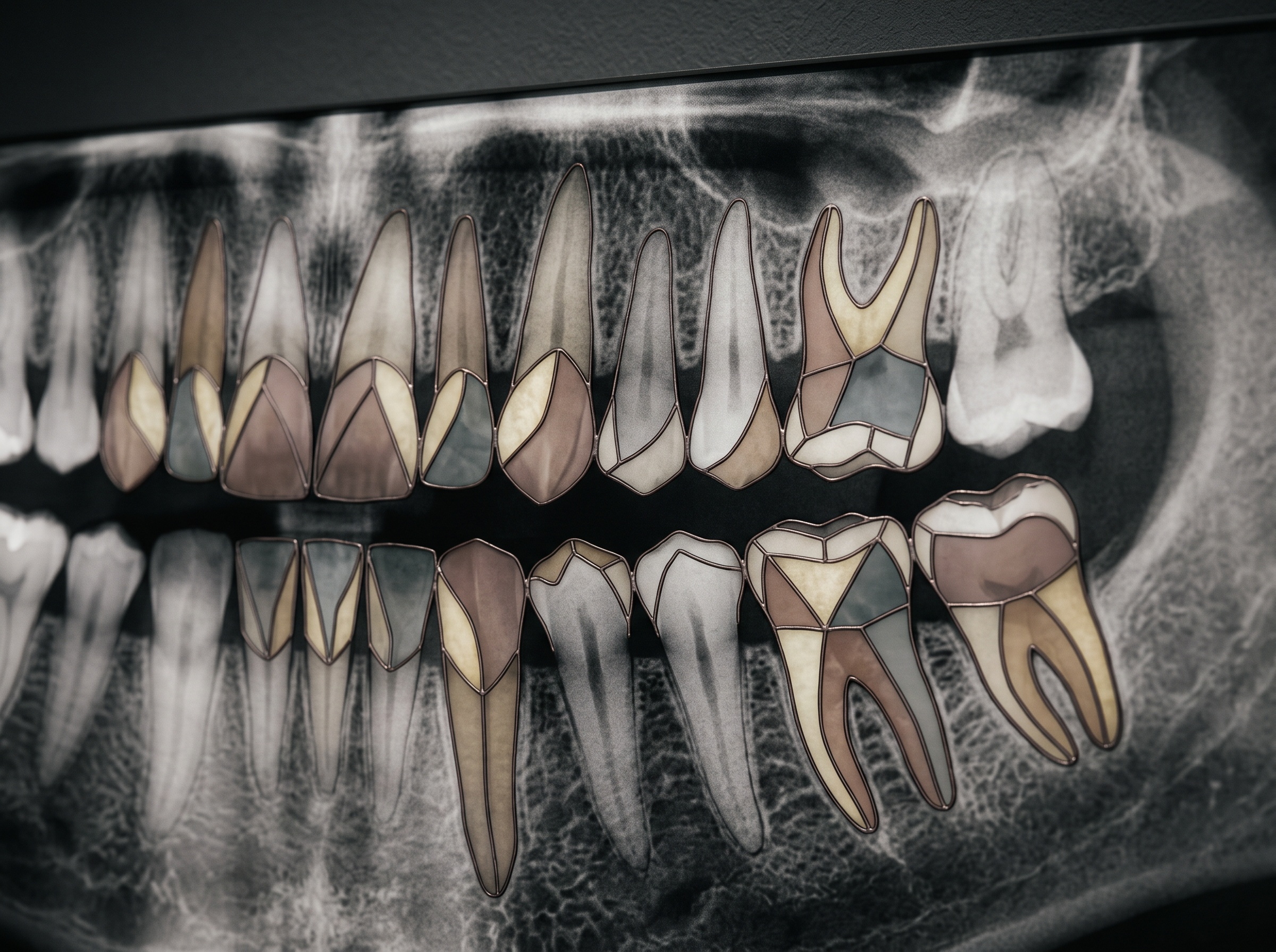
Not all annotation is equal. The cheapest form draws a box around an object and declares, somewhere inside this rectangle, there is a tooth. It is fast and it is coarse, and for a curved, crowded dental arch it is nearly useless, because neighboring boxes overlap and the background bleeds into everything. Pixel-level annotation, by contrast, assigns a meaning to every single point in the image. The result is a segmentation mask: a second image, laid over the first, in which enamel, dentin, bone, sinus, and restoration each occupy their own precisely bounded territory.
This is the difference between pointing at a painting and copying its every brushstroke. The mask does not approximate the tooth; it is the tooth’s silhouette, rendered as data. That precision is what allows a downstream model to measure, to compare, and to notice the subtle asymmetry that a box would have swallowed whole.
The Multi-Focus Problem
Panoramic imaging carries a peculiar burden that intraoral films do not. The machine rotates around the head and reconstructs a flattened arch from a narrow zone of sharpness called the focal trough. Structures that drift outside that trough blur, stretch, or ghost. A single panoramic is therefore a compromise, sharp in some regions and soft in others, and an annotator asked to trace a blurred premolar is really being asked to guess.
A multi-focus dataset answers this by capturing the same anatomy at several planes of sharpness, so that whatever falls soft in one exposure falls crisp in another. For the person doing the labeling, this is a gift: fewer ambiguous edges, fewer coin-flip decisions, a firmer surface on which to draw. The resulting masks are more faithful, and a model trained on them inherits that faithfulness rather than the annotator’s uncertainty.
Annotation as an Act of Interpretation
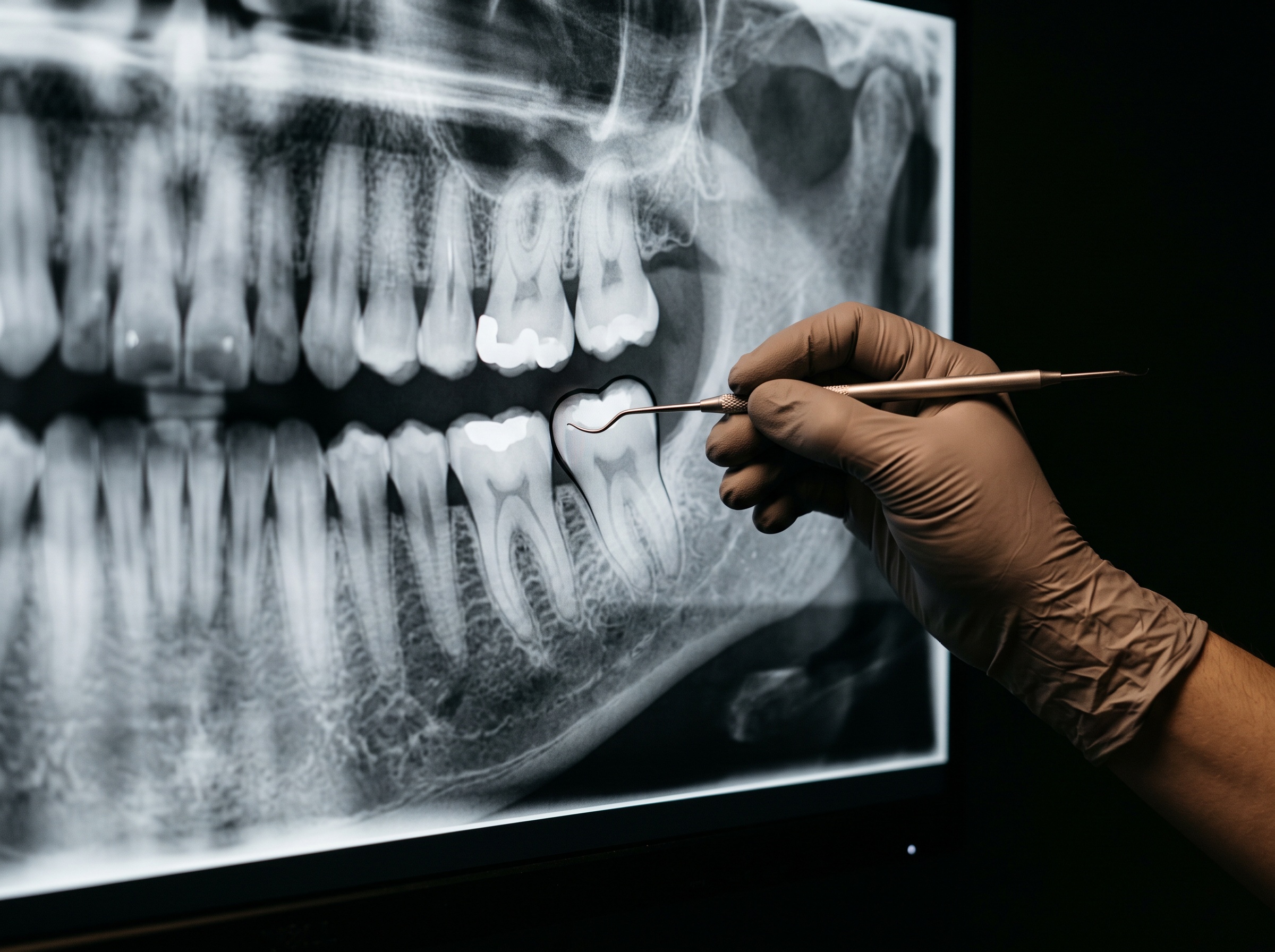
It is tempting to imagine labeling as mechanical tracing, but anyone who has done it knows better. Where does the cementoenamel junction actually sit when caries has undermined it? Is that faint radiolucency a genuine periapical change or the summation shadow of overlapping roots? The annotator is not transcribing a fact; they are making a clinical judgment and freezing it into pixels that the model will treat as ground truth forever.
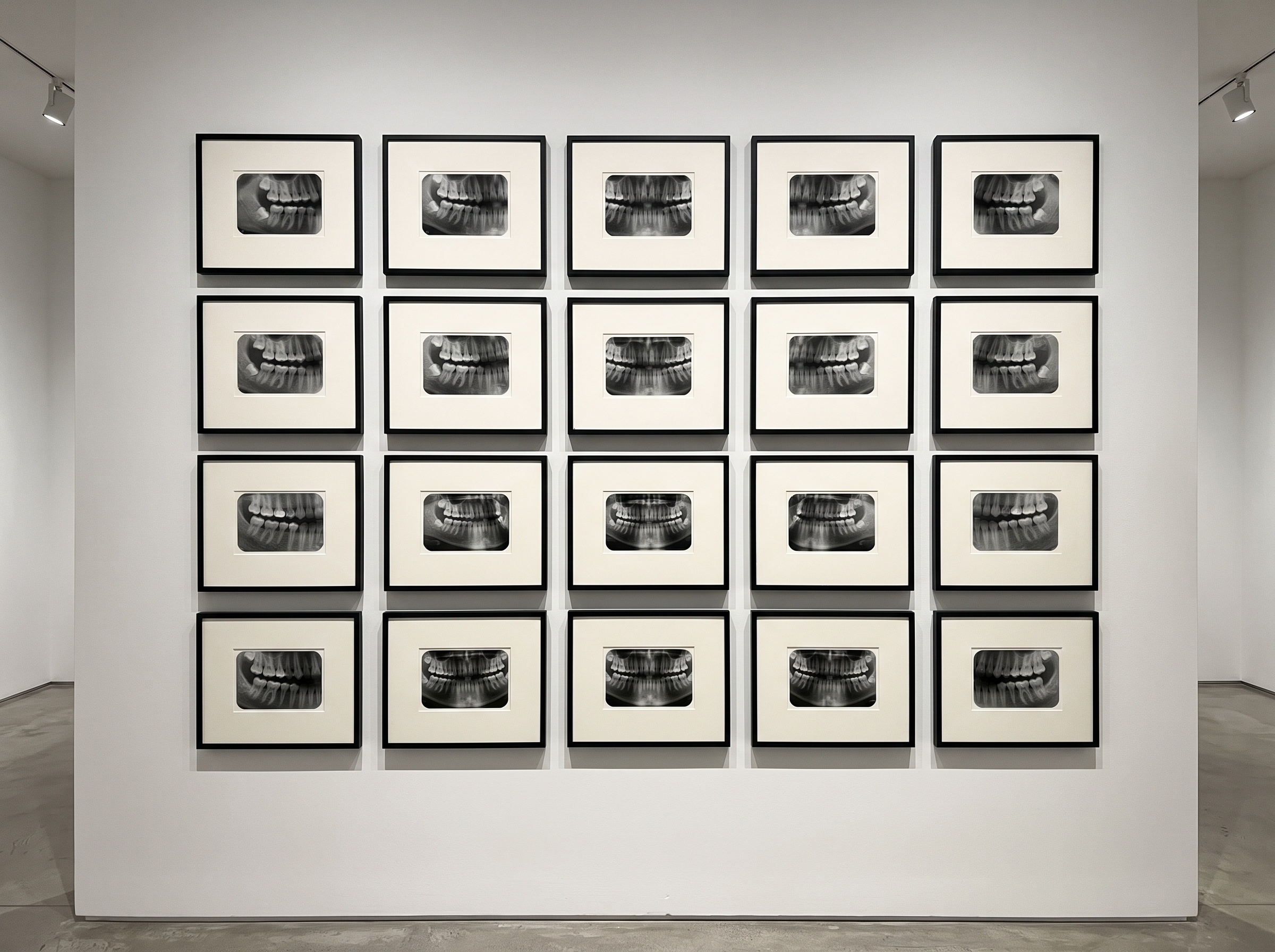
This is why serious datasets rely on multiple readers and measure how often they agree. Disagreement is not failure but information, a map of exactly where the anatomy resists clean interpretation. A well-built dataset preserves that nuance instead of papering over it, and in doing so it teaches the machine not only what is there but where certainty ends.
Why Open Datasets Matter
For years the most capable dental imaging models were trained on private collections, locked inside the companies that assembled them. The consequence was a quiet inequality: a handful of laboratories could build and validate, while independent clinicians and small research groups had nothing to test against. An openly published, pixel-annotated dataset breaks that enclosure. It lets anyone reproduce a result, probe a model for hidden bias, and benchmark a new idea on common ground.
For a practice that values the image as more than a billing artifact, this openness has a deeper resonance. It treats the radiograph as shared cultural property, a record whose careful annotation benefits the whole field rather than a single vendor’s roadmap.
Reading the Mask as an Image
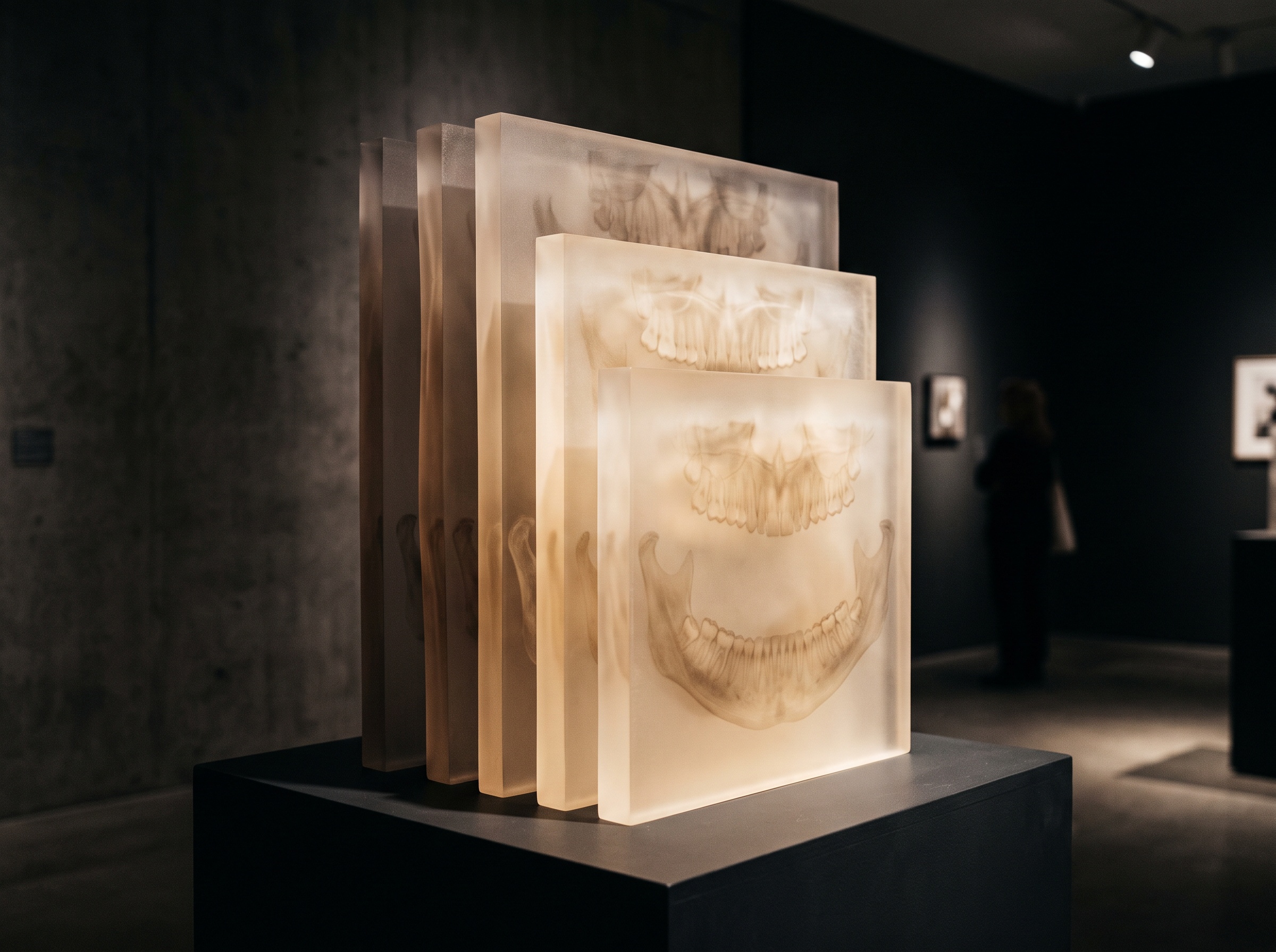
There is an unexpected beauty in the segmentation mask itself. Strip away the grey radiograph and what remains is a mosaic of clean, colored territories, each tooth a distinct pane, the whole arch glowing like stained glass. It is a portrait of understanding rather than of anatomy, a picture of how a trained eye has parsed the scene. Displayed beside the original film, the mask reveals the invisible act of reading that we perform instantly and unconsciously every time we look.
In a gallery devoted to the image as craft, this deserves a frame of its own. The annotation is not a byproduct of the radiograph; it is a second work of art made in dialogue with the first, authored by a clinician’s hand and rendered legible to a machine.
Future Developments
The near future will surely bring models that pre-draw their own masks, leaving the clinician to correct rather than trace from nothing, and that shift will make pixel-level annotation faster and far more common. But it will not make the human judgment disappear; it will concentrate it, moving the expert from draftsman to editor, from the one who draws every line to the one who decides which lines are true. As datasets grow richer and more openly shared, the annotator’s quiet interpretive work will only matter more, because every correction they make ripples outward into countless future readings. The great task ahead is to honor that labor as we honor the radiograph itself, to see annotation not as invisible plumbing but as an imaging craft in its own right, one that decides, quietly and permanently, what our machines will be able to see.
Sources & further reading:
Sorry, the comment form is closed at this time.