


August 24, 2026 One Portrait, Many Verdicts: When a Single Network Reads the Whole Mouth at Once
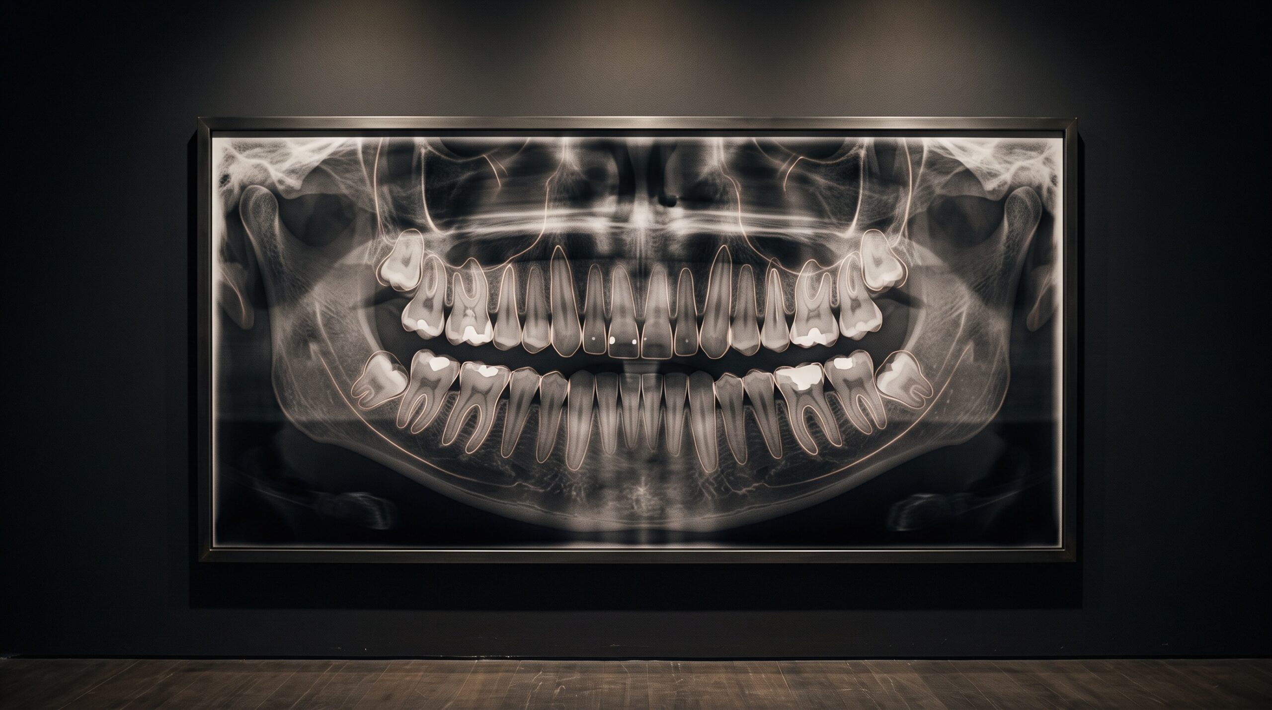
Stand a dental panoramic on a gallery wall and it reads like a portrait: a single sweeping image of the whole mouth, every tooth in its place, the jaws curving edge to edge in one continuous frame. For most of the short history of dental artificial intelligence, however, machines have not been permitted to see it that way. They were trained to answer one question at a time – is there caries here, yes or no; is this a periapical lesion, yes or no – each model a specialist squinting at a single finding. A newer class of networks refuses that narrowness. It looks at the entire portrait and returns not one verdict but many, in a single pass.
From One Question to Many
The distinction is more than a matter of convenience. A great deal of published work, as recent surveys of the field concede, has focused on single-pathology detection or simple binary classification – powerful within its lane, but blind to everything outside it. A mouth does not present one condition at a time. A single panoramic may hold a restoration here, an incipient cavity there, a titanium implant, an impacted third molar riding against a nerve, all coexisting in the same field. A unified, multi-class framework – the kind now emerging under names such as MultiDentNet – is built to name them together, classifying fillings, cavities, implants, and impactions within one diagnostic system rather than handing the image off to a relay of narrow specialists.
This is closer to how a clinician actually reads. No dentist looks at a panoramic asking a single yes-or-no; the eye sweeps the whole arch, holding several suspicions in mind at once and weighing them against one another. A model that assesses many classes simultaneously is not merely faster – it is learning the relational habit of real interpretation, where the presence of one finding quietly changes the probability of the next.
The Portrait Read as Layers
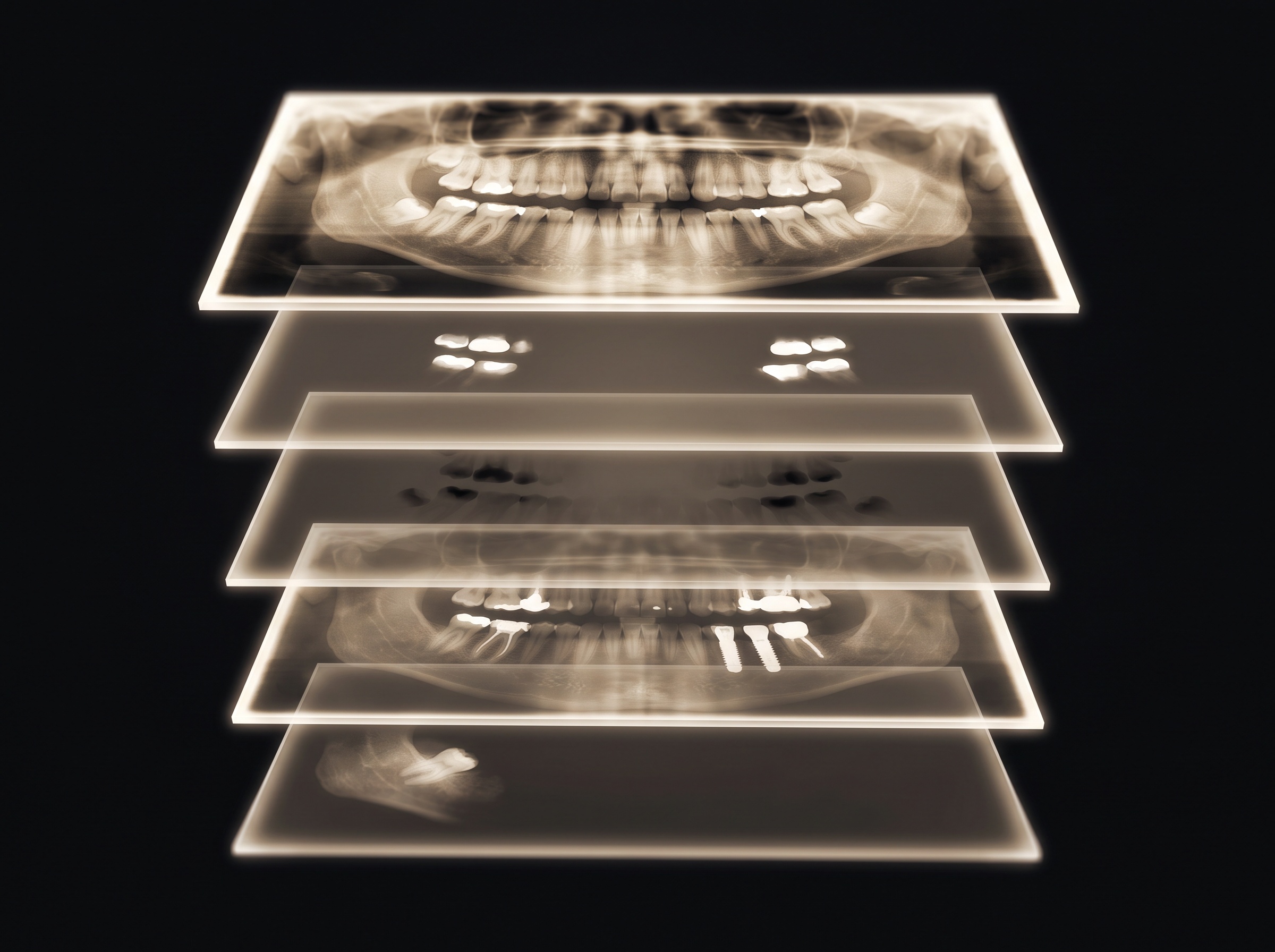
What does it mean, technically, to read the whole mouth at once? The stronger frameworks pair classification with entity segmentation – not just declaring that a condition is present, but tracing precisely where it sits on the image. The panoramic effectively separates into overlaid layers: one isolating the bright signatures of metal restorations, another the low-density shadows of decay, another the unmistakable geometry of an implant. Each finding becomes its own luminous stratum of the same picture.
That spatial grounding matters because a label without a location is a rumor. A network that says “caries present” but cannot point to the tooth has told the clinician almost nothing actionable. One that outlines the lesion, tooth by tooth, produces something that can be checked, trusted, and – crucially – reported. It is the difference between a critic who says a gallery is beautiful and one who can walk you to the single canvas that earns the word.
The Craft Beneath the Machine
None of this intelligence is conjured. It is taught, one careful outline at a time. Every multi-class model rests on a foundation of pixel-level annotation – human experts tracing, on thousands of radiographs, exactly where each finding begins and ends, so the network has a ground truth to imitate. This is the least glamorous and most decisive part of the whole enterprise, and it is genuinely a craft. As anyone who has done it knows, annotating a panoramic radiograph is a discipline of its own, demanding both anatomical fluency and a steady, almost draughtsman’s patience.

The quality of that handwork sets a ceiling the cleverest architecture cannot exceed. A model trained on sloppy borders learns sloppy borders; one trained on inconsistent labels inherits the inconsistency and dignifies it with a confidence score. The romance of deep learning tends to gather around the network, but the honest credit belongs to the annotators, whose quiet precision is the true origin of the machine’s apparent insight.
Where the Machine Is Fooled
A model that learns from human readings also inherits human vulnerabilities. Radiography is full of appearances that lie – shadows and gradients produced by geometry and physics rather than pathology. The classic example is cervical burnout, the radiolucent illusion at the tooth’s neck that mimics decay convincingly enough to deceive an experienced eye. A network trained on images where such artifacts were sometimes mislabeled will faithfully reproduce the mistake, flagging a phantom cavity with the same serene confidence it brings to a real one.
This is why the multi-class ambition must be met with multi-class skepticism. A system that screens for many conditions at once multiplies not only its usefulness but its opportunities to err. The value of these tools lies in triage – surfacing what deserves a second look – not in verdict. The illusions that trap the human reader are precisely the ones most likely to slip past a model that learned by watching us.
Reading in Context, Not in Isolation
The panoramic itself resists tidy interpretation, machine or otherwise. Its every image is assembled from a moving slice of sharpness – the focal trough that determines what lands crisp and what smears into blur – so structures drift in and out of fidelity across the arch. A model must learn to read confidently in the sharp center and cautiously at the distorted edges, a nuance that only richly annotated, geometry-aware training data can instill. The image is not a neutral canvas; it has its own optics, and a network ignorant of them will over-read the periphery.
Even a flawless reading is only half the task. A finding is clinically inert until it is communicated, which is why the output of these systems ultimately has to feed the same discipline that governs human interpretation – the move toward structured reporting that makes a radiograph’s findings legible and consistent. A model that returns a clean, structured list of located findings is not just diagnosing; it is drafting the beginnings of a report.
Future Developments
The trajectory is toward unification without erasure – one network that reads the whole portrait, names every finding, locates each on the image, and hands the clinician a single legible summary rather than a scatter of alarms. The most ambitious frameworks already reach past classification toward preliminary oral-lesion triage, gesturing at a day when a routine panoramic quietly flags the anomaly that warrants urgent human attention. The right ambition is not a machine that pronounces, but one that composes the placard beside the picture – gathering many verdicts into one clear statement and leaving the final reading, as it must remain, to the trained eye standing before the image. Seen that way, multi-class deep learning is less a replacement for interpretation than its newest instrument: a way of seeing the entire mouth at once, and of remembering that the whole was always more than the sum of its isolated questions.
Sources & further reading:
- Advanced Deep Learning Techniques for Classifying Dental Conditions Using Panoramic X-Ray Images (arXiv, 2025)
- Advanced Deep Learning Models for Classifying Dental Diseases from Panoramic Radiographs, Diagnostics 16(3):503 (2026)
Sorry, the comment form is closed at this time.