


August 16, 2026 Cervical Burnout: The Radiolucent Illusion at the Tooth’s Neck That Mimics Caries
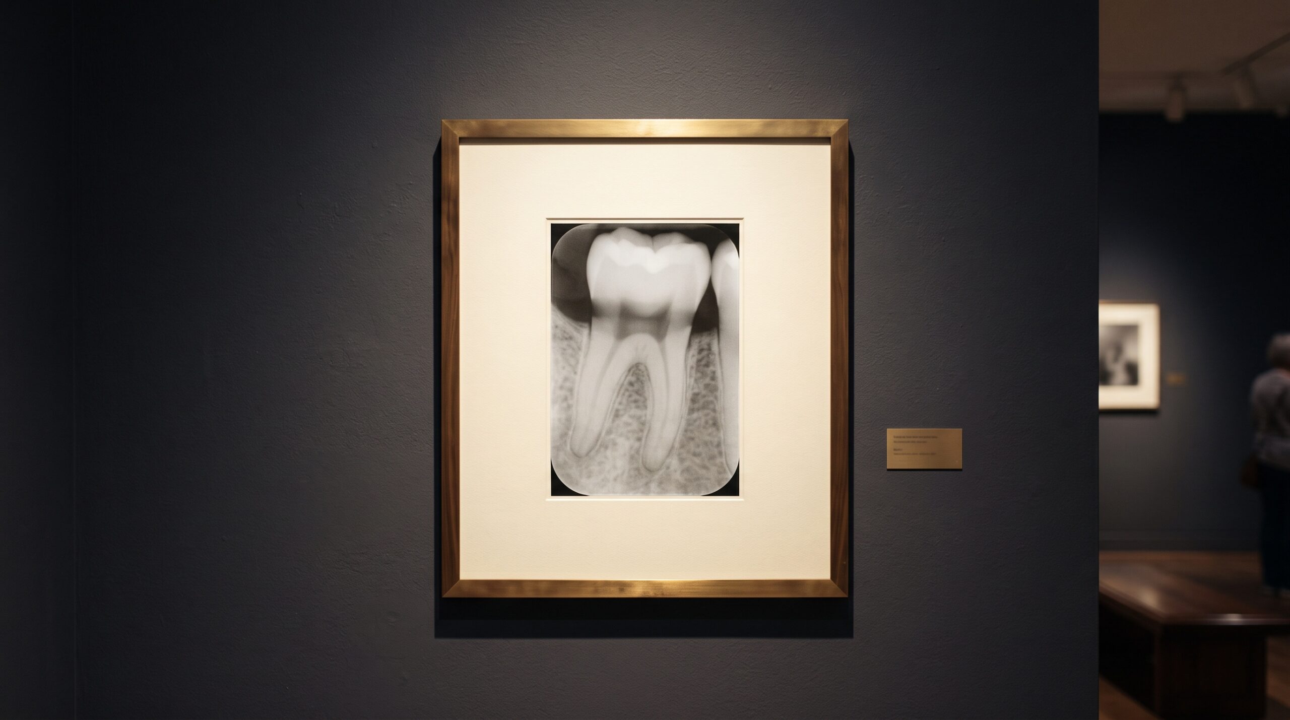
Study a bitewing for long enough and you will meet it: a soft, dusky band running along the neck of a tooth, sitting just below the bright shoulder of the enamel and just above the pale crest of the bone. To an anxious eye it reads instantly as decay — a dark wound gnawing into the root surface, the interproximal shadow of a lesion that demands the drill. And yet, more often than not, there is nothing there at all. What the film has recorded is not a hole in the tooth but a hole in the physics: cervical burnout, one of radiography’s most persuasive illusions, and a small masterclass in the difference between what a beam does and what a disease does.
The Anatomy of a Thin Place
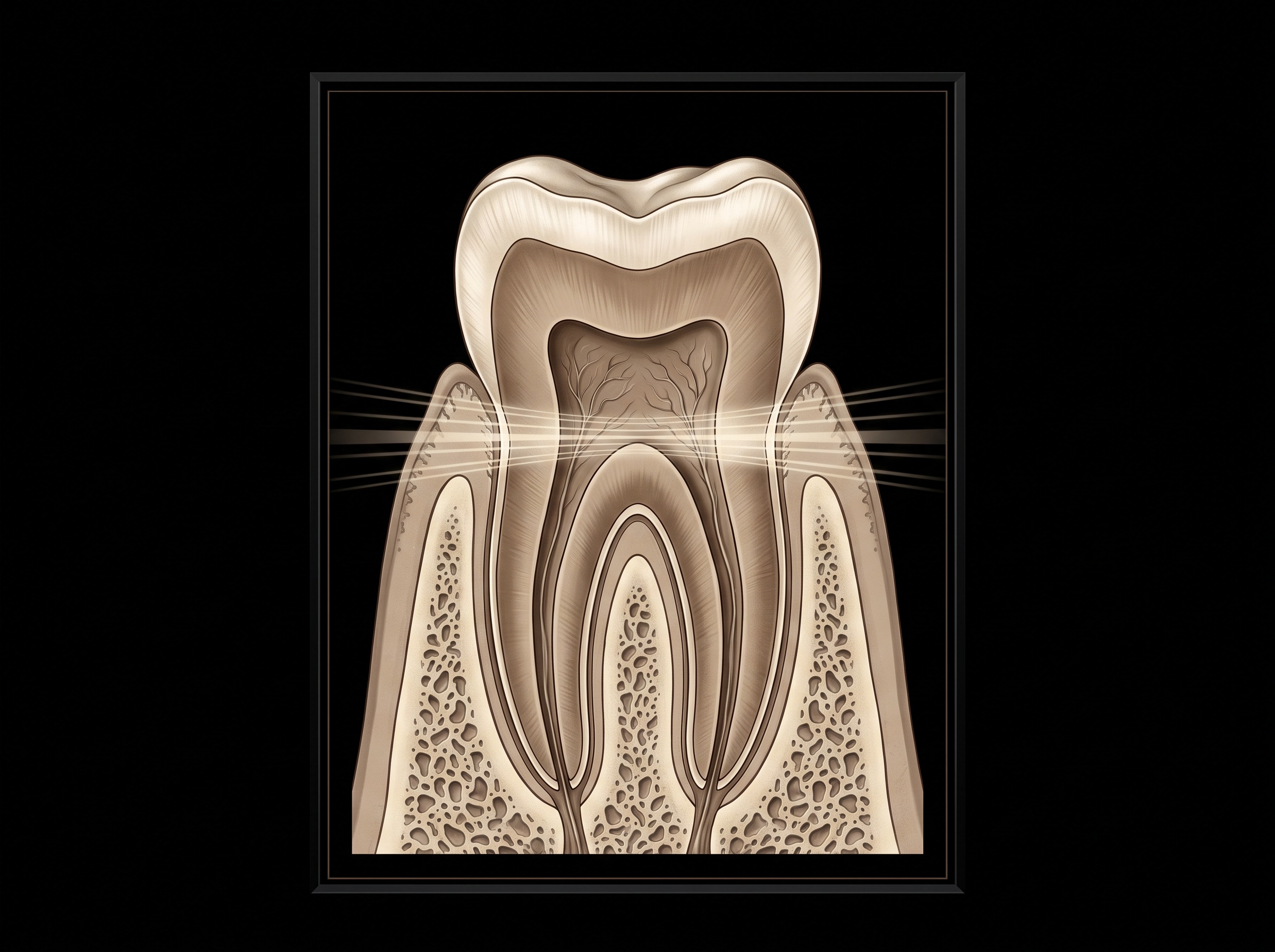
The illusion is born from geometry. A tooth is not a uniform block of mineral; it is thickest and most radiopaque at the crown, where a cap of dense enamel overlies dentin, and it is well defended at the root, where the surrounding alveolar bone lends its own attenuation to the picture. Between these two strongholds lies the cervical region — the neck of the tooth, at and just below the cementoenamel junction. Here the enamel cap has tapered away to nothing, the crestal bone has not yet risen to embrace the root, and what remains is a comparatively slender wedge of dentin and cementum, unshielded on either flank. It is, in the strictest radiographic sense, a thin place: a corridor through which X-rays pass with less to stop them than anywhere else along the tooth’s profile.
Why the Beam Darkens the Neck
Attenuation is the whole story. Every photon that survives its passage through the tissue and reaches the sensor darkens the corresponding point of the image; the more tissue it must cross, the more of the beam is absorbed, and the lighter that region records. Through the enamel-armoured crown, a great many photons are stopped, and the crown prints bright. Through the bone-braced root, likewise. But through the thin cervical wedge, comparatively little is absorbed — the beam pours through almost unimpeded — and a surplus of photons strikes the receptor there, burning that narrow band darker than its neighbours. This is a phenomenon of quantity, of raw penetration, which is precisely why it expresses itself as density rather than as true contrast — the same grammar of exposure explored in the grayscale craft of contrast and density. Push the exposure higher — more kilovoltage, more overpenetration — and the effect deepens, the neck of every tooth in the arch acquiring the same shadowy collar.
The Eye Finishes What the Beam Began
If the physics laid the shadow down, human perception darkens it further. Where a bright region meets a darker one, the visual system exaggerates the boundary, manufacturing an illusory extra-dark fringe hugging the edge of the lighter zone — the Mach band, a quirk of our own contrast-enhancing retina. On a radiograph, the brilliant edge of the enamel meets the dim cervical band exactly along this junction, and the eye obligingly deepens the burnout, lending it a crispness and a menace the raw exposure never contained. It is a sobering reminder that a radiograph is not read by a photometer but by a nervous system, and that the same faculties which make us sensitive diagnosticians also make us suggestible ones — a theme that runs through the ways sharpening, contrast, and noise reduction can deceive on a digital radiograph. The illusion, in the end, is a collaboration between the tube and the observer.
Telling the Illusion From the Lesion
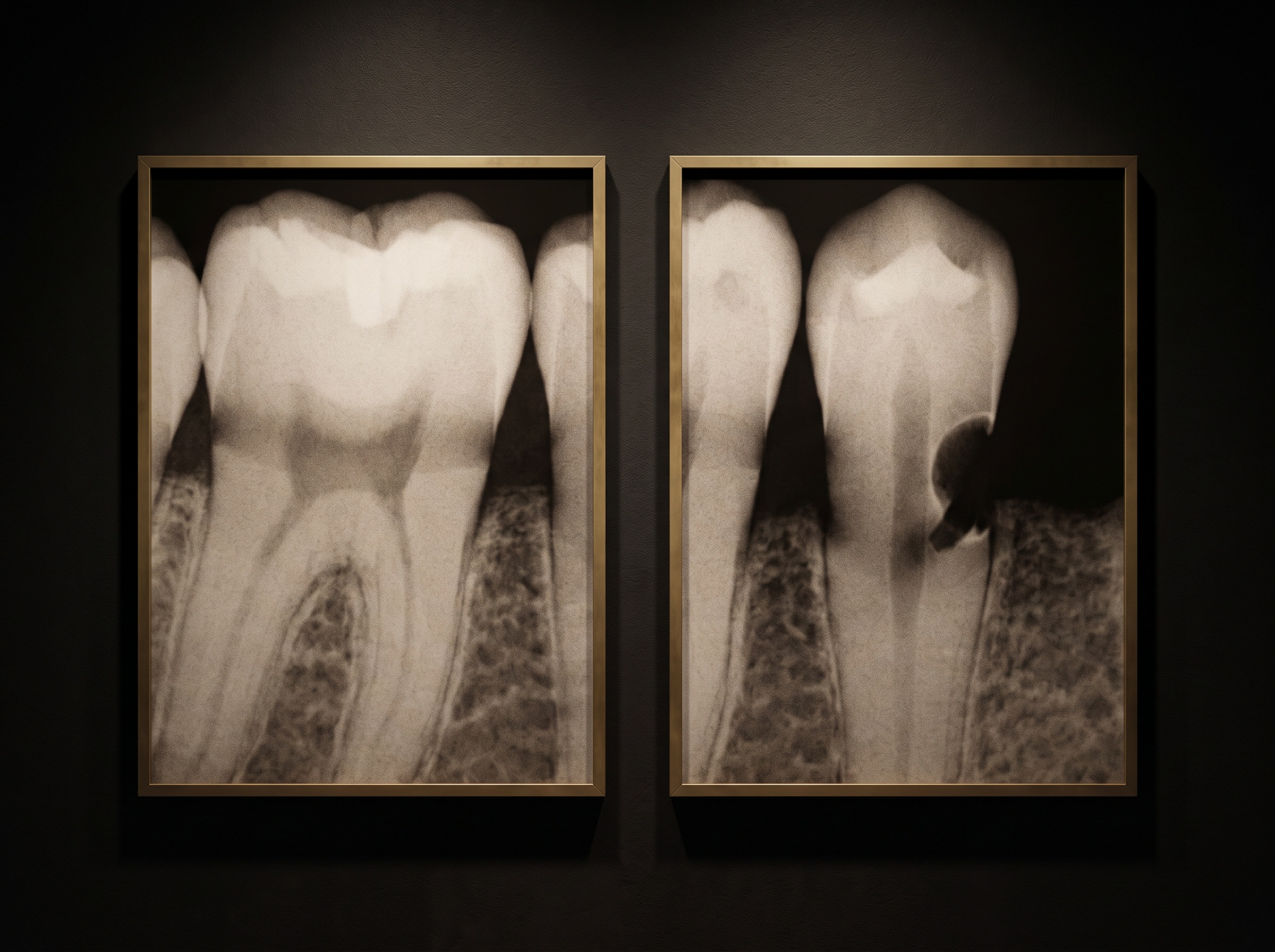
The clinical stakes are real, because cervical burnout occupies exactly the territory — the interproximal cervical surface — where root caries genuinely likes to begin, and a misread can mean either an unnecessary restoration or a missed cavity. Fortunately, the two have different handwriting. Burnout is diffuse and ill-defined, fading gently into the surrounding tooth with no crisp border; it follows the natural contour of the cervical anatomy; and, being a property of shape and exposure rather than of disease, it tends to appear symmetrically, showing up on the same surfaces of contralateral teeth across the arch. True caries, by contrast, is discrete and localized — a small, cupped, comparatively well-defined notch that bites into the root surface, often with a slightly rounded or triangular form, and it does not politely mirror itself on the tooth across the mouth. The disciplined reader looks for the borders: a soft gradient argues for the artifact, a defined margin for the lesion. This is the same close attention to what the geometry imposes versus what the pathology reveals that governs the honest reading of a bitewing.
Working Against the Deception
Because burnout is partly a creature of overexposure, the operator holds several levers against it. Sound exposure technique — resisting the temptation to overpenetrate — keeps the cervical band from deepening into caricature. In the digital darkroom, adjusting the window and the contrast can flatten the illusory fringe and reveal whether a defined margin lurks beneath the gradient, though the same tools, used carelessly, can just as easily manufacture a border that was never there. Bilateral comparison remains the quiet workhorse: a shadow that appears identically on the matching surface of the opposite tooth is speaking of anatomy, not decay. And none of it substitutes for the clinical eye and the explorer at the chair; the radiograph raises the question, but the mouth answers it. The best diagnosticians treat the burnout band not as a verdict but as an invitation to look again, more slowly.
Future Developments
The next reader of the cervical band may not be human at all. As machine-learning caries detectors move from novelty to fixture, cervical burnout becomes their classic trap — a benign radiolucency sitting precisely where the model has learned to expect a lesion, and a fertile source of false positives unless the training data has taught the network the difference between a soft symmetric gradient and a cupped, defined margin. Getting that distinction into the machine is itself a craft, one that depends on the careful, expert labelling behind teaching a model to see. Meanwhile, better beam filtration, exposure-optimising algorithms, and perceptually-aware display pipelines that account for Mach banding all promise to quiet the illusion at its source. But the underlying truth will endure: the neck of a tooth is a thin place, and light — even invisible light — will always find the thin places first. To read a radiograph well is, in part, to know which shadows are wounds and which are only the beam telling the truth about where the tooth is slender. That, too, is a kind of connoisseurship, and it is the art the gallery exists to cultivate.
Sources & further reading:
Sorry, the comment form is closed at this time.