


August 26, 2026 The Radiograph That Talks Back: When Vision-Language AI Learns to Describe a Dental Scan
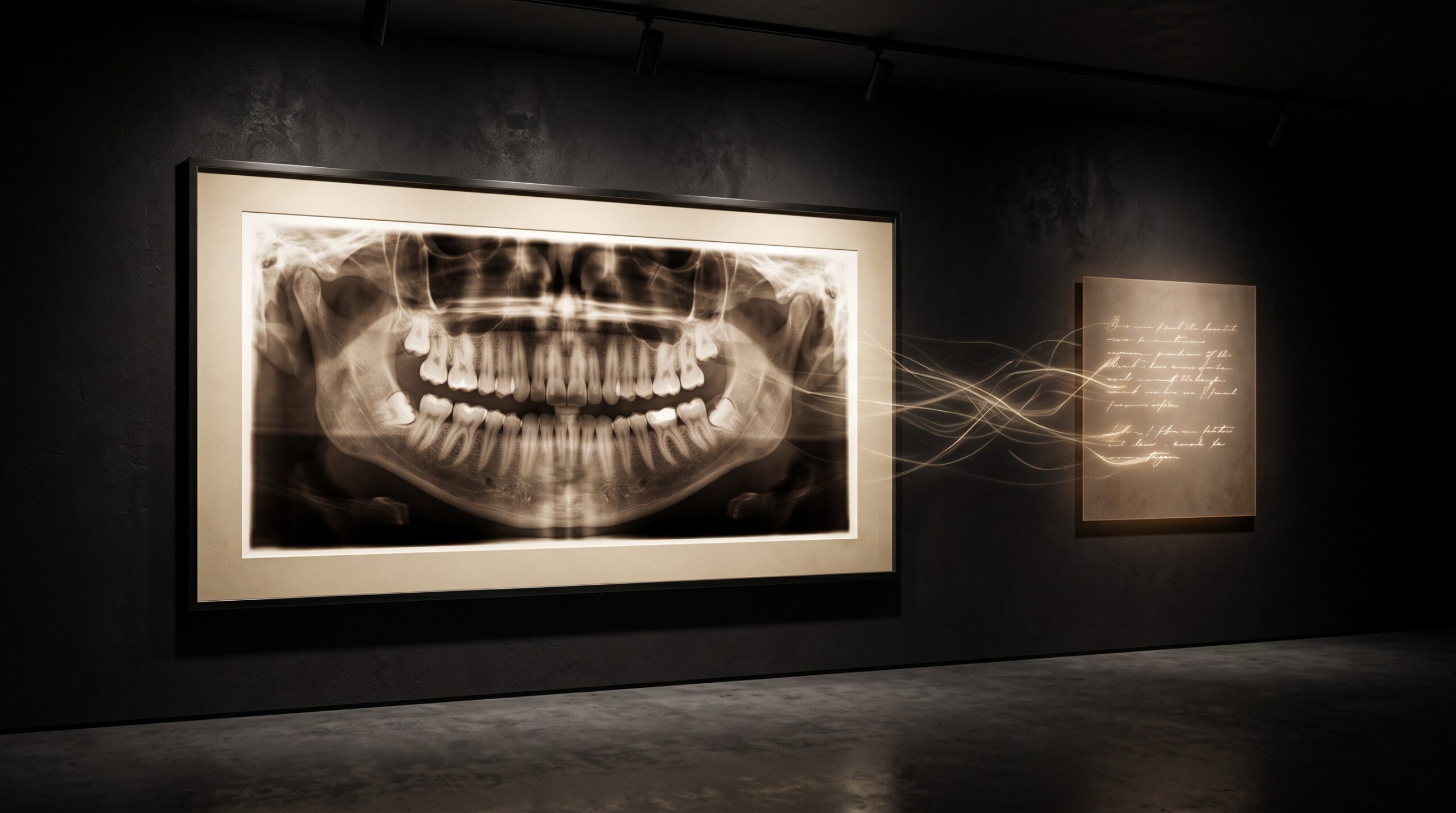
For most of the short history of dental artificial intelligence, the machine has communicated by pointing. It drew a ring around a shadow between two molars, shaded a region of suspected bone loss, outlined a tooth and gave it a number. This is the grammar of detection, and it is a good grammar – precise, spatial, and easy to lay directly over the film. But it is a curiously mute one. The picture was marked, yet nothing was said. A new class of model is changing that. Trained to take in a radiograph and answer questions about it in ordinary sentences, the dental vision-language model does something that still feels faintly uncanny the first time you watch it: it describes the picture back to you.
From Pointing to Speaking
The distinction is not cosmetic. A detection network and a vision-language model are built to produce different kinds of output, and that difference reaches all the way down into what each can be trusted to do. A detector maps pixels to a fixed vocabulary of findings – caries, calculus, periapical radiolucency – and marks where each one sits. A vision-language model, or VLM, maps the same pixels into open-ended language: it can summarize the whole film, answer a follow-up question, note an incidental finding the question did not ask about, and hedge when the evidence is thin. Where the detector’s answer is a set of coordinates, the VLM’s answer is a paragraph. It is the difference between an instrument that flags and a colleague who talks.
A recent preprint, describing a system its authors call DentVLM, is a clean illustration of where this is heading. Rather than being trained to ring a single pathology, it is built for what the authors frame as comprehensive dental diagnosis: caries detection, tooth segmentation and identification, assessment of malocclusion, evaluation of periodontal disease, and more general pathology recognition – all expressed through language rather than through overlays alone. Crucially, it is not confined to one kind of picture.
One Reader for Three Kinds of Picture
Dentistry does not image the mouth in a single medium, and this is where a language-fluent model earns its keep. DentVLM was built to take in panoramic radiographs, intraoral photographs, and cone-beam CT – the flat sweep of the jaw, the close colour portrait of a tooth and its gingiva, and the volumetric depth of a 3D scan. These are radically different data: one is a projection, one is reflected light, one is a reconstructed volume. A detector usually needs a separately trained head for each. A vision-language model can, in principle, hold all three in the same representational space and speak about them in one register – describing a stain on an intraoral photograph and a radiolucency on a panoramic with the same vocabulary of clinical description.
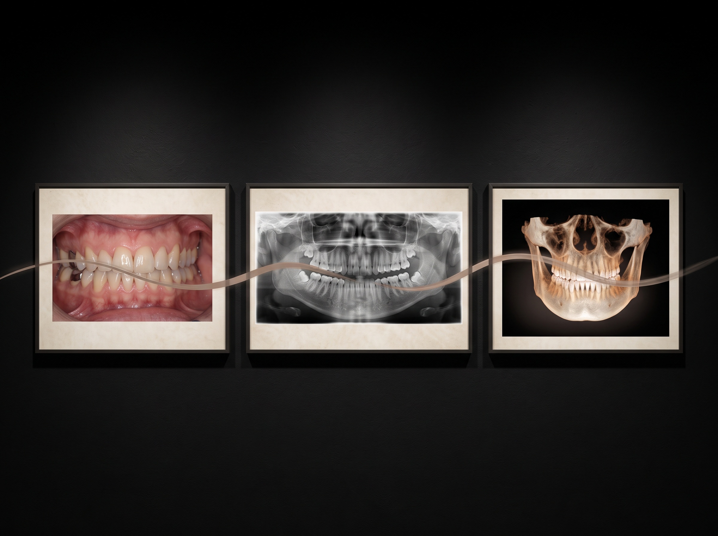
That unifying instinct is showing up elsewhere in the literature too, in work that pretrains across cone-beam CT and intraoral scans together so that a model learns the correspondences between them. It is the same conviction seen from another angle in our piece on a single network that reads the whole mouth at once: the field is tired of narrow, one-finding tools and is reaching for models that take in the entire picture and account for all of it. The vision-language model simply adds the final faculty – the ability to render that whole reading as prose.
How Good Is the Talking?
It is fair to be sceptical of a machine that produces fluent clinical sentences, because fluency is exactly what a language model can counterfeit. The relevant question is not whether the description reads well but whether it is right. Here the DentVLM work does the sensible thing: it benchmarks the specialist model against the capable generalists – GPT-4o, Gemini, and open models such as Qwen2-VL and LLaVA – on dental-specific tasks, and reports that domain adaptation matters. A general-purpose vision-language model, impressive as it is on everyday images, tends to founder on the particular grammar of a radiograph; a model tuned on dental data reads it far more reliably. That is an unglamorous but important result. It says the path forward is not a single omniscient model but focused adaptation to the strange visual dialect of dental imaging.
The Danger of a Confident Sentence
A described finding carries a risk a ring does not. When a detector marks the wrong spot, the error is visible and local – the clinician looks, sees nothing, dismisses it. When a language model asserts, in a well-formed sentence, that there is early interproximal caries on the distal of the second premolar, the error is smoother and more persuasive. This is the confabulation problem, and it is the central discipline of the field: a description is only as safe as its grounding, its ability to tie each claim back to the specific pixels that justify it. A trustworthy dental VLM must be able to point while it speaks – to say here, and this is why – so that a clinician can check the sentence against the film rather than take it on faith.
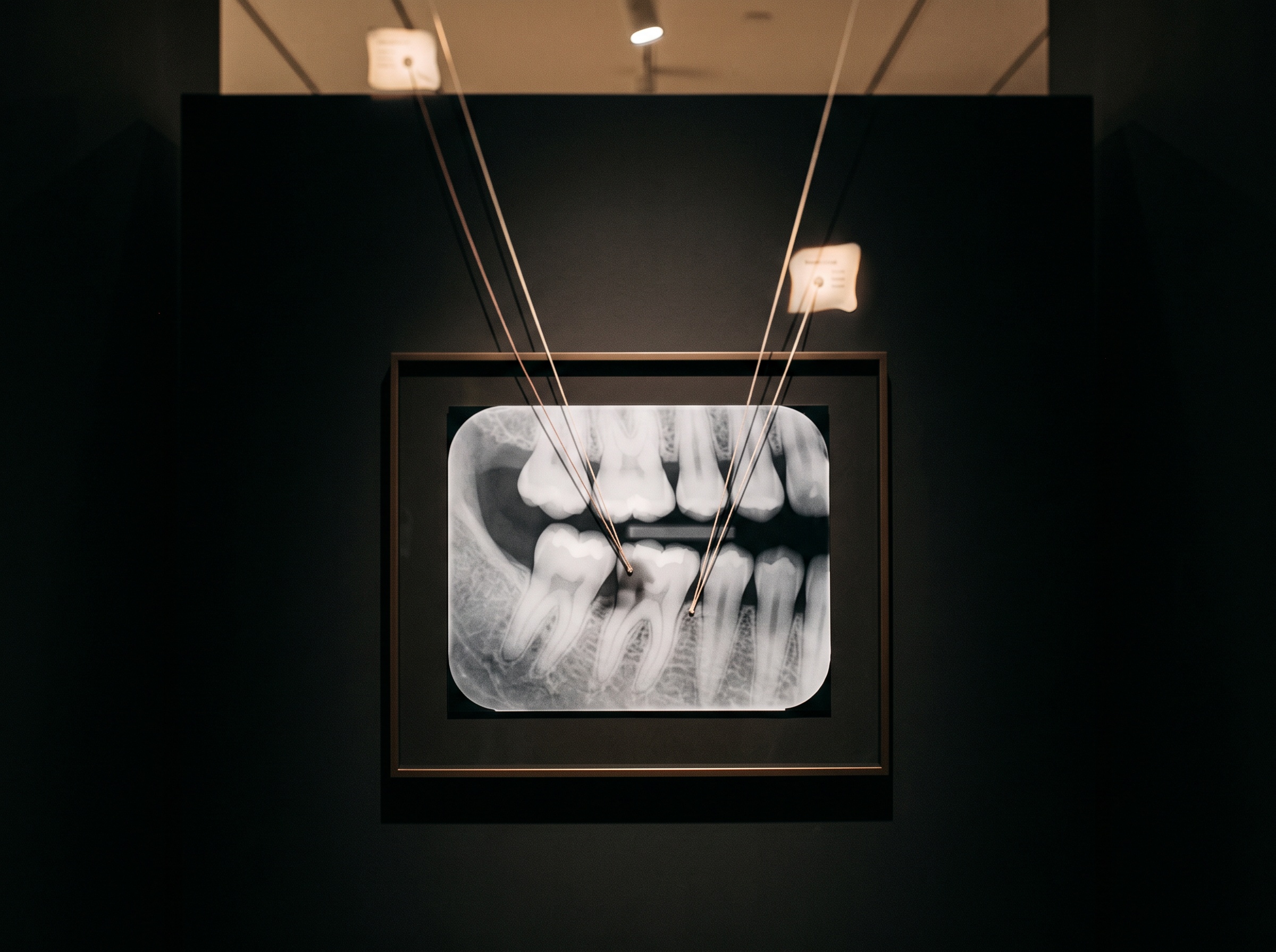
This is precisely why the shift from detection to description does not retire the older discipline of structured reporting that makes a radiograph legible; it inherits it. The most useful descriptions are not free-flowing essays but disciplined ones – findings named consistently, uncertainty stated, negatives noted – which is structured reporting arrived at from the other direction. And it keeps the clinician firmly in the position that FDA-cleared detection that draws its findings onto the film has already established: the machine assists, surfaces, and suggests; the person decides.
Why This Matters for the Image Itself
There is a quieter consequence for anyone who thinks of imaging as craft. When the only consumer of a radiograph was a human eye or a detection overlay, image quality was judged by legibility to a person. When the consumer is also a model that will describe the picture, quality acquires a second audience. Compression that a clinician would forgive, a border the eye would mentally complete, a contrast setting flattering to the human reader – these can quietly change what the model says. The picture and its caption are now coupled. A radiograph is no longer only something to be seen; it is something to be paraphrased, and the fidelity of the paraphrase depends on the fidelity of the image. The old imaging virtues – honest exposure, clean geometry, uncompressed capture – become, once again, the foundation on which everything downstream is built.
Future Developments
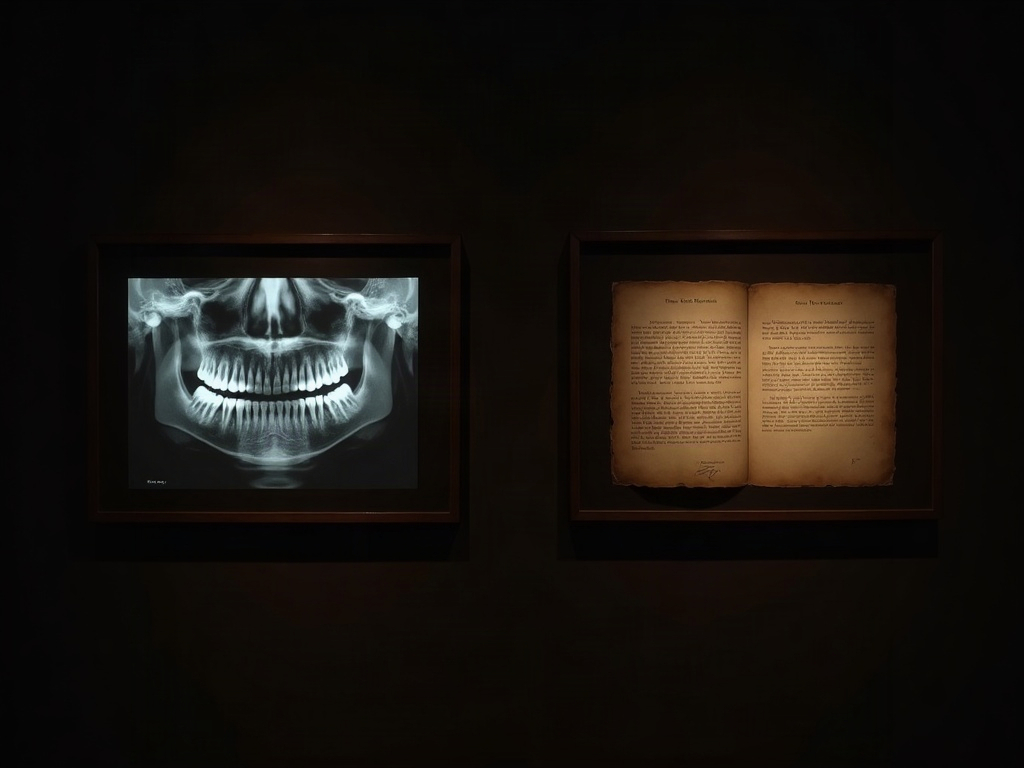
The near horizon is a scan that arrives already narrated: a draft description generated at the moment of capture, grounded to the exact regions it refers to, offered to the clinician as a first sentence to accept, edit, or overrule – never as a verdict. The harder and more valuable frontier is calibration – teaching these models the humility to say I am not sure, to fall silent on a corner of the film where the evidence runs out rather than manufacture a confident clause. A model that describes brilliantly but never doubts is a liability; one that describes well and knows the edge of its own knowledge is a genuine colleague. The portrait of the mouth is learning to speak. The work ahead is teaching it, as we teach every good clinician, when to hold its tongue.
Sources & further reading:
- DentVLM: A Multimodal Vision-Language Model for Comprehensive Dental Diagnosis and Enhanced Clinical Practice (arXiv preprint)
- Multimodal Contrastive Pretraining of CBCT and IOS for Enhanced Tooth Segmentation (arXiv preprint)
- Dental Tribune: A new era in US dental imaging – AI-driven, safe and patient-centric solutions
Sorry, the comment form is closed at this time.