


November 3, 2026 The Scanner Loses Its Box: What a 106-Gram Wand Signals About Who Gets to Capture in 3D
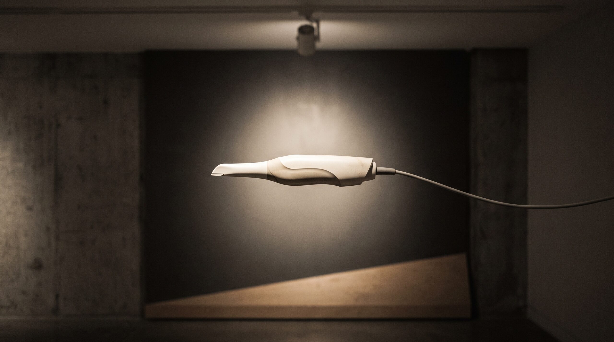
Most of the instruments in this gallery earn their place through what they reveal. A scanner earns its place through something humbler: how little the patient and the clinician have to notice it while it works. The newest of these, SHINING 3D’s Aoralscan Elf, weighs 106 grams and connects to the computer through a single cable, with no separate power box sitting on the cart. Those are small numbers, and it would be easy to file them under comfort. But a scanner light enough to be forgotten and cheap enough to be ubiquitous is not just a nicer wand. It is a quiet argument about who, in the profession, now gets to make a three-dimensional record of the mouth at all.
What the Numbers Actually Describe
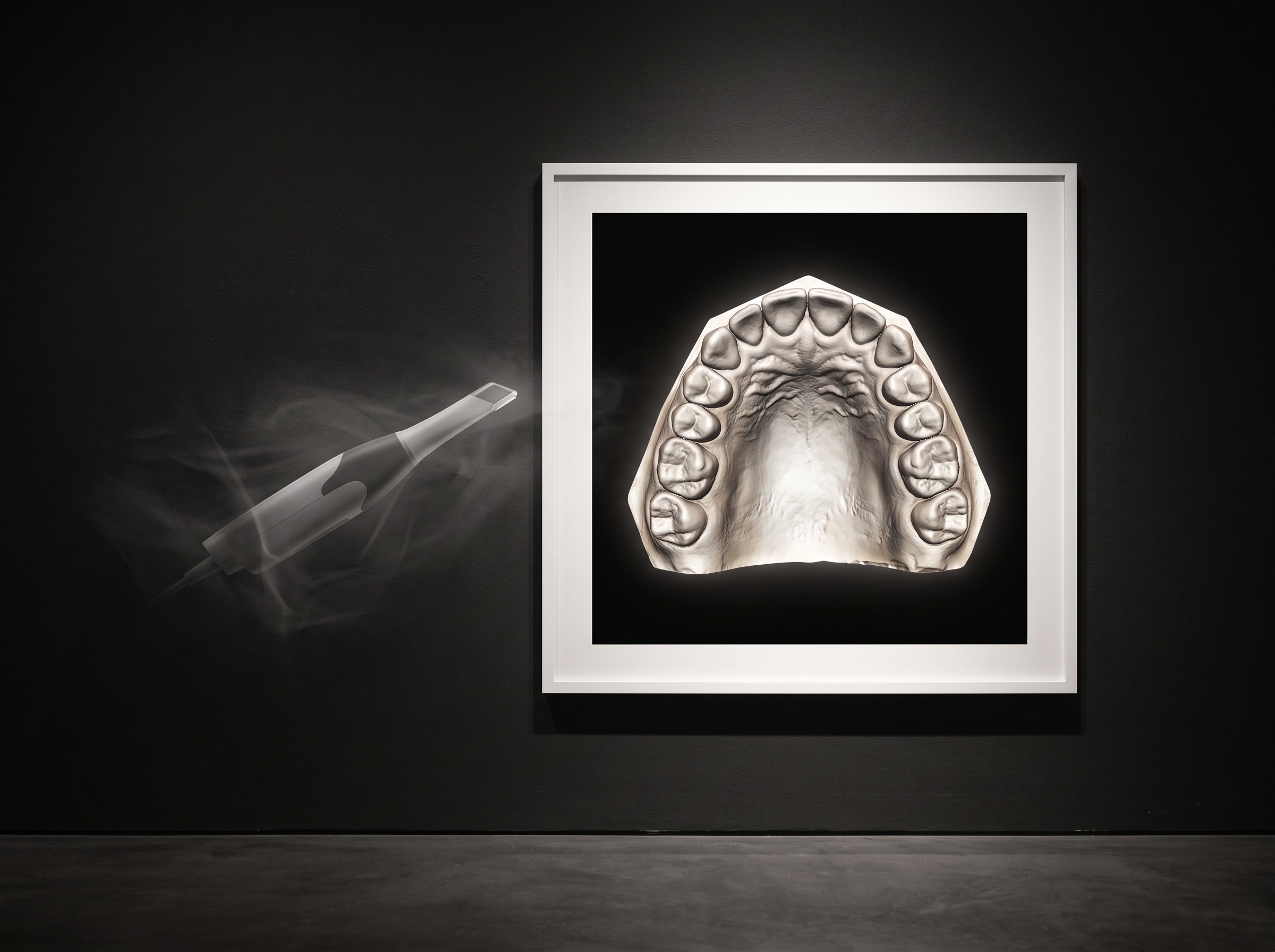
Read the specification sheet as a still life and the character of the device emerges. The body is a long, slim wand of roughly 245 by 27 by 30 millimetres, and the whole thing weighs about 106 grams, a fraction of the heft of earlier scanners from the same family that ran closer to 375 grams. It draws its power and sends its data down one USB Type-C connection rated at five volts and three amps, which is why there is no external box on the trolley and no second cable to drape across the operatory. Capture itself is non-contact structured light: the wand projects a known pattern of light onto the teeth and reads how that pattern deforms across every surface, and three interchangeable tips, from a wider field down to a small 12 by 9 millimetre window, let the operator trade coverage for access in a tight posterior corner. None of this is exotic. It is the maturation of a technology, rendered small.
Weight Is a Clinical Variable, Not a Comfort
It is tempting to treat grams as a matter of fatigue, but weight quietly governs the quality of the record itself. A heavy wand held at arm’s length over a reluctant patient produces micro-tremor, and tremor during a structured-light sweep is simply noise the software must stitch over, more retakes, more gaps bridged by inference rather than data. A featherweight instrument that the hand barely registers holds a steadier path across the arch, and a steadier path is a cleaner mesh. We traced this physiology of the hand in detail when we looked at how the featherweight scanner rewrites the ergonomics of capture, and it remains true: ergonomics is not a lifestyle feature bolted onto imaging, it is upstream of accuracy. The same holds for the other great enemy of the intraoral scan, the wet field. A lighter wand that moves confidently still has to contend with saliva and blood, which is why the discipline of moisture control and isolation remains the operator’s responsibility no matter how refined the hardware becomes.
The Box That Went Away
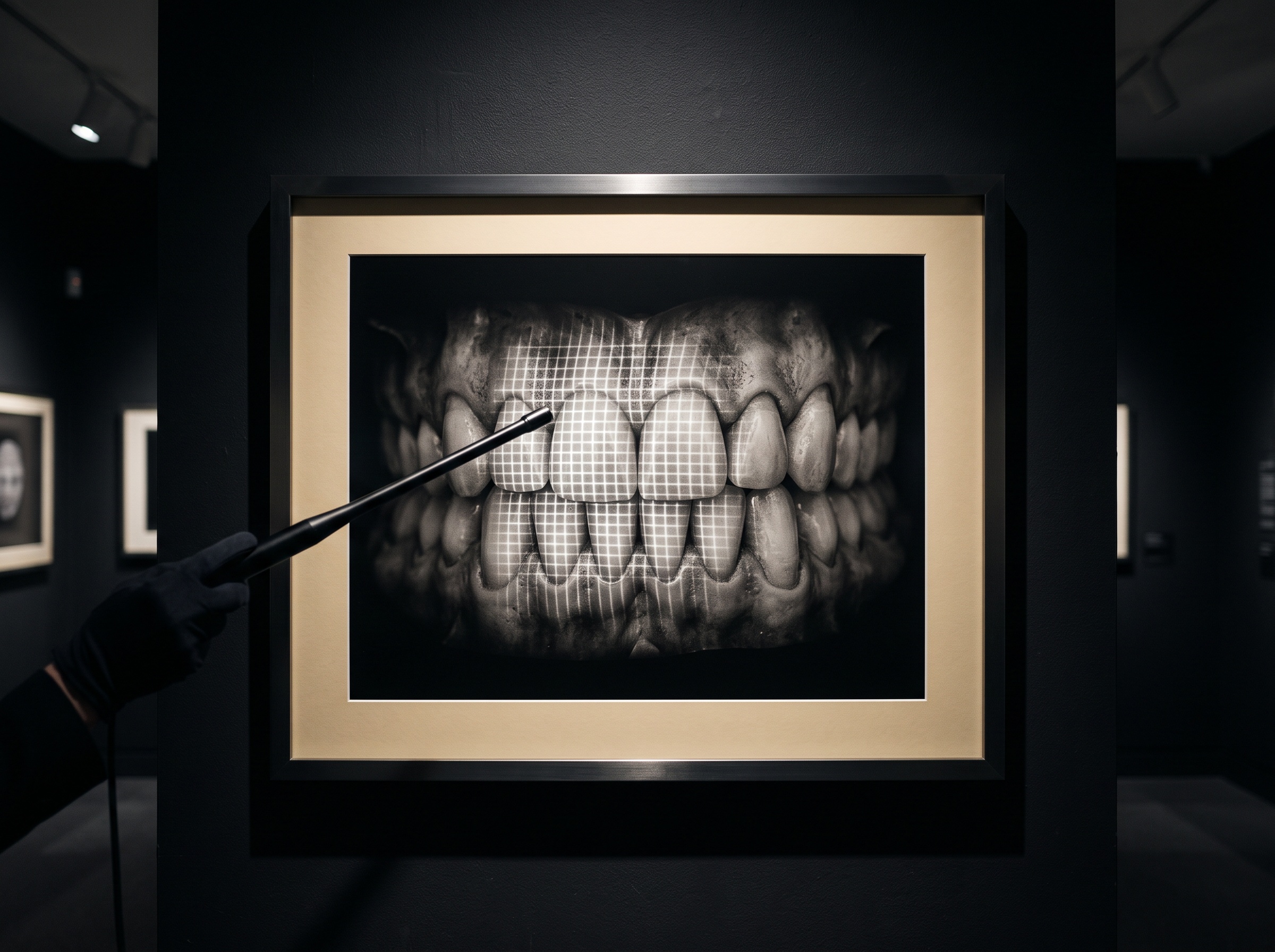
The detail most worth dwelling on is the one that is absent: the power box. For years an intraoral scanner was a system, a wand tethered to a dedicated unit with its own supply, its own footprint, its own place in the room. Collapsing that system into a single Type-C cable powered off the computer does more than tidy the cart. It lowers the threshold of what a scanning setup is. A device that runs off a laptop port can move between operatories, travel to a satellite clinic, or sit in a practice that never had the bench space or the budget for a full cart-mounted system. The engineering story is about power delivery over a modern connector; the professional story is about the footprint of entry shrinking until it almost disappears.
Who Gets to Capture
This is the part that matters beyond any one product. When a mid-tier scanner lands near twelve thousand dollars rather than the far higher sums the first generation commanded, and when it needs nothing but a laptop and a cable, the population of clinicians who can realistically own one widens. The solo practitioner, the rural clinic, the practice that digitised its radiography years ago but never its impressions, all move within reach of a true three-dimensional record. That shift changes the baseline of the profession. The intraoral scan stops being the signature of a flagship practice and becomes, like the digital radiograph before it, simply part of how a mouth is documented. And once capture is routine, the three-dimensional record becomes the substrate everything downstream draws on, from the scan-to-mockup workflows that turn a digital impression into a previewed smile to the archival comparison of one visit against the next. Democratise the capture and you democratise everything it feeds.
Access Is Not the Same as Fidelity
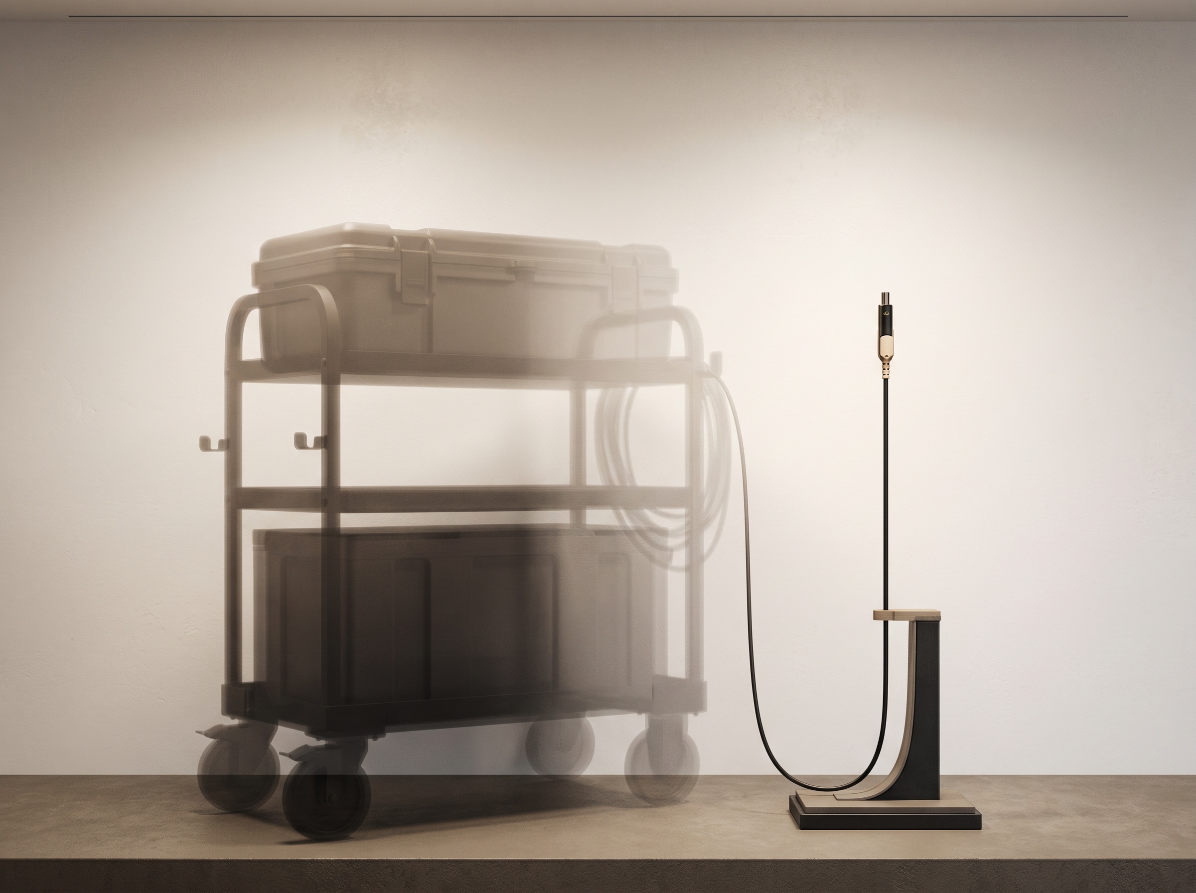
A fair gallery note resists the easy triumph. Making capture cheap and light does not make it good by itself, and there are honest trade-offs in a value-tier instrument. This particular scanner, for instance, does not support the intraoral photogrammetry workflow used to verify full-arch implant frameworks, the kind of high-stakes dimensional check we examined under stereophotogrammetry and the verification of vertical dimension. A tool that lowers the barrier to everyday scanning is not automatically the tool for the most demanding full-arch case, and pretending otherwise does the patient no favour. The art, as always, is matching the instrument to the question. What a light, affordable, single-cable scanner does well is make the ordinary three-dimensional record ordinary, and that is a large achievement precisely because it is unglamorous.
Future Developments
The trajectory this device points toward is one of disappearance. The scanner is shedding its weight, its power supply, its second cable, and in time much of its price, until the act of capturing a mouth in three dimensions carries about as much ceremony as taking a photograph. What remains, and grows, is everything that happens to the data afterward: the analysis, the comparison across time, the design and the verification that treat the scan as the primary record rather than a convenience. The instrument is becoming invisible so the image can become central. At this gallery we have always held that the finest tool is the one that gets out of the way of the picture, and a 106-gram wand on a single cable is simply that principle, made literal, and handed to far more of the people who do the work.
Sources & further reading:
No Comments