


October 5, 2026 Keeping It Dry: How Moisture Control Governs Intraoral Scan Accuracy
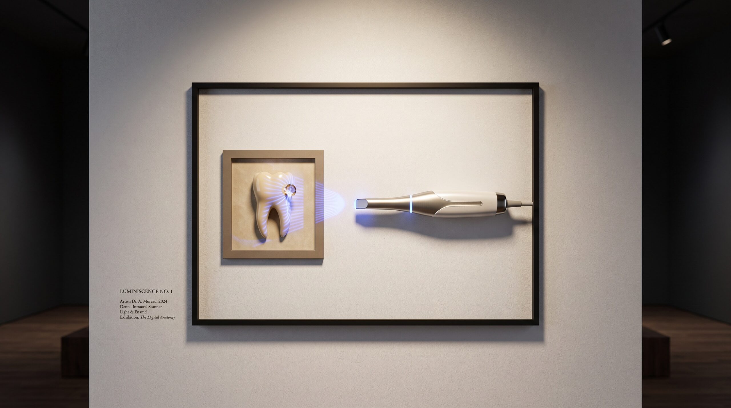
There is a quiet irony at the centre of the digital impression. We have built an exquisitely sensitive optical instrument — a wand that reads a projected pattern of light bouncing back off a surface and reconstructs it, frame by frame, into a three-dimensional model accurate to tens of microns — and we point it at the single wettest, most restless surface in all of clinical medicine. The mouth secretes, pools, fogs, and bleeds, and it does all of this precisely where the scan matters most. The failure mode of intraoral scanning is rarely dramatic. It is not a crash or an error message. It is a model that looks beautiful on the monitor and fits badly in the mouth, because somewhere along a margin a film of fluid stood in for tooth, and the software, having no way to know the difference, measured the fluid.
Light Cannot Tell Tooth From Water
To understand why moisture is so corrosive to accuracy, it helps to remember what the scanner actually sees. It does not see teeth. It sees light — a structured pattern it casts onto a surface and then watches deform, inferring depth from the distortion. That inference assumes the surface it is reading is the surface you care about. A bead of saliva, a thin glaze of crevicular fluid, a smear of blood: each is a genuine optical surface. It reflects light, it has a shape, and it has the added mischief of being specular — mirror-like — scattering the projected pattern in ways the algorithm tries valiantly to stitch into geometry. The result is not noise that the software flags and discards. It is false geometry, confidently rendered, indistinguishable on screen from the real thing. The whole discipline of reading a scan as a made object — described in what really governs the accuracy of an intraoral scan — begins with accepting that the instrument is faithful to whatever surface happens to be there, and that keeping the wrong surface out of the frame is the operator’s job, not the optics’.
The Margin Is Where It All Goes Wrong
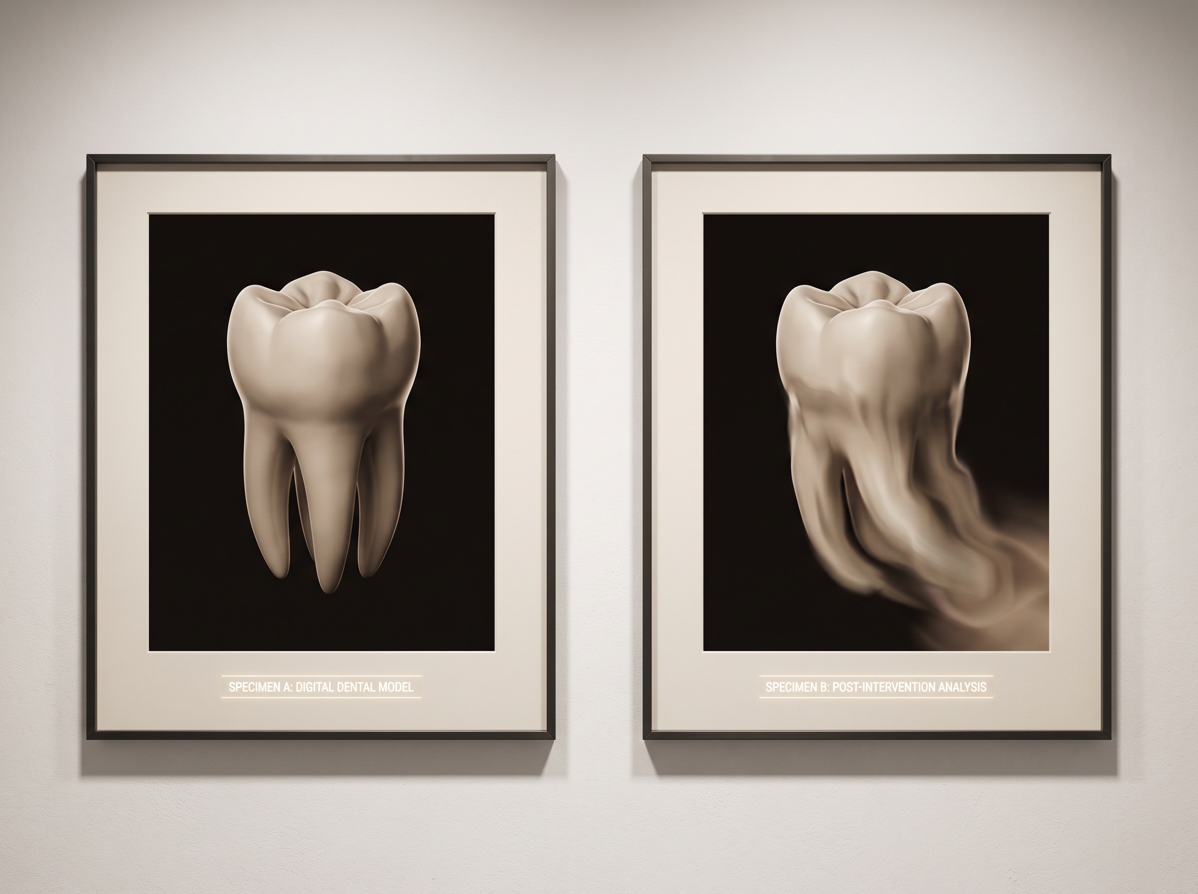
Not every part of a scan is equally vulnerable. A broad occlusal table tolerates a little moisture; the geometry is forgiving and there is plenty of dry enamel for the algorithm to lock onto. The margin is another matter entirely. The finish line of a crown preparation, the delicate junction where tooth meets gingiva, is both the most important feature in the entire impression and the one place the mouth most aggressively refuses to stay dry. The gingival crevice is a reservoir. It weeps crevicular fluid continuously, and it refills within seconds of being dried. A margin that was clean when you began a quadrant can be awash by the time the wand returns to it. Because this is exactly the region where a restoration’s fit is decided — where a gap of a few tens of microns separates a crown that seats from one that leaks — a moisture error here is not a cosmetic blemish on the model. It is the difference between a restoration that lasts and one that fails slowly from the edge inward.
A Dry Field Is Equipment and Sequence, Not Luck
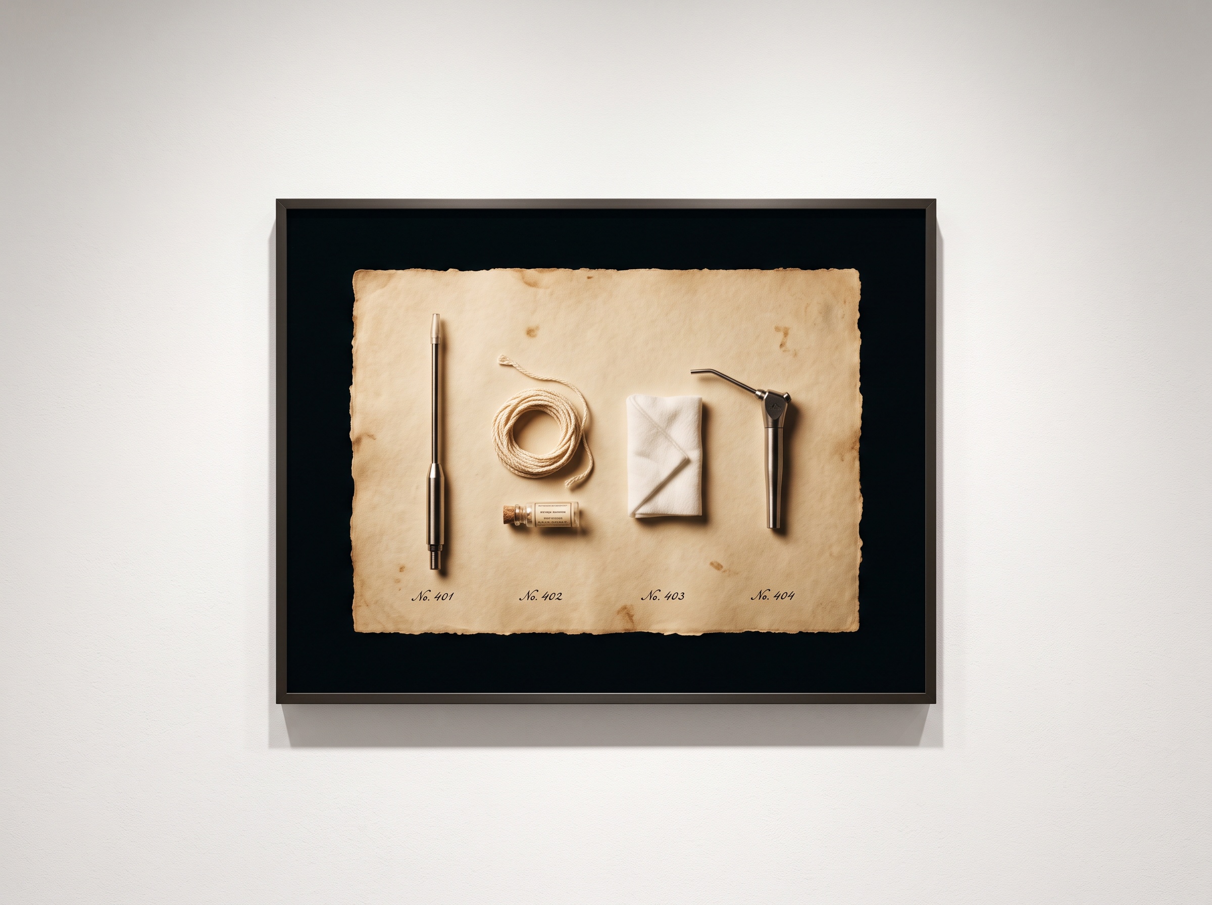
The good news is that moisture control is an entirely solvable problem, and the solution is unglamorous: the same isolation discipline that good operative dentistry has always demanded, applied with the scan in mind. Effective suction that actually keeps pace with flow, not a tip parked in the wrong vestibule. Retraction that opens the crevice and holds it open long enough to capture the margin dry — cord, paste, or a cordless technique, chosen for the case. A measured burst of air to clear and, where needed, to lift the gingiva away from the finish line. And, quietly important, sequence: scanning the area you have just isolated before it refloods, rather than isolating everything and hoping it holds. The operator who treats the dry field as a transient window to be used immediately, rather than a state to be established once and assumed permanent, captures clean margins where another operator captures smears. The scanner is only ever as good as the second of dryness it is given.
The Enemies You Do Not See: Fog and the Warm Tip
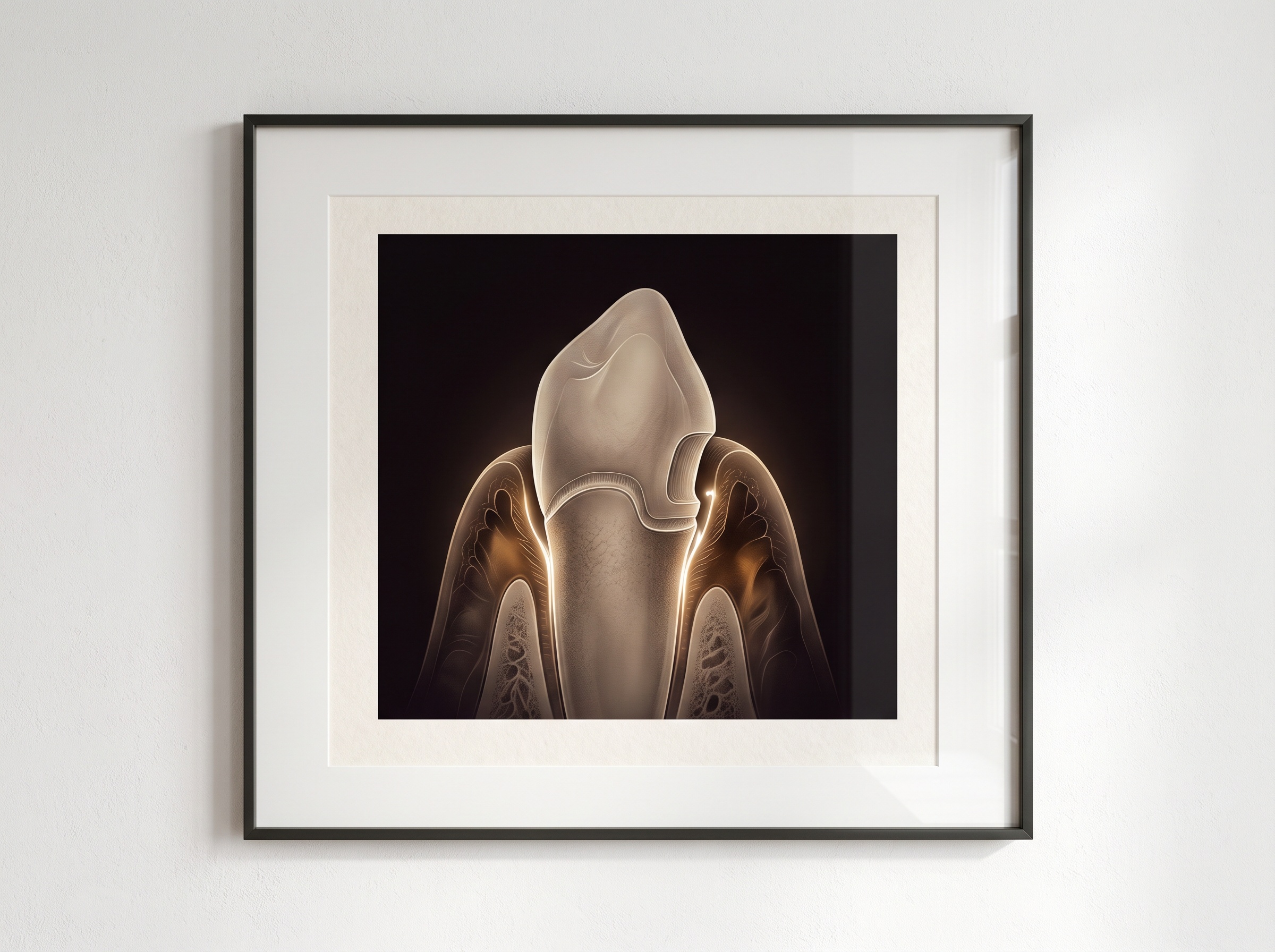
Not all moisture is in the mouth. A cold scanner tip introduced into a warm, humid oral cavity fogs instantly, exactly as a pair of glasses does on a winter doorstep — and a fogged tip casts its structured light through a veil of condensation that blurs and shifts the pattern. This is why most systems warm the tip, and why a tip that has cooled between patients or during a long pause is a hidden source of soft, driftless error. The same optical honesty that matters when a mirror fogs during photography matters here; the discipline of a clear optical path, familiar from shooting fog-free occlusal and intraoral images, applies with equal force to the scanner. Breath, too, contributes — the patient’s exhalation and the clinician’s own can lay a film on tip or tooth. None of these announce themselves. They do not pool visibly like saliva. They simply degrade the signal the scanner is trying to read, and the model absorbs the degradation as a subtle loss of fidelity you will only discover when the restoration arrives.
Reading the Scan for What the Field Left Behind
Because moisture errors render as plausible geometry, the defence is not only prevention but inspection — knowing how to interrogate a finished scan for the signatures of a wet field. A margin that looks unnaturally smooth or rounded, lacking the crisp transition a dry finish line should show. A small bulge or web of tissue-coloured mesh bridging a crevice that ought to be open. A patch of surface that stitched oddly, where the system clearly fought to reconcile frames. Understanding how the scanner assembles its picture — how structured light becomes a stitched mesh built frame by frame — is what lets a clinician look at a questionable region and recognise it as fluid artefact rather than true anatomy. The practitioner who inspects the margin at full magnification before dismissing the patient, rather than trusting the seductive clean render on the chairside screen, is the one who rescans the one area that needed it instead of discovering the problem at try-in.

Future Developments
The trajectory of the technology is toward tolerance — scanners and algorithms that are less easily fooled by the mouth’s wetness. Research is advancing on fluid-aware reconstruction, where the software learns to recognise the optical signature of saliva or blood and refuses to build geometry from it, flagging the region for recapture rather than silently rendering it. Wavelengths and illumination strategies less perturbed by thin liquid films are being explored, as are tip designs that resist fogging more robustly. One can imagine, before long, a scan that pauses and says, in effect, this margin is wet — dry it and come back. Yet even the most forgiving instrument will have a limit, and the limit will always sit at the margin, in the crevice that refills in seconds. For the foreseeable future the decisive variable will remain human: the operator who establishes a dry field, uses it at once, and reads the result with suspicion. The scanner has made the impression digital. It has not yet made the mouth dry, and until it does, keeping it dry remains the quiet craft on which every accurate scan still depends.
Sources & further reading:
- Accuracy of intraoral scanners and the influence of moisture (Journal of Prosthetic Dentistry)
- Digital impressions: isolation and field control best practices (International Journal of Computerized Dentistry)
No Comments