


February 5, 2026 Trueness and Precision: What Really Governs the Accuracy of an Intraoral Scan
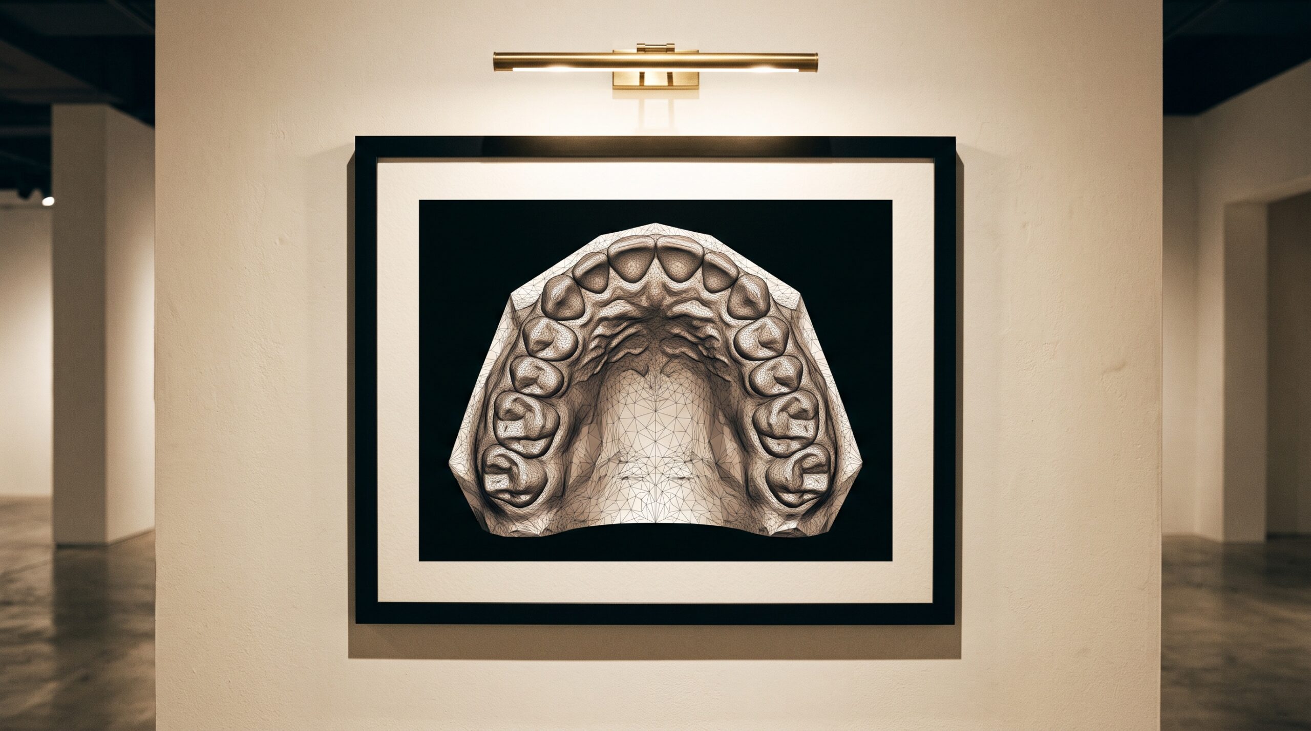
On the monitor, a digital impression is a seductive thing. It rotates cleanly in space, rendered in confident colour, every cusp and margin apparently rendered to perfection, and the eye — trained by a lifetime of screens — reads that smoothness as truth. Yet a scan that looks perfect and a scan that is accurate are two entirely separate claims, and the quiet distance between them is exactly where a crown returns from the lab a fraction proud, where a full-arch framework rocks, where the beautiful model on screen was subtly, invisibly wrong. To photograph the mouth in three dimensions is to inherit an old problem of measurement, dressed in new light.
The vocabulary that dissolves the confusion is borrowed from metrology, and it is worth speaking precisely. Accuracy, in the formal sense set out in ISO 5725, is not one property but two: trueness and precision. A scanner can possess either without the other, and only when it holds both can its model be trusted to carry a restoration from screen to seat.
Two Virtues That Are Often Confused
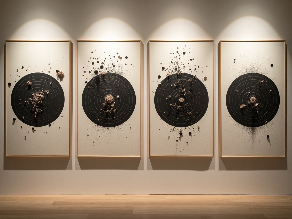
Trueness is closeness to reality — how faithfully the digital surface matches the actual geometry of the tooth and arch. A scanner of high trueness produces a model whose dimensions, on average, sit right on top of the real ones. Precision is a different virtue entirely: repeatability. Scan the same quadrant five times and a precise instrument returns five models that agree closely with each other — whether or not they agree with the truth.
The distinction is not academic, because the two failures look nothing alike and demand different remedies. A scanner that is precise but not true errs the same way every time — a consistent bias, correctable once it is understood. A scanner that is true but not precise scatters around the correct answer, so any single scan is a gamble even though the average is sound. The instrument you actually want is the rare one that is both: tightly clustered, and clustered on the truth. Everything that follows in a digital workflow — the fit of the margin, the passivity of a framework, the occlusion that needs no adjustment — rests on that unglamorous pair of words.
How the Scanner Sees
An intraoral scanner does not take a photograph in any ordinary sense. It projects light — a structured pattern of lines, or the shifting focal planes of a confocal system — onto the tissue and reads how that light is deformed or brought to focus by the surface it meets. From each momentary view it recovers the depth of a small patch of the mouth: not a picture, but a cloud of three-dimensional points. The wand is, in effect, taking thousands of tiny partial measurements a second, each one a single honest glance at a sliver of enamel.
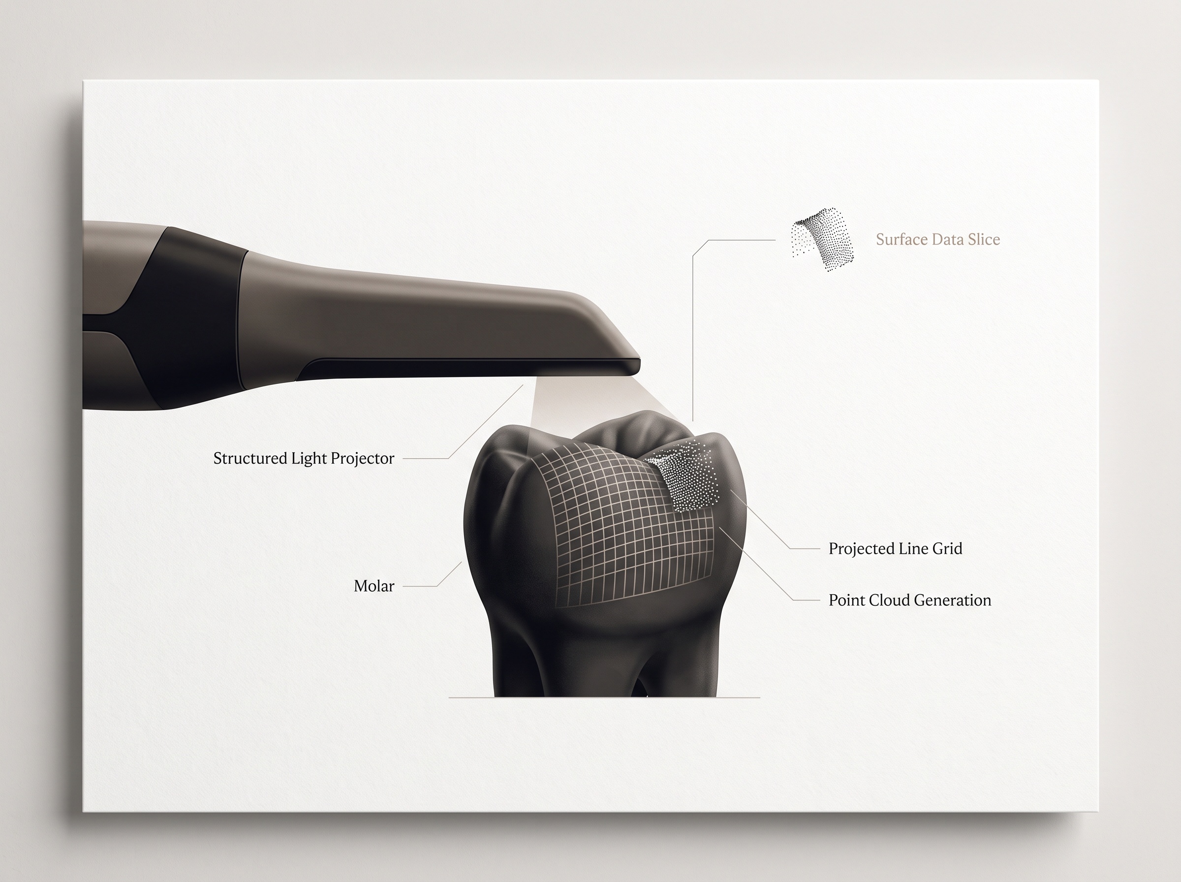
No single glance sees the arch. The complete model is assembled from that torrent of overlapping frames, and it is in the assembly — not the individual capture — that the real character of a scanner is written. Understanding how those fragments are joined is the key to understanding where accuracy is won and lost.
The Stitch Is Where Error Lives
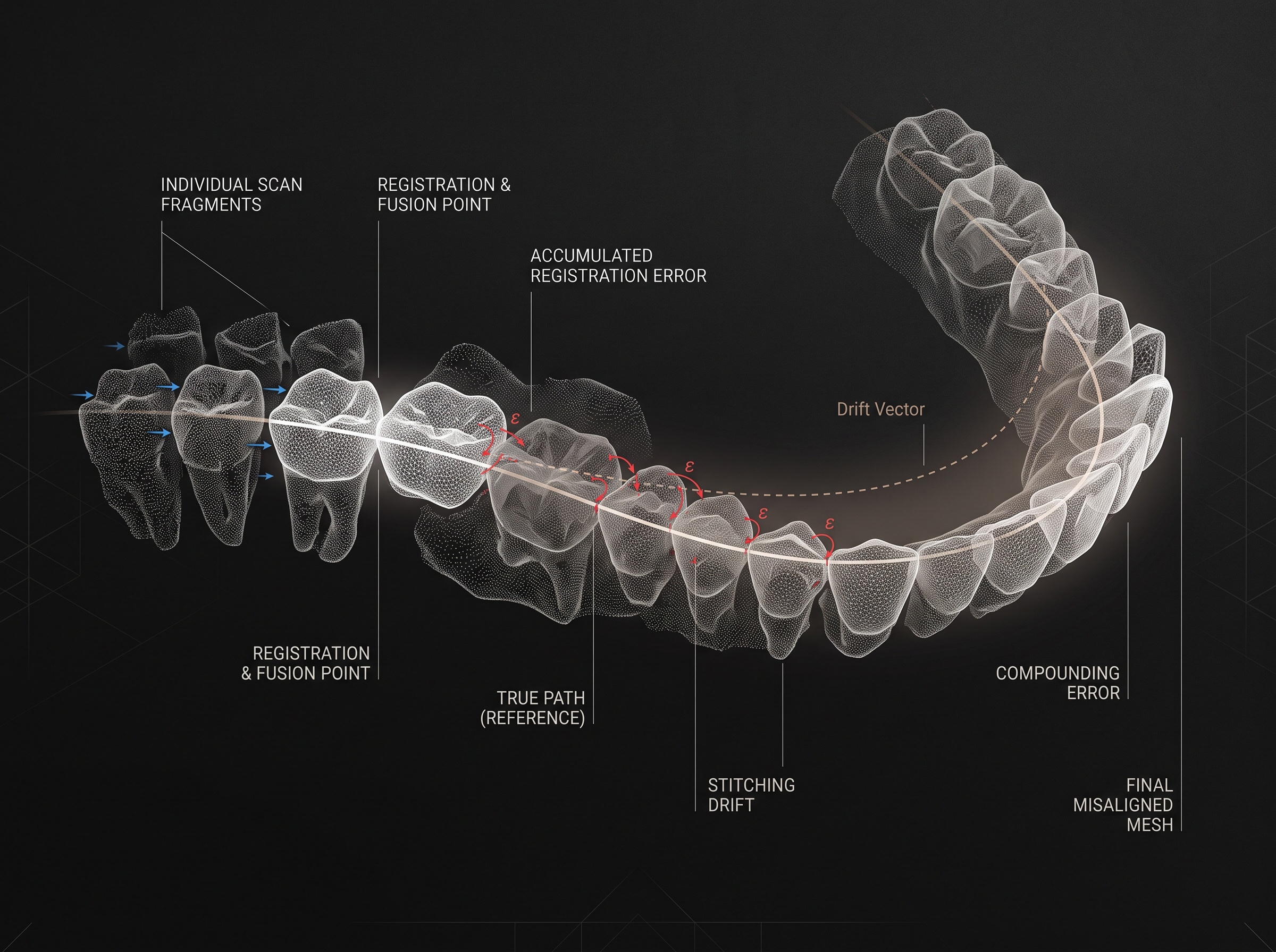
To build one continuous surface, the software must decide how each new frame aligns with the ones already captured — a process called registration, or stitching. It searches for the overlap, rotates and slides the incoming fragment until its shared geometry matches, and fuses it into the growing mesh. Done thousands of times, this is what turns a swarm of local glances into a single arch you can rotate on screen.
But every registration carries a tiny uncertainty, and here lies the central truth of digital impressions: those small errors do not simply average away — they can accumulate. Each frame is aligned to the previous, so a minute rotational error early in the sweep propagates and compounds along the span, a slow drift that grows with distance from where the scan began. This is why a single prepared tooth or a short quadrant is, for a good scanner, almost trivially accurate, while the full arch remains the genuine frontier. Scan a lone molar and there is little room for drift to build; scan canine-to-canine-to-molar across the whole vault and the far end can wander measurably from the truth even as the model looks seamless. The literature comparing scanners is remarkably consistent on this point: differences between systems that vanish over a single unit become pronounced, and clinically decisive, across a complete arch.
The Operator Is Part of the Instrument
It is tempting to treat accuracy as a specification stamped on the scanner, but the person holding the wand is inseparable from the result. Because the model is stitched from overlapping views, the path the operator traces — the scan strategy — directly shapes how error accumulates. A disciplined, unbroken sweep that keeps generous overlap and returns cleanly to close its loops gives the registration algorithm the redundancy it needs to stay honest. A hurried, skipping motion that lifts away and rejoins, that leaves thin overlap or scans reflective saliva and mobile soft tissue, invites the stitch to guess — and a guess, once fused, becomes indistinguishable from measurement.
So two clinicians, the same scanner, the same mouth, can produce models of measurably different accuracy. The wet, moving, translucent reality of the oral cavity only sharpens this: enamel scatters projected light beneath its surface, blood and saliva throw specular glare, the tongue and cheek intrude. The instrument’s trueness and precision are a ceiling; technique decides how close to that ceiling a given impression actually rises. In digital dentistry the operator does not merely use the measuring device — for the duration of the scan, they are a component of it.
Reading a Scan as a Made Object
There is a discipline in learning to look at a digital impression not as a finished truth handed down by the machine, but as a constructed image with a provenance — assembled, stitched, and susceptible in known places. The finest digital clinicians develop an eye for the suspect regions: the far reaches of a full-arch sweep, the edentulous stretch with little geometry to grip, the deep interproximal shadow the light could not fully reach. They verify rather than assume, and they respect the difference between a model that is precise enough to look consistent and one that is true enough to fit.
Seen this way, the rotating arch on the monitor rejoins the tradition this gallery keeps returning to: it is a portrait built from thousands of individual glances, and like every portrait it carries the hand of its making. To read it well is to hold its beauty and its provenance in the same gaze — to admire the seamless surface while remembering, precisely, how it was assembled and where it might have quietly drifted from the truth.
Future Developments
The trajectory of the field is a direct assault on the stitch. Scanners increasingly fold in principles of photogrammetry and global registration — solving all the frames against one another at once rather than chaining each to the last — so that error is distributed and closed rather than allowed to accumulate down the arch. Machine-learning registration is beginning to recognise and reconcile tissue that older algorithms found ambiguous, and to flag, in real time, the regions where overlap grew thin and confidence fell. The most telling change may be cultural rather than optical: instruments that report their own uncertainty, showing the clinician not just a surface but a map of where that surface can be trusted. As trueness and precision both climb and the full arch is finally tamed, the digital impression will come closer to what it has always promised — a measurement honest enough to build upon, and beautiful enough to be worth looking at.
Sources & further reading:
- Trueness and precision of intraoral scanners: a review (International Journal of Computerized Dentistry / PubMed)
- Accuracy of intraoral scanners for full-arch scanning (Journal of Prosthetic Dentistry, ScienceDirect)
- ISO 5725-1: Accuracy (trueness and precision) of measurement methods and results
Sorry, the comment form is closed at this time.