


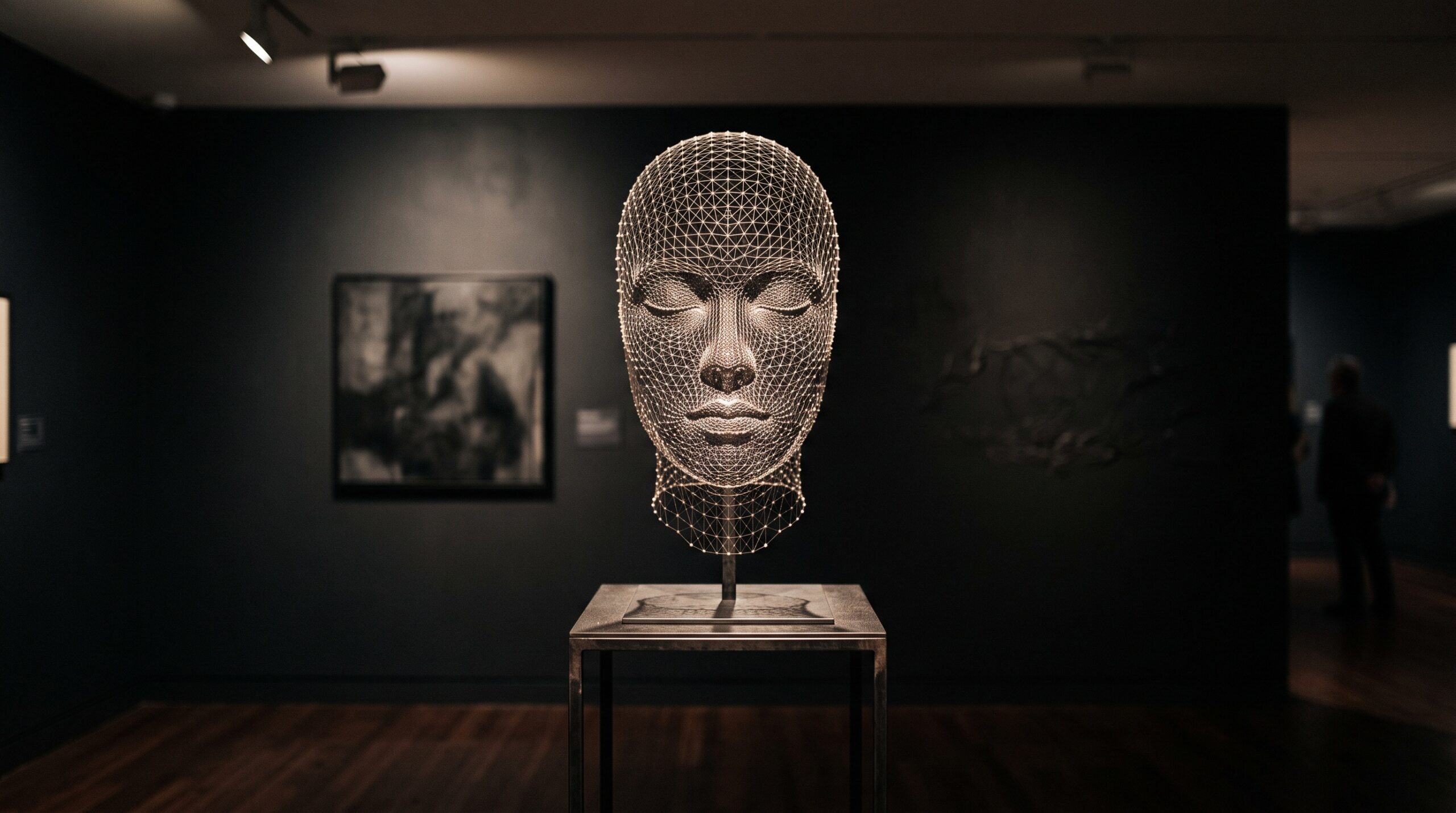
October 18, 2026 The Face as Proof: Stereophotogrammetry and the 3D Verification of a Restored Bite
Every portrait in a gallery is a negotiation between two faces: the one the sitter wore and the one the artist chose to record. Dentistry’s most ambitious restorations stage the same negotiation, only in three living dimensions and across the lower third of a real human face. When a clinician rebuilds a bite worn flat by years of erosion, the jaw is reopened by a handful of millimeters — and those millimeters do not stay politely inside the mouth. They push the chin down and forward, lengthen the lower face, re-drape the lips, and reset the quiet geometry by which we read a face as balanced or collapsed. For decades the profession could plan that transformation with great precision and then judge its result the oldest way imaginable: by looking. A new clinical report suggests that era of looking is giving way to an era of measuring.
The collapse no one photographs
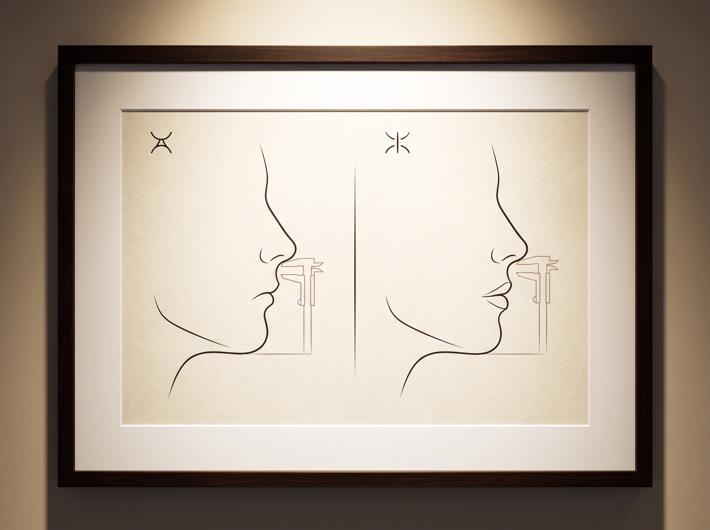
Start with what wear actually does. Generalized erosive tooth wear — from acid, from grinding, from the slow chemistry of a modern diet — does not merely shorten teeth; it shortens the face. As the posterior teeth lose height, the mandible over-closes to find contact, and the vertical dimension of occlusion, the established distance between the jaws when the teeth meet, quietly diminishes. The outward signs are the ones patients notice without understanding: a chin that seems to creep toward the nose, deepening folds at the corners of the mouth, a lower face that has lost its proportion. It is one of the few truly three-dimensional problems in dentistry, and for most of its history it has been documented in resolutely two-dimensional ways.
Rebuilding it is a material-science undertaking as much as an aesthetic one. The contemporary answer is a minimally invasive, digitally driven full-mouth rehabilitation — typically with thin, bonded lithium-disilicate restorations that reopen the bite while preserving enamel rather than grinding teeth down to stumps. The plan is conceived digitally, previewed, and milled. But a plan is a promise, and a promise about a face needs a way to be checked.
Why the old proof was really a guess
Consider how that check used to be made. The bite was opened on an articulator, a mechanical surrogate for the jaws; the soft-tissue effect was anticipated from profile photographs and cephalometric tracings; and the final verdict was delivered by the clinician’s and patient’s eyes in a mirror. None of this is worthless — skilled operators achieve beautiful results this way every day — but notice what it is not. A two-dimensional photograph flattens a change that is inherently volumetric. A cephalogram measures bone, not the living envelope of lips and cheeks that actually meets the world. And the eye, as we have explored before on these pages, is a magnificent but unreliable instrument of record. The result could be admired. It could not easily be proven, nor the planned change compared, millimeter for millimeter, against the delivered one.
Stereophotogrammetry: capturing the face as a surface
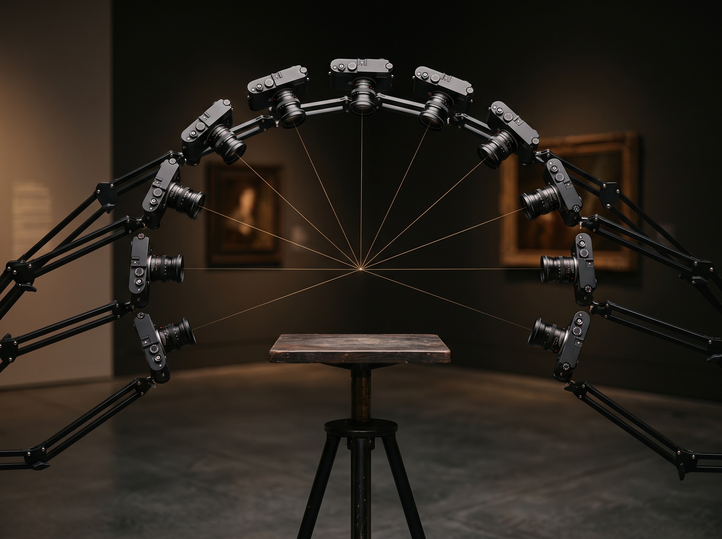
The instrument closing that gap is stereophotogrammetry, and it is worth understanding on its own terms rather than as a black box. The technique is old in principle and borrowed from far outside dentistry — it is how cartographers once built elevation maps from overlapping aerial photographs, and how film studios capture an actor’s face for a digital double. Several calibrated cameras, arranged around the subject, fire at the same instant from known positions. Software then finds the same point on the skin in multiple images and, by triangulating the angles, computes that point’s exact location in space. Do this for hundreds of thousands of points and the result is not a picture of a face but a measurable surface — a dense three-dimensional mesh faithful to a fraction of a millimeter.
The simultaneity is the quiet genius of it. A face is never still; a laser scanner that sweeps across it over several seconds is defeated by the smallest swallow or breath. Capturing every point in a single flash freezes the living surface honestly. This is the same impulse toward richer, more dimensional capture that runs through so much of modern imaging — from efforts to record the smile in motion as four-dimensional data to the drive to fuse the face, the bone, and the teeth into a single virtual patient. Stereophotogrammetry contributes the one layer those models most need and most struggle to capture well: the true outer skin.
Three captures, one superimposition
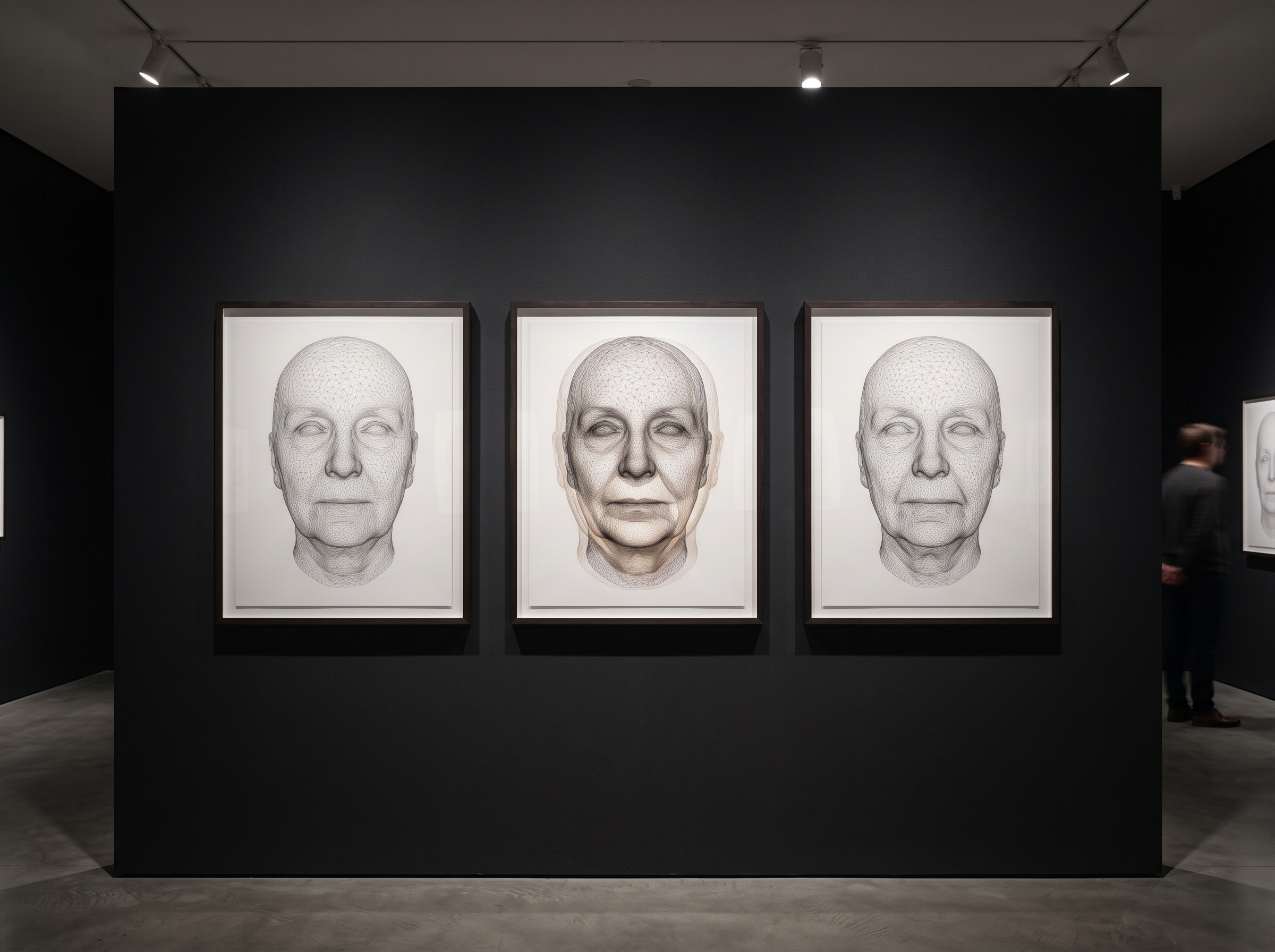
Here the method becomes a verification engine, and the recent report in the Journal of Esthetic and Restorative Dentistry is instructive. The face was captured three times: before any treatment, at the planning stage once the new vertical dimension had been designed, and after the rehabilitation was delivered. Three surfaces of one face, each a precise record of a moment. The analytical move is to register them — to align the three meshes on the stable regions that do not change, such as the forehead and the bridge of the nose, so that any divergence elsewhere is real change and not merely a different head position.
Once aligned, the lower face can be interrogated quantitatively. How far did the chin point actually travel? What was the linear change at the lips and the volumetric change across the lower third? Most tellingly, how closely did the delivered face match the planned one — the validation of accuracy between planification and final result that, the authors note, had simply not been reported before for a soft-tissue facial reconstruction of this kind. The reported finding is the one the field has been waiting to hear: the fully digital approach could restore the vertical dimension predictably while keeping the face in harmony, with the before-and-after measurements landing close together. It is a conservator’s overlay study, applied to the human face.
What it means to prove an aesthetic result
It is tempting to file this under mere documentation, but something more interesting is happening. Dentistry has always had an uneasy relationship with the subjective nature of beauty; the most consequential outcomes — does the smile look right, does the face look balanced — have been the least measurable. Stereophotogrammetry does not make beauty objective, and it should not pretend to. What it does is give the aesthetic judgment a measurable spine. The clinician still decides what a harmonious lower face should be; this is, after all, the same design sensibility that animates previewing a restored smile before a single bur touches enamel. But now the gap between intention and outcome can be audited rather than assumed, and the record of that outcome belongs to the gallery’s oldest values: provenance, measurement, and proof that the thing on the wall is what it claims to be.
Future Developments
The near future points toward making this invisible. Multi-camera rigs are shrinking, and the same triangulation is beginning to run on the depth sensors already built into ordinary tablets and phones, which will carry stereophotogrammetric capture out of the research clinic and onto the everyday operatory counter. As facial surface capture is routinely fused with the CBCT skeleton beneath and the intraoral scan within, the three-millimeter decision to open a bite will be previewed on a complete, measurable virtual face and then verified against that preview at delivery — the plan and its proof, bound together. The deeper shift is cultural rather than technical. For a century the restored face has been something clinicians asked patients to admire. It is becoming something they can measure, compare, and stand behind. The gallery learned long ago that a beautiful object is worth more when its history can be proven; dental imaging, having rebuilt the face so skillfully for so long, is finally learning to prove it.
Sources & further reading:
No Comments