


September 22, 2026 Imaging the Joint That Moves: MRI, CBCT, and the Craft of Picturing the Temporomandibular Joint
The temporomandibular joint is the one structure in the mouth that refuses to sit still for its portrait. Every other subject a dental image captures – a tooth, a root, a slice of bone – is content to be photographed once, from one direction, in a single state. The joint just in front of the ear, where the lower jaw meets the skull, is not. It is part hard bone and part soft cartilage; it works as a matched left-and-right pair that must be read together; and, above all, it is defined by movement. Those three properties are exactly the ones no single imaging method can honor at once. To picture the joint well is to accept, from the outset, that you will need more than one instrument – and to know precisely what each one keeps and what it quietly throws away.
Two Tissues, One Joint
The difficulty begins with anatomy. The rounded head of the lower jaw, the condyle, rides in a shallow socket in the skull, and between the two sits a small pad of fibrocartilage – the articular disc – that cushions the bones and guides their glide. A picture of the joint that shows only the bone has left out the very tissue that most often fails; a picture that shows only the disc has no frame of hard landmarks to place it against. The joint is a duet of a hard part and a soft part, and the modalities that image it divide neatly along that same seam. One machine is fluent in bone and mute about cartilage. The other reads soft tissue eloquently and treats dense bone as a near-silence. Choosing between them is not a matter of which is better; it is a matter of which question is being asked.
The Bone the Cone-Beam Sees
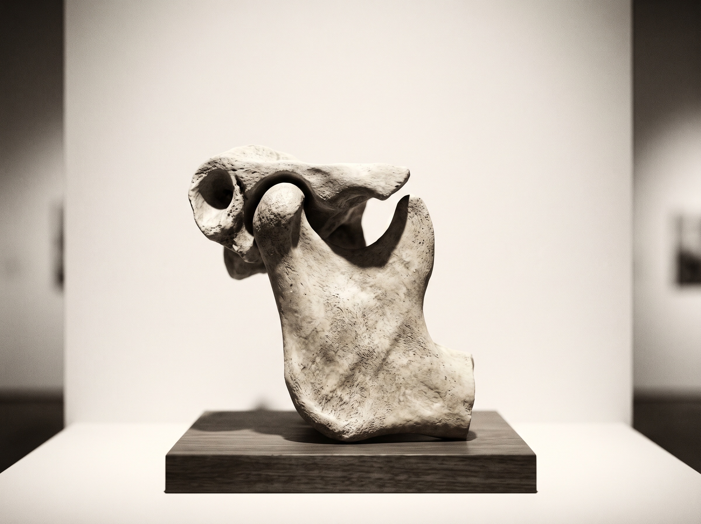
Cone-beam CT is the instrument of the bone. It renders the condyle, the socket, and the slope of the eminence it climbs during opening in high, isotropic spatial detail, and it does so in three dimensions that can be resliced along the joint’s own axis rather than the head’s. In a joint under stress it shows the sequel of that stress written into the bone: flattening of the once-rounded condyle, surface erosions, small subchondral cysts, the bony spur of an osteophyte, the slow remodeling of a joint adapting to a load it was not built for. This is the same volumetric advantage that lets cone-beam imaging expose the hidden three-dimensional anatomy a flat radiograph cannot. But bone is a slow witness. By the time the condyle has flattened, the disorder that flattened it is often years old, and the disc that failed first is nowhere in the picture – cone-beam CT is simply blind to it. The bone tells you what happened. It rarely tells you what is happening.
The Disc That Only MRI Can Show
For the disc itself there is, in routine practice, only one instrument: magnetic resonance imaging. MRI shows the little pad of cartilage directly – its shape, its position over the condyle, and, most consequentially, its displacement. In the healthy joint the disc sits atop the condyle like a cap; in the disordered joint it slips forward, and the moment it does becomes visible as a dark, biconcave form pulled ahead of where it belongs. MRI also reads the joint’s inflammation in a way bone imaging cannot: a bright rim of fluid on a T2-weighted sequence betrays an effusion, and edema in the condylar marrow signals a bone still actively changing rather than merely scarred. It accomplishes all of this without ionizing radiation – no small matter for a disorder that falls disproportionately on younger patients who may be imaged more than once. Where cone-beam CT records the aftermath in bone, MRI catches the soft tissue in the act.
Open and Closed: Imaging a Joint in Two Positions
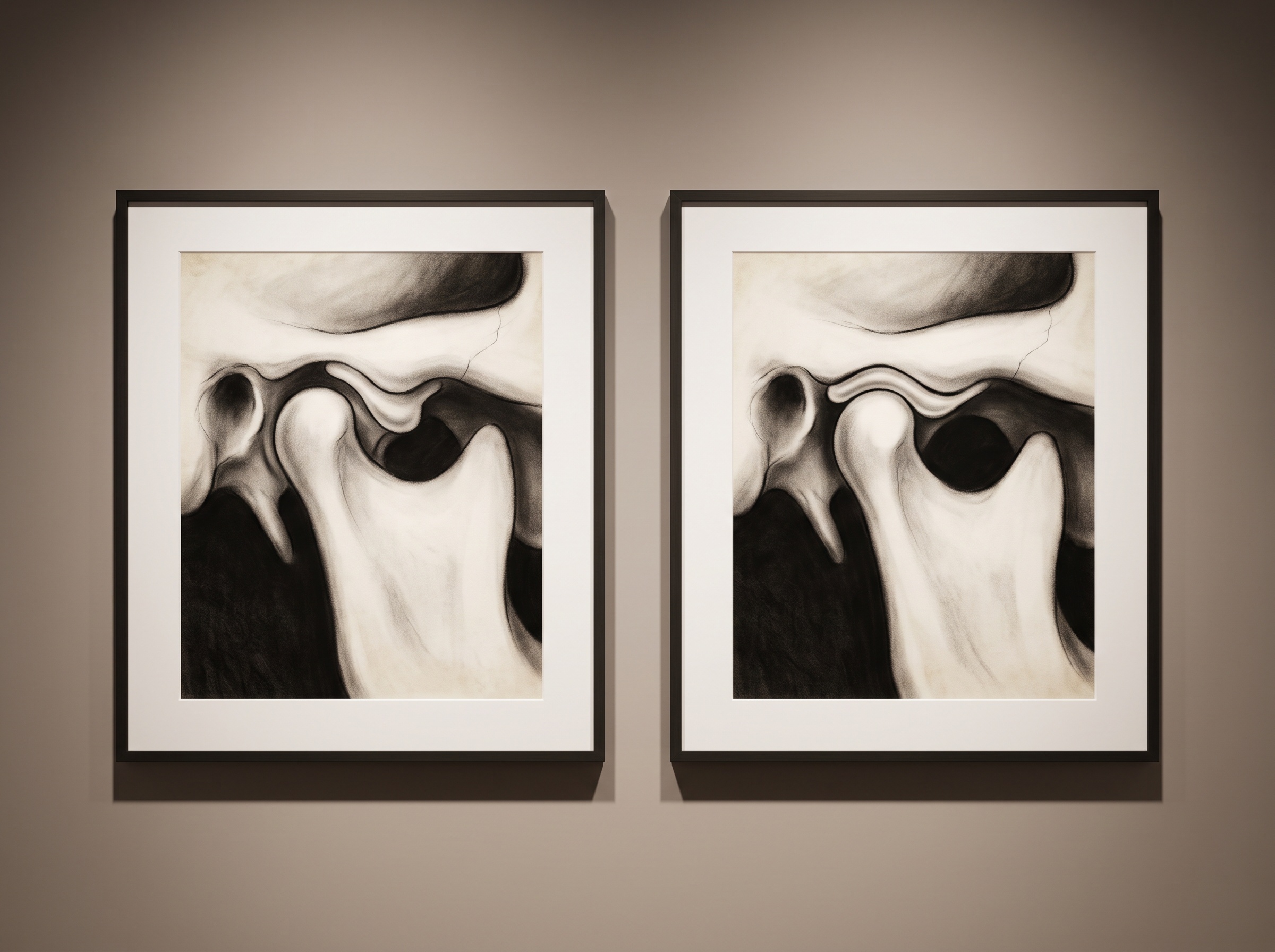
Even MRI, though, freezes what is fundamentally a motion. A single frame of a displaced disc raises the question that matters most to the patient: does it stay displaced, or does it snap back onto the condyle when the mouth opens? So the joint is imaged twice – once with the teeth together and once with the mouth held at maximum opening – and the pair is read as a small before-and-after. If a disc that sat forward in the closed view is recaptured onto the translated condyle in the open view, that is displacement with reduction, the anatomy behind the familiar opening click. If it stays stubbornly ahead in both, that is displacement without reduction, the anatomy of a jaw that will not fully open. Two static frames, deliberately chosen, are made to yield an inference about movement – the same act of assembling motion from stillness that animates four-dimensional capture of a smile as it moves. It is a workaround, and an elegant one, but it is still a workaround: two photographs standing in for a film.
The Craft of Choosing
Because the two instruments answer different questions, the first act of imaging the joint is not exposure but selection. A clinical picture dominated by mechanical noise – clicking, locking, limited opening – is a soft-tissue question, and it points to MRI. A picture of degenerative change, crepitus, a suspected fracture or a bony asymmetry is a hard-tissue question, and it points to cone-beam CT. Reaching for the wrong one wastes a scan and, in the case of CBCT, spends radiation to photograph a tissue it cannot see. Above both sits the older discipline: imaging the joint at all only when the result will change what is done for the patient, and preferring the radiation-free study when it will answer the question just as well. That is the same restraint argued in weighing a cone-beam scan’s dose against what it reveals – and it applies with particular force to a joint whose sufferers are so often young.
Future Developments
The workaround is already giving way. Dynamic, or cine, MRI films the joint through its actual opening and closing rather than sampling two frozen ends, turning the inferred movement into a movement genuinely seen. New pulse sequences – zero-echo-time and ultrashort-echo-time imaging – coax a signal out of dense bone that ordinarily reads as silence on MRI, raising the prospect of a single radiation-free scan that renders the condyle and the disc together, healing the seam that has always forced two machines on one joint. And AI disc-segmentation is beginning to trace the faint soft-tissue pad automatically, standardizing a measurement that has long depended on the reader’s eye. The trajectory is the one imaging always follows here: from a still that must be interpreted toward a moving picture that can simply be watched, and from two partial views toward one honest whole. At PatientGallery we hold that an image of the body is a made thing, composed with intent. The joint that moves has always demanded two portraits and a leap of inference between them. The work now is to close that gap – to let the joint, at last, be pictured in the one state that defines it.
No Comments