


September 15, 2026 Two Energies, One Scan: How Spectral CBCT Tells Bone From Metal
A conventional cone-beam scan, for all its three-dimensional richness, measures only one thing at each point in the volume: how strongly that speck of tissue weakened the X-ray beam passing through it. That single figure – the attenuation – becomes the grey you see. It is an extraordinary amount of information to pull from a shadow, but it hides an ambiguity that no amount of resolution can resolve. Absorption is not identity. A thin sliver of very dense enamel and a faint trace of metal can weaken the beam by exactly the same amount, and so cast exactly the same grey. The image, looking only at brightness, has no way to tell them apart.
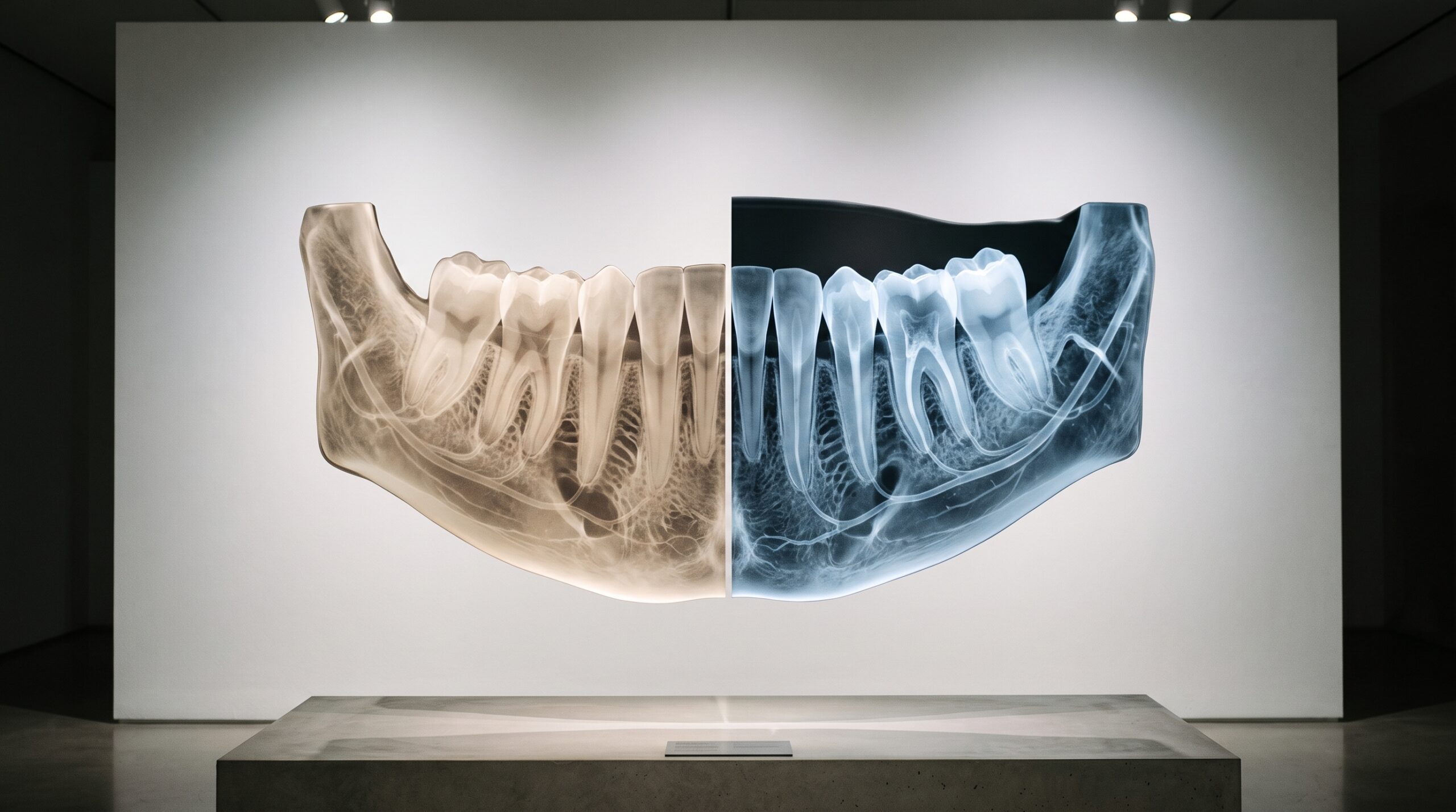
Spectral, or dual-energy, cone-beam CT is the imaging system’s answer to that ambiguity. Instead of interrogating the tissue with a single X-ray energy and recording one number, it reads two energies at once – a low-energy view and a high-energy view of the same anatomy – and compares how the material responded to each. From that comparison it can work backwards, not merely to how much a voxel absorbed, but to what the voxel is actually made of. It is the difference between seeing how bright something is and seeing its substance.
One Number for Two Materials
The root of the problem is that a single attenuation value is a coarse summary. Bone, enamel, dentine, soft tissue, gutta-percha, titanium, and the alloys of an old crown occupy an enormous range of composition, yet the moment each is reduced to one grey number, materials that are chemically worlds apart can land on the same tone. This is why the very densest structures and the metallic ones are so easily confused, and why a conventional scan cannot reliably say whether a bright region is thick cortical bone or a fleck of restorative material. The ambiguity is compounded by the fact that a cone-beam scanner’s numbers are not even stable, calibrated values – as we have explored in why a CBCT’s grey values are not true Hounsfield units, the same tissue can read differently depending on where it sits in the field. One number was never going to be enough to name a material.

Two Energies, Two Fingerprints
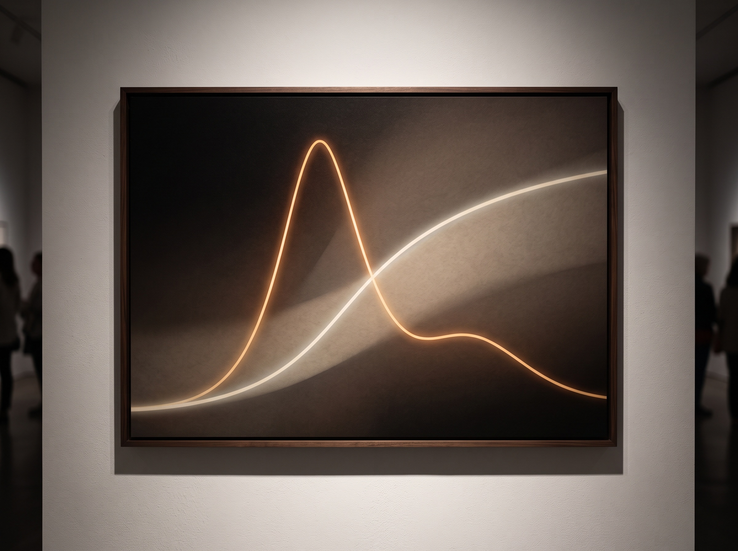
The escape from that ambiguity lies in a quiet fact of physics: X-ray attenuation is not fixed – it changes with the energy of the beam, and it changes differently for different materials. Two interactions compete inside the tissue. The photoelectric effect, which depends steeply on a material’s atomic number, dominates at low energies and makes high-atomic-number substances like metal absorb dramatically. Compton scattering, which tracks a material’s electron density, dominates at higher energies and flattens those differences out. Because these two effects rise and fall at different rates, a material traced across two energies produces a distinctive slope – a fingerprint. Enamel behaves one way as the energy changes; a titanium implant behaves another. A single energy cannot see that slope. Two energies can, and in that slope lies the material’s identity.
From Two Views to a Map of Materials
Capturing those two views can be done several ways: rapidly switching the tube voltage between low and high, using two X-ray sources at once, layering a detector so its top and bottom sense different energies, or – most elegantly – using an energy-resolving detector that sorts each arriving photon by its energy as it lands. However the two datasets are acquired, the reconstruction can then recombine them mathematically into new kinds of image the raw scan never held. One is the material-decomposition map, which separates the volume into its constituents – bone here, soft tissue there, high-atomic-number metal isolated on its own – so the scan reads as a chart of substances rather than an undifferentiated field of grey. Another is the virtual monochromatic image, a synthetic reconstruction of how the anatomy would look if it had been imaged at a single chosen energy, tuned to whatever the clinician most needs to see.
Taming the Metal Streak
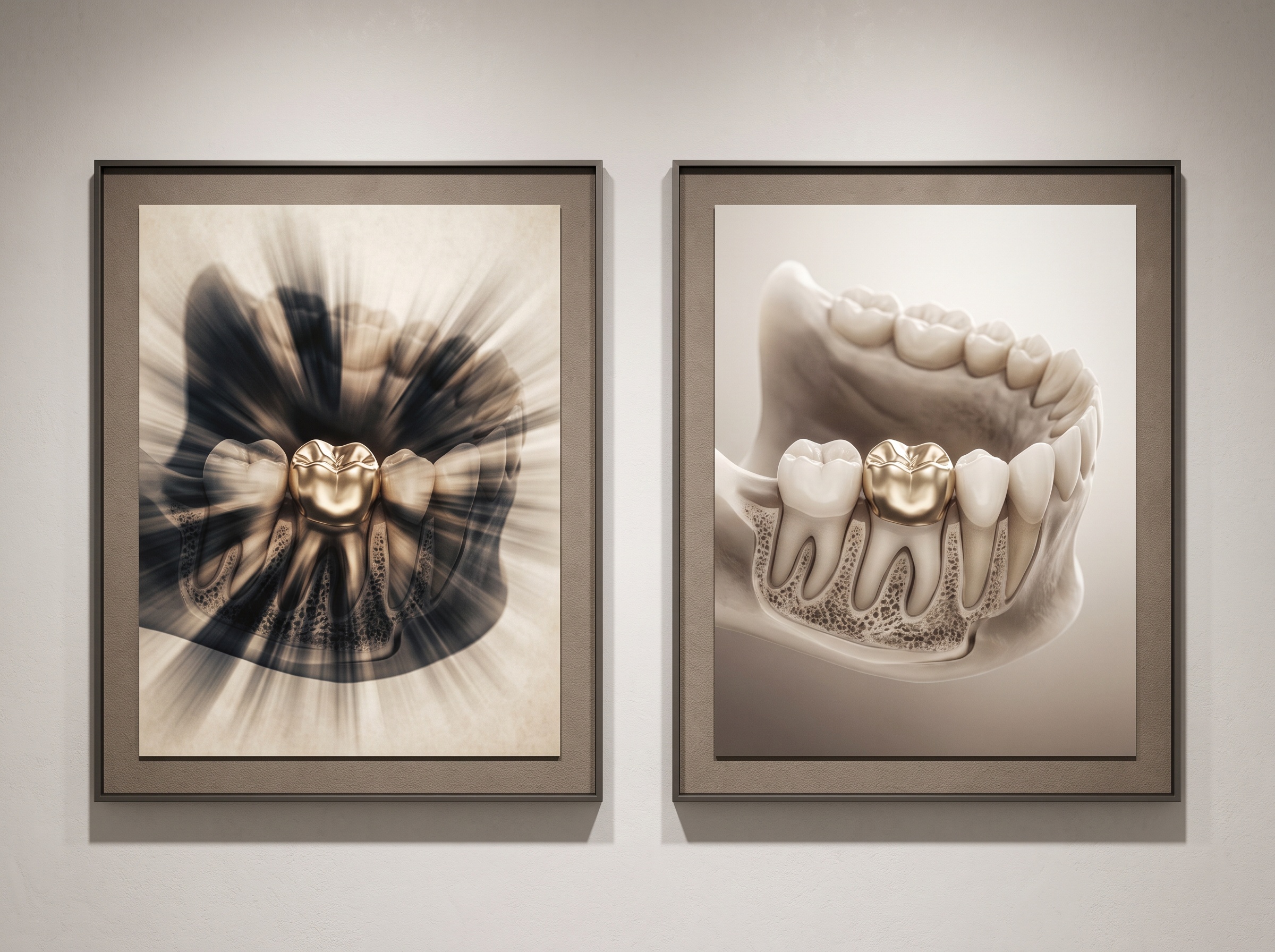
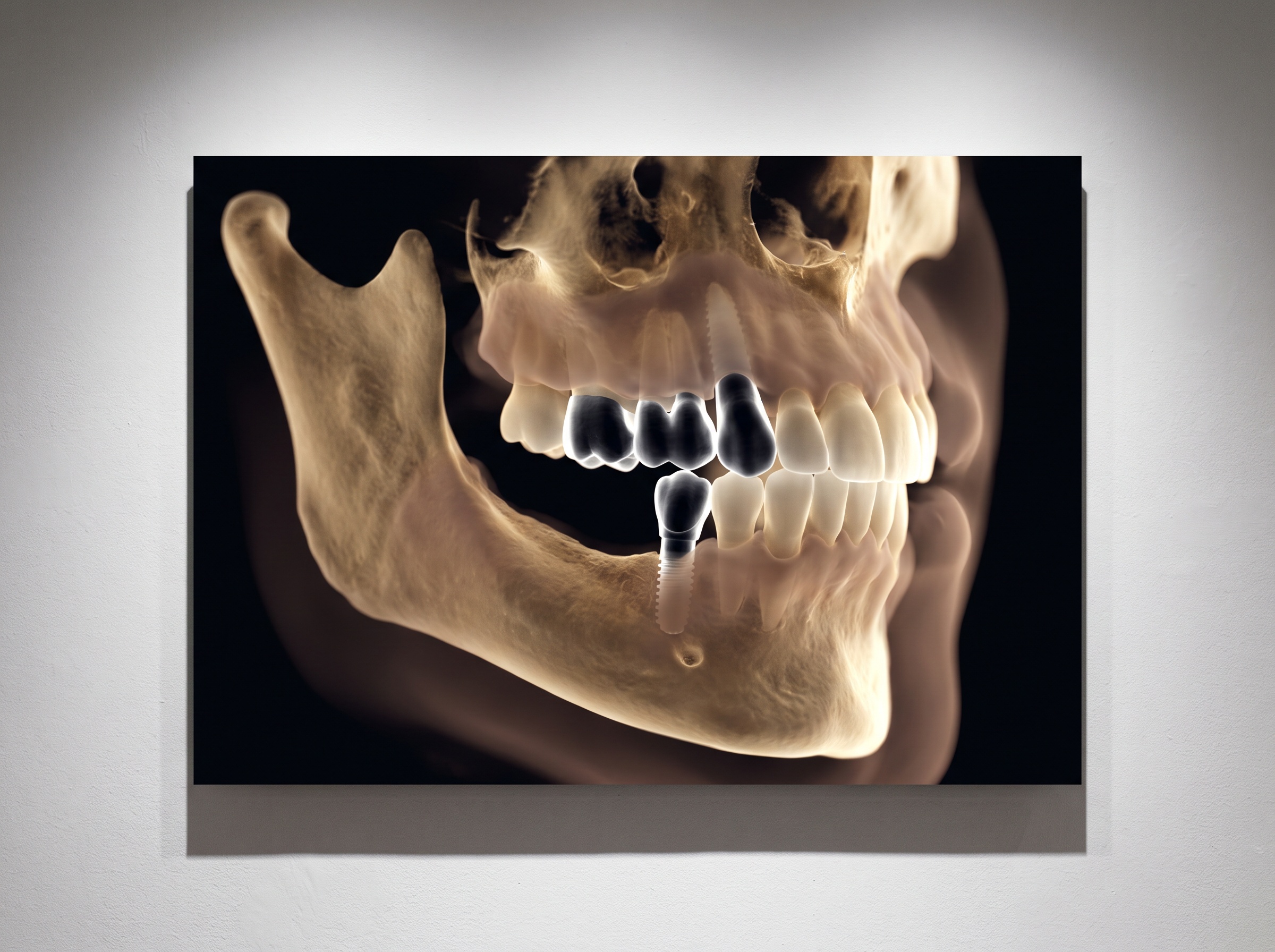
For dentistry, the most immediate gift of that flexibility is relief from metal. Dense restorations, posts, and implants are the bane of the cone-beam volume, throwing beam-hardening and metal-streak artefacts across exactly the region a clinician most wants to read – the bone at an implant threshold, the canal beside a post, the margin under a crown. Because these artefacts are themselves an energy effect, born of the beam being preferentially stripped of its low-energy photons, spectral imaging can push back against them at their source. Reconstructing a high-keV virtual monochromatic image mimics a harder, more penetrating beam that the metal cannot distort so violently, and the radiating streaks that once buried the surrounding anatomy soften and recede. The metal remains; the ruin it spread across its neighbourhood does not.
The Honest Limits
PatientGallery’s admiration for a new capability has never meant pretending it is finished, and spectral cone-beam CT is emphatically still arriving in dentistry rather than settled into it. The hardware to acquire two clean energies is more complex and more costly than a conventional scanner, and dedicated dental spectral systems remain far from routine. Dual-energy processing can raise image noise, so the material information is bought at a price that must be managed against dose rather than assumed to be free. Material decomposition is itself a reconstruction, built on assumptions about which materials are present, and like any reconstruction it can be misled – a wrong assumption produces a confident but incorrect map. The promise is real, but it is a frontier, not a solved problem.
Future Developments
The clearest path forward runs through the detector. The energy-resolving photon-counting detector, which sorts photons by energy as an intrinsic part of how it works, makes spectral imaging natural rather than bolted-on, and as that physics migrates from medical CT into dental cone-beam systems, the two-energy read may become simply what a scan is, not a special mode invoked for hard cases. Material maps will likely be surfaced automatically, with software labelling bone, soft tissue, and metal and suppressing artefact before a clinician has scrolled a slice. Further out lies the quieter ambition beneath all of it: a volume that is quantitative and honest about substance, in which a bright voxel announces not merely how much it absorbed but what it is – so that the scan is read, at last, by material rather than by shadow. That is the deeper shift spectral imaging represents. For a century the radiograph asked one question of every point in the body: how dark is your shadow? Spectral CBCT has begun, quietly, to ask a better one: what are you made of?
Sources & further reading:
No Comments