


September 29, 2026 Same Lingual, Opposite Buccal: The Tube-Shift Rule and the Craft of Finding Depth in a Flat Radiograph
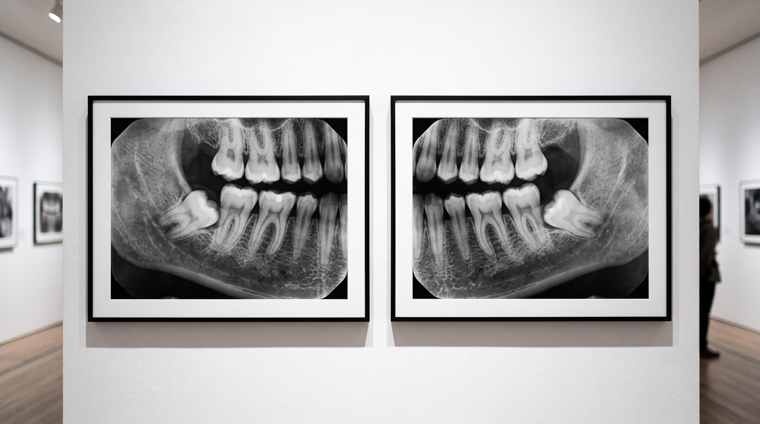
A periapical radiograph is a beautiful lie of omission. It gathers a living, three-dimensional mouth — teeth set at angles, roots curving toward cheek or tongue, a canine buried somewhere in the thickness of bone — and presses all of it flat onto a single plane. What survives is exquisitely detailed in two dimensions and utterly silent about the third. Every point along the beam’s path is stacked into one shadow, so a structure sitting near the lip and a structure sitting near the palate can land on the same spot of the image, indistinguishable. The craft of radiography has always included ways to recover what this flattening quietly erases, and the oldest, most elegant of them needs no software and no second machine: you simply take the picture twice, from two slightly different angles, and read how the shadows move.
That technique has a name that generations of students have chanted into memory — SLOB: Same Lingual, Opposite Buccal — and behind the mnemonic lies a small, durable piece of visual reasoning that belongs as much to the history of seeing as to dentistry.
The Problem of the Flattened Mouth
Projection radiography works by casting shadows. A diverging beam of x-rays passes through tissue and lands on a sensor, and denser structures absorb more, printing darker on the exposed image. The geometry is honest but pitiless: it preserves height and width and destroys depth. Two objects lying along the same ray — one buccal, toward the cheek; one lingual or palatal, toward the tongue — superimpose. On the film they may appear to touch, to overlap, even to be the same object. For most routine work this loss is tolerable. For the questions that actually change a treatment plan — where exactly is that impacted canine, does this root lie inside or outside the sinus wall, is that radiopaque fleck sitting in the soft tissue of the cheek or embedded in bone — the missing dimension is the entire question.
Same Lingual, Opposite Buccal
The rule exploits parallax, the same cue that lets two eyes judge distance. Take a first exposure. Then move the x-ray tube head horizontally — shift it mesially or distally — and take a second, keeping the sensor and patient as stable as possible. Objects closer to the sensor (lingual or palatal, since the sensor sits behind the teeth on the tongue side) will appear to move in the same direction as the tube shifted. Objects farther from the sensor and nearer the source (buccal, toward the cheek) will appear to move in the opposite direction. Same direction as the tube: lingual. Opposite direction: buccal. Watch which way the object of interest slides relative to a fixed reference — a known root apex, a stable landmark — and the third dimension resolves itself. A canine that drifts the same way you moved the tube is palatal; one that swims against you is buccal.
The vertical version obeys the same logic when the tube is shifted up or down instead, and the broader principle — that displacement reveals depth — is often called the buccal object rule or, in its clinical shorthand, the tube-shift technique.
Clark and the Century-Old Instinct
The insight is not new. It was articulated for dentistry early in the twentieth century — Clark’s description of localizing objects by shifting the tube dates to 1909 — barely more than a decade after Roentgen’s discovery and the first dental radiographs. That timing matters. Before there was any hope of reconstructing a volume, before the mathematics of tomography existed even as a dream, clinicians had already reasoned their way to a method for seeing in depth using nothing but two flat pictures and an understanding of how shadows behave. It is a reminder that imaging has always been as much interpretation as capture — that the craft lives not only in the instrument but in the mind trained to read what the instrument cannot directly say.
Reading the Shift at the Chair
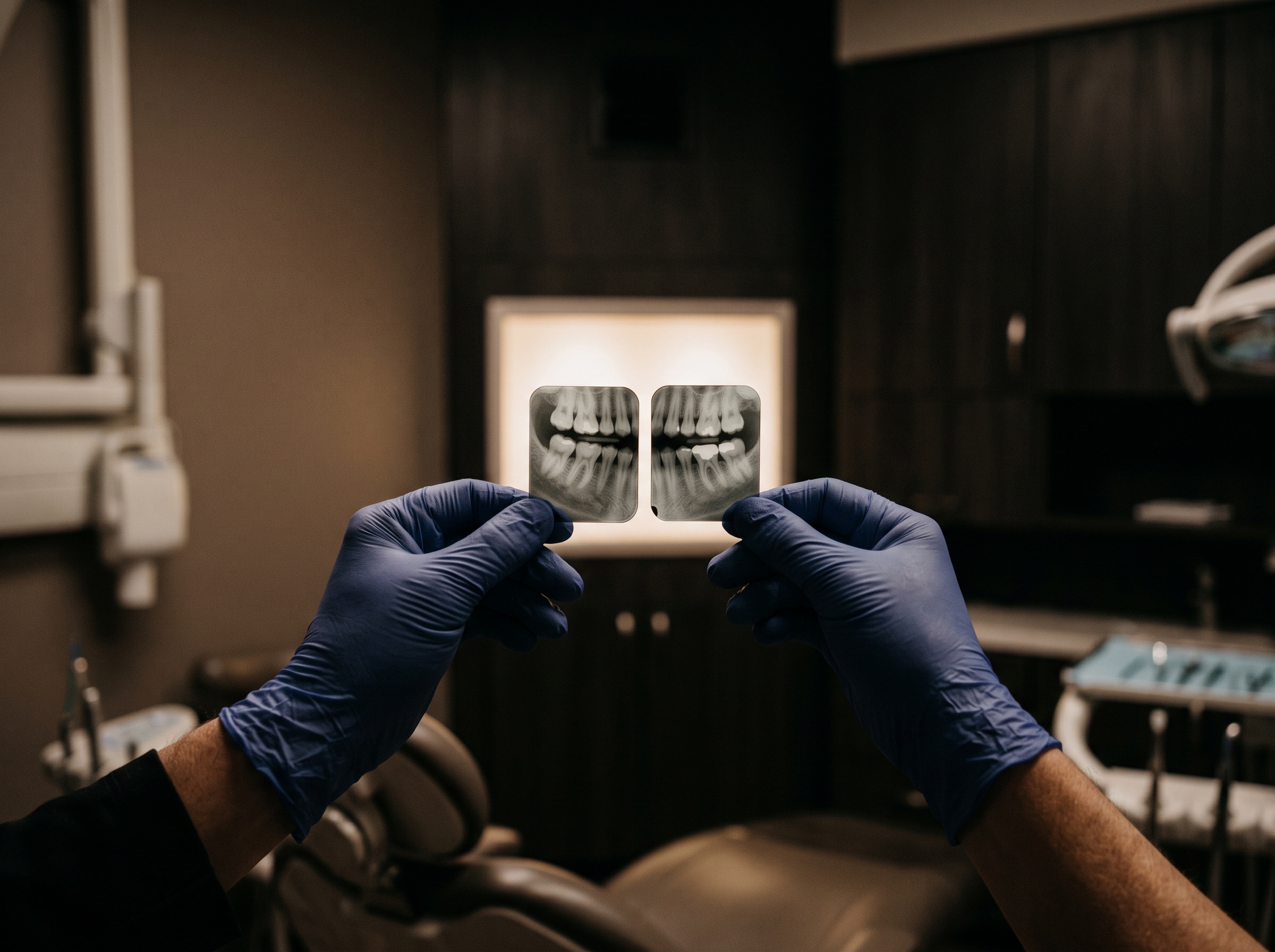
In practice the technique is quieter and more forgiving than its formal statement suggests. The clinician does not measure angles; the clinician compares. Two films are set side by side, and the eye tracks a single relationship: how does the object move against its neighbor? A useful discipline is to choose a stable reference before looking — the apex of an adjacent erupted tooth, a clear point of the cortical outline — and to note the direction of the tube shift explicitly, because the whole reading collapses if you forget which way you moved. Good technique also means changing only the horizontal angulation between exposures and holding everything else still, so that the displacement you see is depth and not the confounding noise of a patient who turned their head. Done cleanly, the answer arrives almost instantly, the way stereo vision snaps into place.
Where the Rule Earns Its Keep
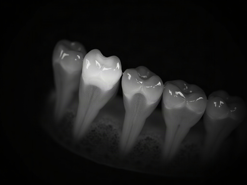
The classic case is the impacted maxillary canine, the tooth that fails to descend and hides in the bone of the palate or the vestibule. Whether it sits buccal or palatal dictates the surgical approach and the direction of orthodontic traction, and getting it wrong is costly. Two angled periapicals, read by SLOB, have guided that decision for a hundred years. The rule serves equally in separating superimposed root canals in a multi-rooted tooth, in deciding whether a supernumerary tooth lies labial or lingual, in localizing a broken instrument fragment or a foreign body, and in judging whether a radiopacity floats in soft tissue or is anchored in bone. In each, the question is the same: near the sensor or far from it? And the answer costs one extra exposure and a moment of clear thinking.
The Limits of Two Pictures
Parallax localization is elegant, but it is inference, not measurement. It tells you a direction — buccal or lingual — not a distance in millimeters. It depends on a clean horizontal shift and a cooperative patient, and it grows unreliable when landmarks are scarce or the anatomy is genuinely ambiguous. When the true three-dimensional relationship must be known precisely — the exact position of an impacted tooth against the roots it threatens, the course of the inferior alveolar nerve beside a wisdom tooth — cone-beam computed tomography answers directly, reconstructing an actual volume you can turn and slice. The new professional guidance emphasizing that imaging is most effective used in moderation is, in part, a call to reach for that higher dose only when a simpler, lower-dose method genuinely cannot answer the question. Often, the two-film shift can.
Future Developments
It would be easy to file the tube-shift rule under history, a clever workaround made obsolete by volumetric imaging. That reading misses something. CBCT has not retired SLOB so much as reframed it: the third dimension the rule struggled to infer is now, when truly needed, simply rendered. But the instinct the rule trained — the habit of asking what a flat image is hiding, of refusing to take a single projection as the whole truth — is exactly the literacy a clinician needs to use a three-dimensional dataset well. The most advanced imaging still produces flat slices that a human must assemble into understanding, and the same reasoning about depth, superimposition, and point of view governs how honestly they are read. The picture keeps changing. The craft of seeing past its surface, first practiced with two small films and a shifted tube, endures.
Sources & further reading:
- New ADA recommendations confirm dental imaging most effectively used in moderation
- The Hidden Architecture of a Tooth: What Cone-Beam CT Reveals That a Flat Radiograph Cannot
No Comments