


October 22, 2026 Counting Every Photon: How Photon-Counting CT Is Quietly Arriving in Oral and Maxillofacial Imaging
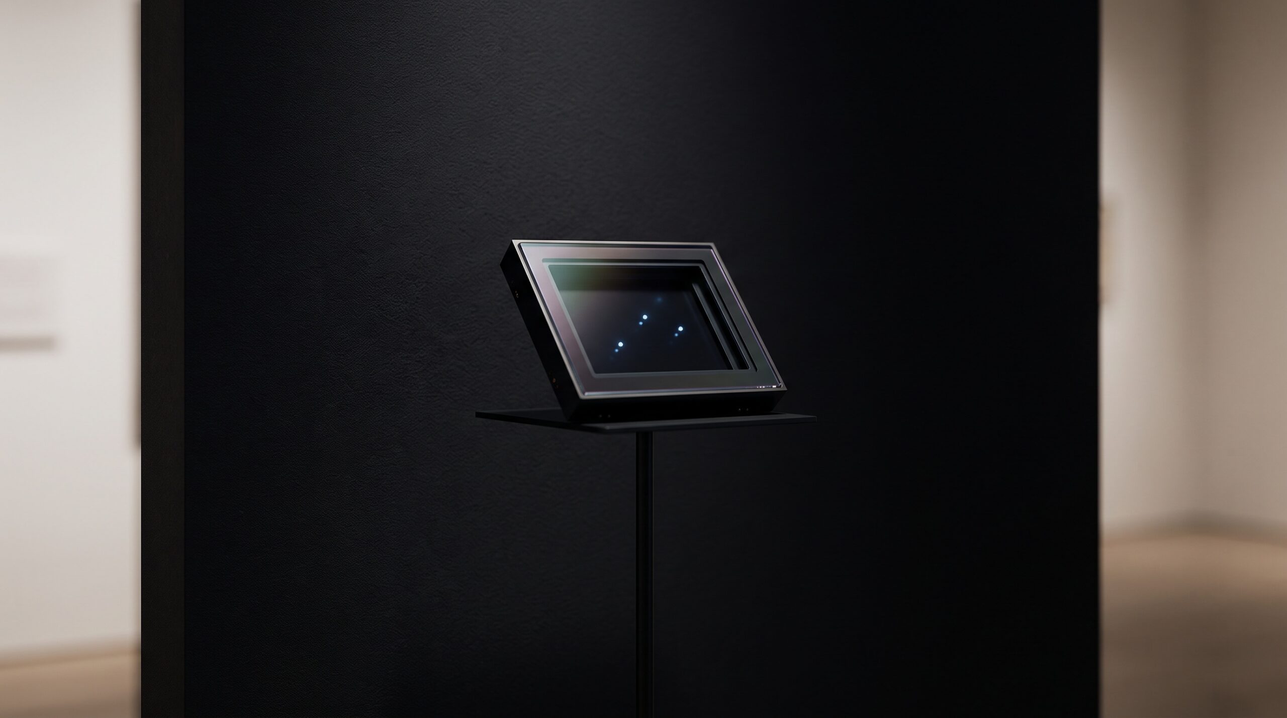
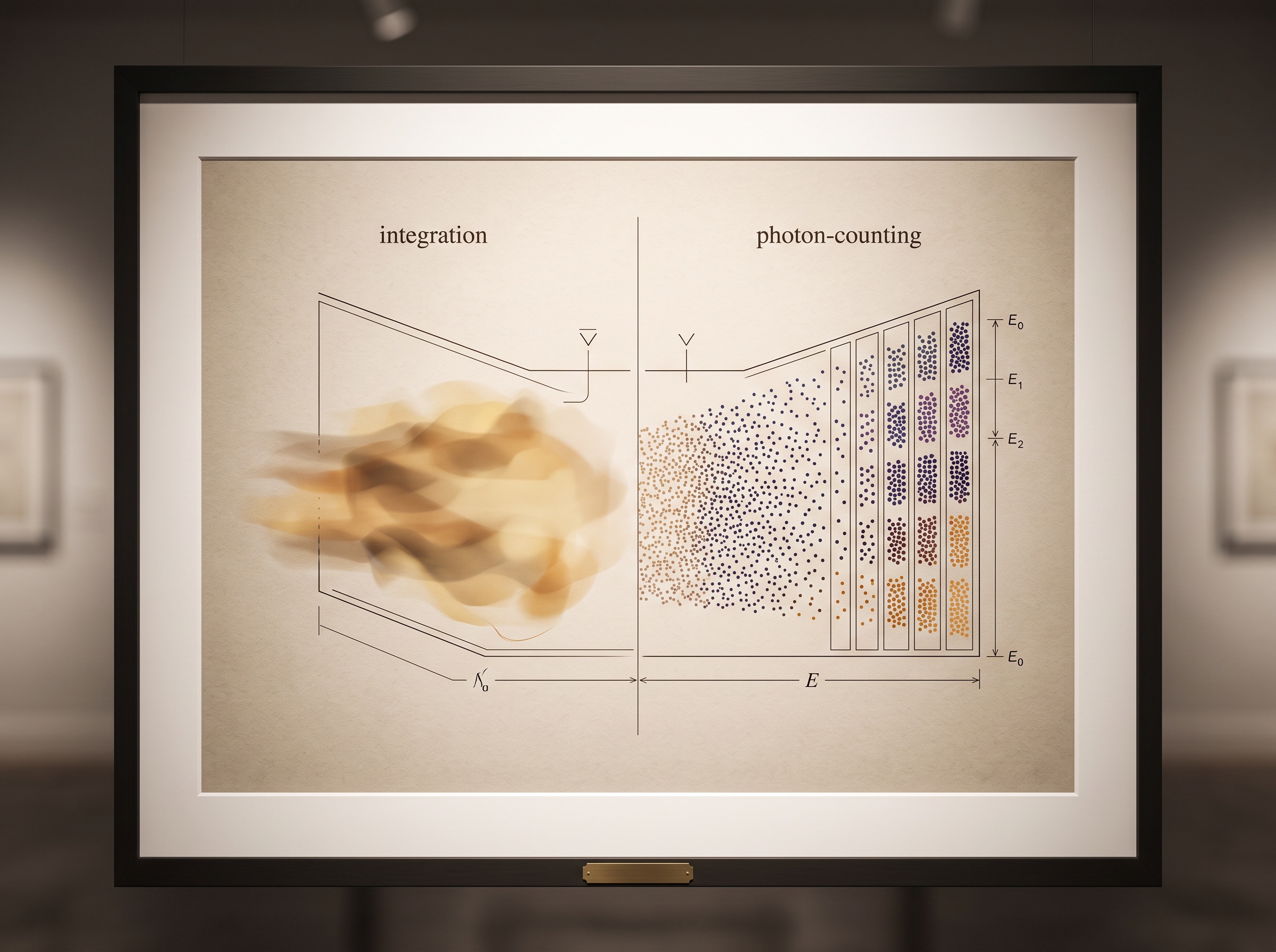
Almost everything we celebrate in dental imaging happens either side of the detector: the cleverness of the source that makes the beam, the artistry of the software that reconstructs and reads it. The detector itself we treat as a passive pane of glass the X-rays happen to strike. Yet for more than a century that pane has done something quietly lossy. Conventional CT and CBCT detectors are energy-integrating: they catch a flood of X-ray photons, let each one deposit its energy into a scintillator, and report only the total light that pooled up over the exposure. How many photons arrived, and the energy each one carried, is averaged away into a single brightness. A different kind of detector, now crossing from medical radiology into the oral and maxillofacial world, refuses that compromise. It counts the photons one at a time and reads the energy of each. The first careful dental studies of 2026 are in, and they suggest that this small change in how a photon is registered reaches the two hardest problems in our field — metal and dose — in a way no amount of downstream cleverness ever could.
The detector that stopped averaging
A photon-counting detector replaces the scintillator-and-photodiode sandwich with a slab of a direct-conversion semiconductor, typically cadmium telluride or cadmium zinc telluride. When an X-ray photon lands, it liberates a cloud of charge in the crystal, and that charge is swept directly to pixelated electrodes as a sharp electrical pulse. Two things follow from this that an integrating detector simply cannot do. First, each pulse is a discrete event, so the detector counts photons rather than summing a wash of light — and because electronic background noise produces pulses too small to clear the counting threshold, that noise is rejected outright rather than baked into the image. The detector has, in effect, a true zero. Second, the height of each pulse is proportional to the photon’s energy, so the detector can sort arriving photons into energy bins: it reads not just how many came, but the color of each. The same lineage that gave us the leap from film to the direct-capture sensors described in our look at how CCD, CMOS, and phosphor-plate sensors compare reaches its logical end here — a detector that registers the individual quantum of light and remembers its energy.
Why this matters most where metal lives
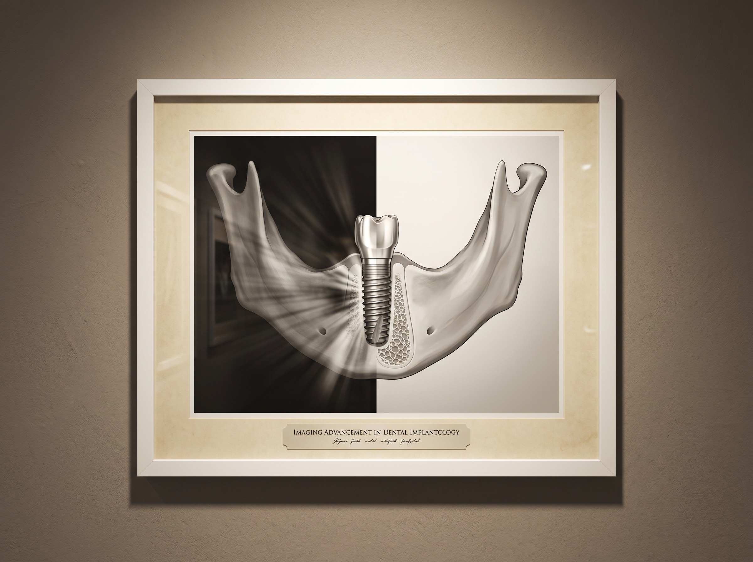
Dentistry is, radiographically, a landscape of metal. Implants, posts, crowns, amalgams and orthodontic hardware all scatter and harden the beam, throwing the dark and bright streaks every clinician knows and dreads — precisely across the peri-implant bone, the canal, the margin a surgeon or endodontist most needs to see. Photon-counting detectors attack this on two fronts at once. Because they are inherently spectral, a single scan can be reconstructed into virtual monoenergetic images: the machine computes how the anatomy would have looked had it been imaged at a chosen single energy, and pushing that virtual energy high dramatically suppresses the beam-hardening streak around metal. Layer a dedicated metal-artifact-reduction algorithm on top and the halo around a titanium implant can be lifted to the point that the surrounding trabecular bone becomes legible again. A 2026 ex vivo study from the oral and maxillofacial group in Zurich put exactly this to the test on dental implants and found that combining high-energy virtual monoenergetic imaging with artifact reduction markedly improved image quality and diagnostic confidence around the hardware. It is the same spectral logic we explored in dual-energy CBCT and material decomposition — only here the energy information is captured natively, photon by photon, rather than assembled from two separate exposures.
The dividend hiding in the noise floor
The second hard problem is dose, and the gain here is more subtle but no less real. Because electronic noise is rejected at the threshold rather than integrated into the signal, a photon-counting detector keeps producing usable images in exactly the regime where integrating detectors drown: the low-dose scan, and the large patient whose tissue attenuates the beam until only a weak, noisy signal survives to the far side. A December 2025 phantom study across a range of body sizes found photon-counting CT delivered superior dose efficiency compared with conventional detectors, and that the advantage widened as the phantom grew — the enhanced efficiency translating into either lower noise at the same dose or the same image at a lower dose. For maxillofacial imaging the implication is pointed: the craniofacial skeleton sits deep behind dense, variable soft tissue, and the patients who most need generous diagnostic clarity are often the ones whose anatomy most degrades it. A detector that squanders no signal to its own noise is, in practice, a detector that lets us ask for less radiation and still receive an honest picture.
One scan, many images
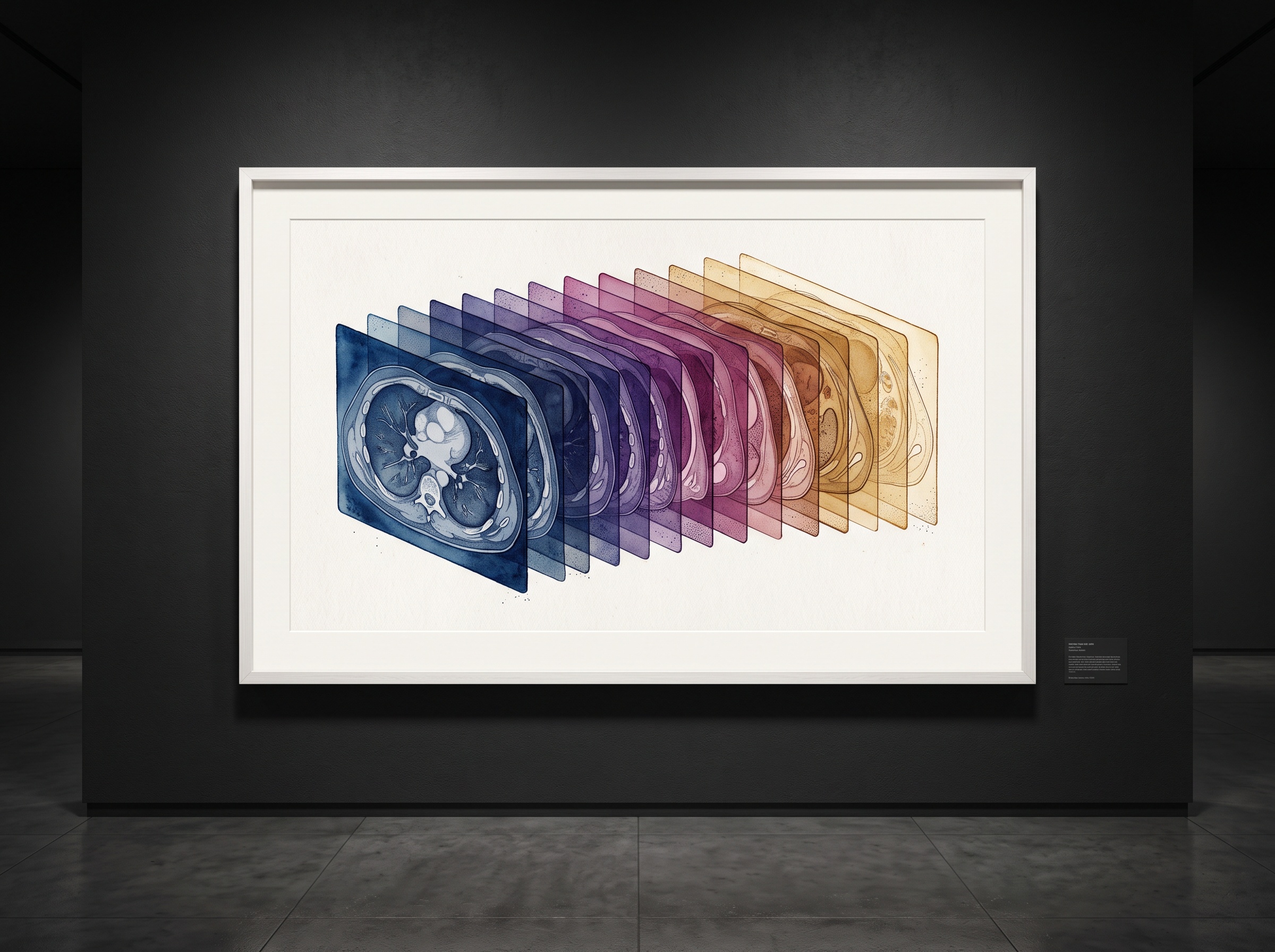
There is a quieter, almost aesthetic consequence worth dwelling on. Because the energy of every photon is recorded, a single photon-counting acquisition is not one image but a latent family of them. From that one scan the reconstruction can synthesize virtual monoenergetic images across a whole span of energies, iodine or calcium maps, and material-specific reconstructions — the operator choosing, after the fact, the energy at which the anatomy reads most clearly, rather than committing to it at the moment of exposure. It is a shift from capturing a picture to capturing the information from which many pictures can be drawn, and it rhymes with everything happening on the reconstruction side, where the deep-learning methods now rebuilding low-dose volumes turn sparse, noisy data into clean images. Spectral capture and intelligent reconstruction are converging on the same promise: all of the diagnostic content, far less of the dose and the artifact that used to be the price of it.
The honest ledger
None of this has arrived in the operatory yet, and it is worth being plain about why. Clinical photon-counting systems today are large, expensive hospital CT scanners, not chairside units; the technology reaches dentistry first through oral and maxillofacial radiology referred to those machines, and much of the dental evidence so far is ex vivo or phantom work rather than large patient trials. The detector crystals are demanding to manufacture uniformly and at scale, charge-sharing between tiny pixels and pulse pile-up at high flux are real engineering frictions, and the spectral reconstructions carry a learning curve of their own. These are the growing pains of a modality mid-crossing from the bench to the clinic — the same awkward adolescence every imaging advance passes through. What is not in doubt is the direction. The question is no longer whether counting photons beats pooling them, but how quickly the counting detector shrinks toward the chair.
Future Developments
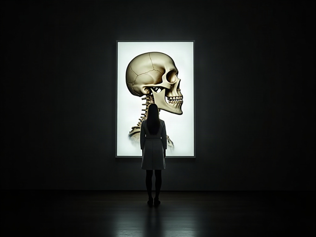
The horizon here is a cone-beam machine built around a photon-counting detector: the dose economy and the spectral, metal-cutting advantages of this technology brought down to the compact, upright, dentition-focused geometry our field actually uses, rather than borrowed from a whole-body hospital gantry. Pair a counting detector with the artifact-aware, learning-based reconstruction already reshaping how volumes are built, and one can imagine a routine implant or endodontic scan that chooses its own virtual energy to erase the streak, rejects its own electronic noise to spare the dose, and hands the clinician not a single fixed image but a spectrum to interrogate. For a gallery that treats the image as the art, there is a fitting lesson in looking this far back along the chain. We have spent twenty years perfecting the light we send and the software that reads what returns, while the surface that catches it quietly threw half the story away. Teach that surface to count — and to remember the color of what it counts — and the picture it returns is cleaner, quieter, and more truthful. Reinvent the detector, and you reinvent the image, at the very place it is finally received.
Sources & further reading:
- Optimization of dental implant imaging using photon-counting detector computed tomography in oral and maxillofacial surgery: an ex vivo study
- Obese patient imaging: improved dose efficiency with photon-counting CT
- Potential Benefits of Photon-Counting CT in Dental Imaging: A Narrative Review
No Comments