


November 17, 2026 The Scanner That Also Looks for Trouble: AI-Integrated Intraoral Scanners and Oral Lesion Screening
The intraoral scanner entered the operatory with a narrow brief. It was there to measure: to replace the alginate tray and the setting putty with a stream of optical frames stitched into a precise three-dimensional model of a prepared tooth, an arch, a bite. Its virtues were all geometric — accuracy to the margin, repeatability, a file a laboratory could mill against. The soft tissue it swept across along the way, the gingiva and the mucosa of the cheek and floor of the mouth, was scenery. The wand recorded it, colored it, and moved on.
A line of recent research asks whether that scenery was ever really incidental. Every scan already carries color and surface texture across the entire field it passes over, not just the teeth. If a model could be taught to read that soft-tissue data the way a trained eye reads mucosa — noticing a patch whose color, surface or contour departs from the healthy field around it — then the instrument built to measure a crown could, in the same pass, quietly screen for the lesion no one came in to find.
From geometry to tissue: what the scan already holds
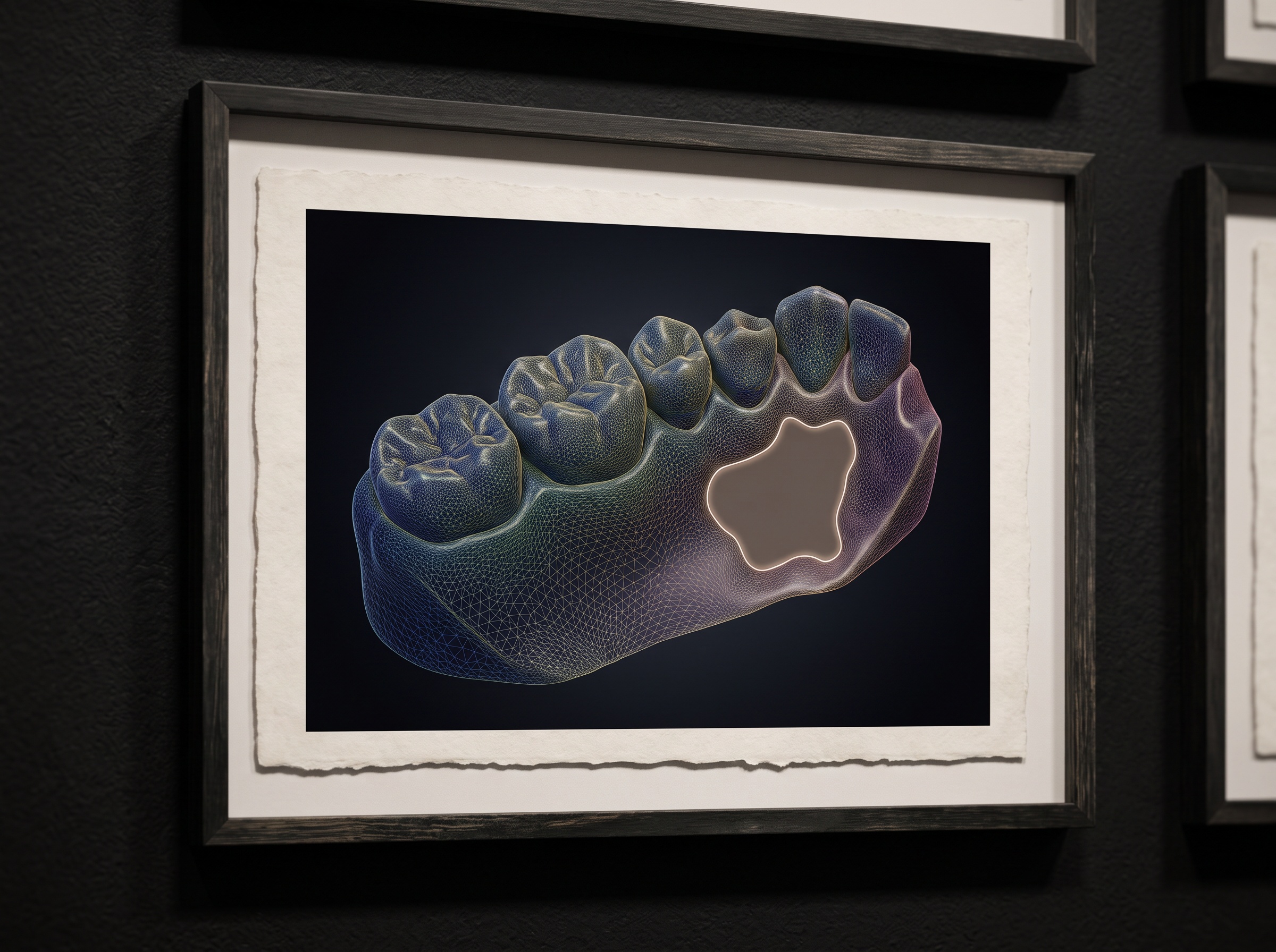
The premise is less exotic than it sounds. A modern scanner captures a dense color map draped over a precise mesh. Within that record is exactly the information a clinician uses in a visual oral examination: the hue of the mucosa, whether a surface is smooth or pebbled or ulcerated, whether an area is raised, and how sharply it differs from its neighbors. The scanner was simply never asked to attend to it. Artificial intelligence changes the economics of attention — a model can examine every square millimeter of captured mucosa with the same patience, on the first scan and the thousandth, without the fatigue that erodes a human survey near the end of a long day.
This is a different proposition from the dedicated optical adjuncts that have long served oral-cancer screening. A device built around autofluorescence, for instance, adds a deliberate step and a separate instrument whose only job is to make mucosal change visible; we traced the craft of that approach in how autofluorescence reveals oral cancer before it shows. AI-integrated scanning makes no such demand on the appointment. It rides on a scan the clinician is taking anyway, which is both its great advantage and the root of its caution: it was not purpose-built for soft-tissue diagnosis, and it must be understood accordingly.
Teaching a model what “normal” looks like
A screening model earns its usefulness by first becoming fluent in the ordinary. Healthy oral mucosa varies enormously — by pigmentation, by site, by the frictional keratosis of a cheek-biter, by the normal vascularity of the floor of the mouth. A model that cannot hold that range as unremarkable will cry wolf at every freckle and every linea alba, and a screen that flags everything screens nothing. The hard engineering is therefore not in spotting the dramatic lesion but in learning the wide, unglamorous territory of normal well enough that a genuinely anomalous patch stands out against it.
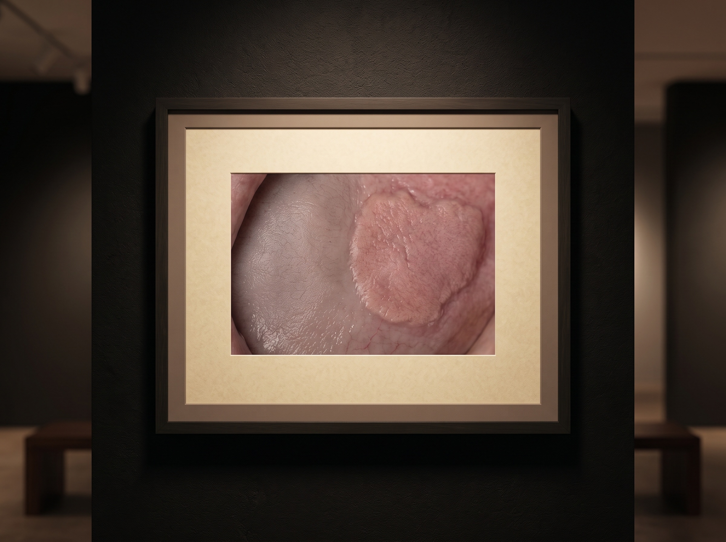
The clinical targets that matter most are the potentially malignant disorders — the leukoplakias, the erythroplakias, the lichenoid changes — precisely because they are the ones a hurried glance can miss and the ones in which early detection changes outcomes. These are also, inconveniently, the subtle ones: a faint white patch, a velvety red area, a texture that reads as difference more than as disease. That the capability research lives in the imaging literature rather than the marketing brochure is telling; the ambition here is early, equivocal signal, not obvious tumor.
The quiet advantage of the repeated scan
There is one thing an intraoral scanner can do that a one-off screening visit cannot, and it may prove the most valuable of all. Because the same patient is scanned repeatedly over months and years — for a new restoration, for aligner progress, for a routine record — the device accumulates a longitudinal archive of each mouth. Change, not just appearance, becomes readable. A mucosal area that is stable for two years and then shifts in color or grows is a fundamentally different signal from a static patch, and it is one that only a tool embedded in ordinary, recurring care can detect.
This is the same logic that makes any AI reading of dental images powerful — the model’s real leverage is consistency across time and across modality, a point we developed in how one algorithm reads the intraoral image, panoramic and CBCT so differently. For soft-tissue screening the temporal axis is not a bonus feature. Surveillance of potentially malignant disorders is largely a matter of watching for change, and a scanner that quietly builds that record turns every impression into a frame in a longer observation.
Screening is not diagnosis — and the difference is the whole point
None of this makes the scanner a pathologist. A flag is a prompt, not a verdict, and conflating the two would be the technology’s most dangerous failure mode. The honest framing is screening: a tool whose job is to raise the right question — look here — and whose success is measured by how reliably it catches what a human might overlook while not burying the clinician in false alarms. A diagnosis of an oral lesion still rests where it always has, on clinical examination and, when warranted, biopsy. The AI’s contribution ends at attention.

That boundary also dictates how the output should be designed and received. A flag that fires too readily trains the clinician to ignore it; one calibrated too conservatively defeats the purpose. And because the device is placed in the hands of general practitioners rather than oral-medicine specialists, the screening signal has to be legible to a busy clinician and tied to a clear next step — examine, document, refer. The value of opportunistic screening is realized only if the flag reliably returns the question to a human, which is also the spirit in which we have described the broader move of AI-driven oral-disease screening reaching beyond the specialist clinic.
Future Developments
What makes this direction compelling is leverage. Oral cancer’s survival statistics are dominated by how late it is usually found, and the people most at risk are not always the ones who sit for a dedicated screening exam — but many of them do, sooner or later, sit for a scan. An instrument already diffusing through everyday practice, already pointed at the mouth, already capturing color and texture across the whole field, is an unusually good place to put an extra set of eyes. The near future is likely to bring better-calibrated models, richer training sets spanning the full diversity of healthy mucosa, and scanners that log soft-tissue observations alongside their geometry as a matter of course. The scanner will still be, first, a measuring instrument. But the gallery it builds was always more than teeth, and teaching it to notice the tissue at the edges of the frame is how an image taken to fit a crown might, once in a long while, catch something that matters far more. Imaging has always been both science and art; here it quietly becomes vigilance as well.
Sources & further reading:
- Could AI-integrated intra-oral scanners improve oral lesion screening? (research overview)
- British Dental Journal / Nature Portfolio — oral cancer early detection and screening
No Comments