


October 21, 2026 The Gallery Leaves the Clinic: SMARTeeth++ and the $3.1M Push to Carry Oral Imaging to the Patient
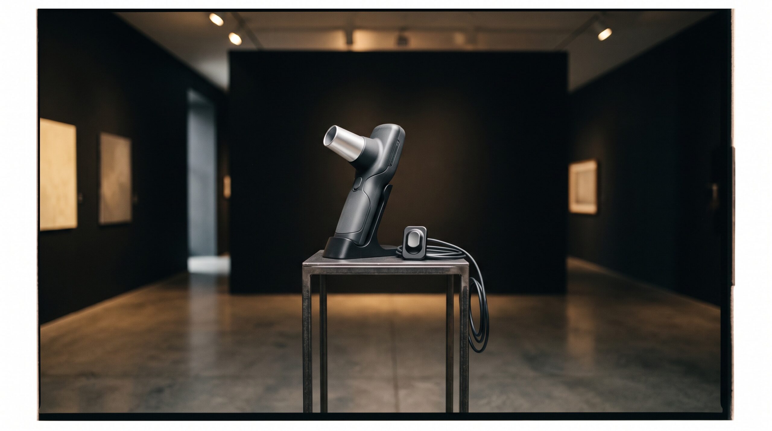
For as long as there have been dental radiographs, the picture has been made in one kind of place: a controlled room, with a mounted source, a trained hand, and a patient held still in a chair built for the purpose. The operatory is where imaging happens because the operatory is where the conditions are right. A new, five-year award of roughly $3.1 million from the National Institute of Dental and Craniofacial Research proposes to break that rule on purpose. Researchers at the University of Rochester’s Eastman Institute for Oral Health are building a system called SMARTeeth++ designed to carry oral imaging out of the clinic entirely — into an obstetrician’s exam room, a community health visit, and eventually the palm of the patient’s own hand. It is an artificial-intelligence platform at heart, but the part worth dwelling on, in a gallery that treats the image as the art, is what happens to the image itself when you take it out of the only room that was ever built to make it well.
A screening that comes to you
The premise of SMARTeeth++ is that most oral disease is caught late not because it is hard to see but because the person who could see it never gets looked at. So the platform inverts the geometry of care: instead of waiting for the patient to arrive at the imaging, it brings the imaging to the patient. It combines three things that usually live in three different places — oral imaging captured with portable equipment, an artificial-intelligence model trained to read that imaging, and the patient’s own medical and dental history — and fuses them into a single screening that can be performed wherever the patient already is. The goal is not a prettier radiograph. It is to detect and measure the earliest stages of tooth decay and gum disease in people who are nowhere near a dental chair, and to do it with a machine’s patience and consistency rather than a scarce specialist’s time.
What the picture has to do now
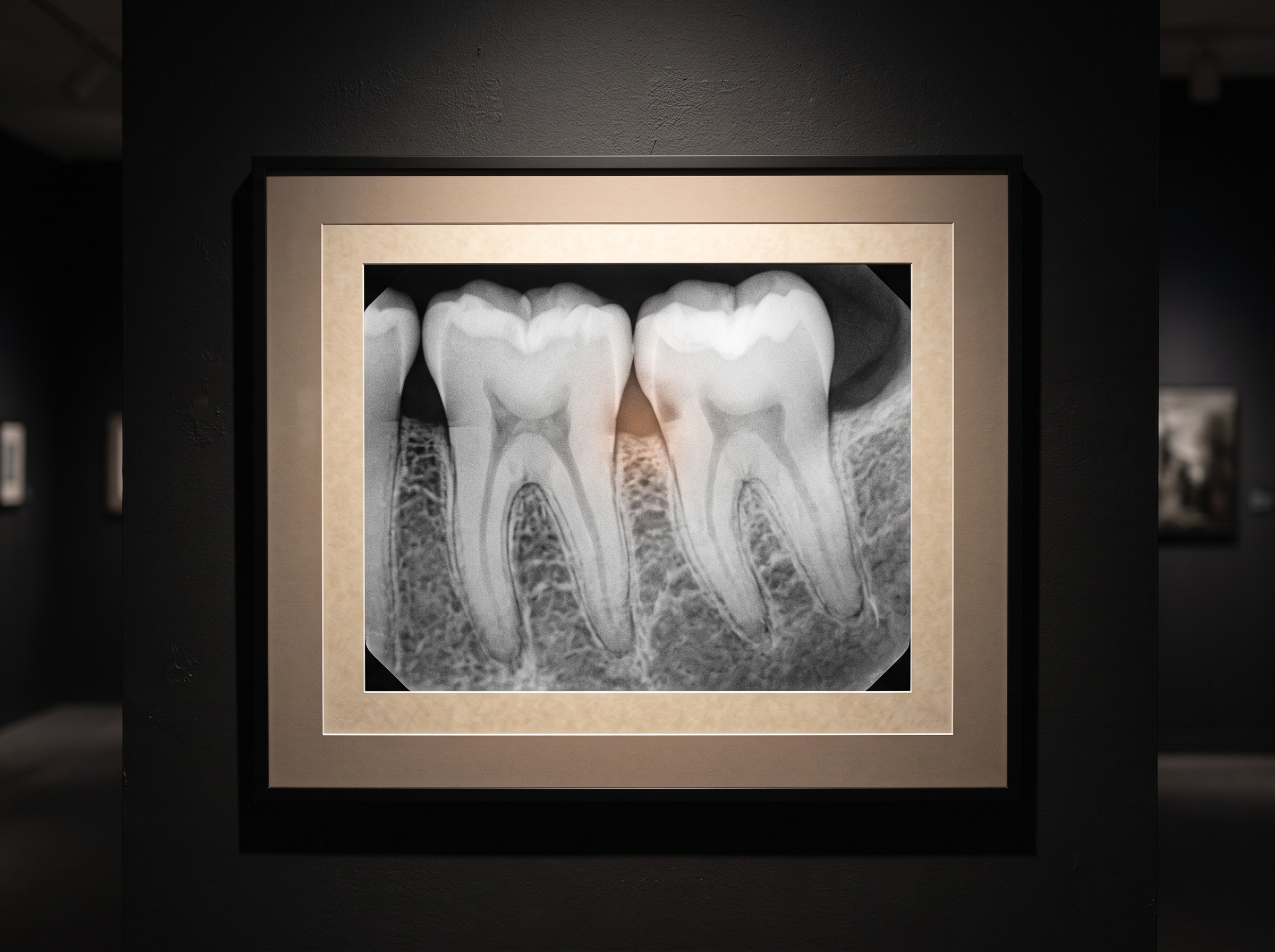
Move capture out of the operatory and you ask the image to do something it has never had to do. In the clinic, image quality is engineered: the geometry is fixed, the exposure is controlled, the positioning is practised, and a human quietly rejects and retakes anything that comes out wrong. Strip those supports away — a portable sensor in an unfamiliar hand, imperfect angulation, uncontrolled light for any photographic component — and the burden of making the picture legible shifts from the conditions of capture onto the image and the model that reads it. This is why a screening platform is, underneath, an image-quality problem. The AI can only measure what the picture faithfully records, and a model trained on pristine clinic radiographs will stumble on the messier ones the field produces. The quantitative reading of an image — the kind of fine texture and density analysis that radiomics performs — is only as trustworthy as the capture beneath it, and a platform meant for the real world has to be robust to a picture made under conditions no operatory would tolerate.
The 71.7% in the obstetric chair
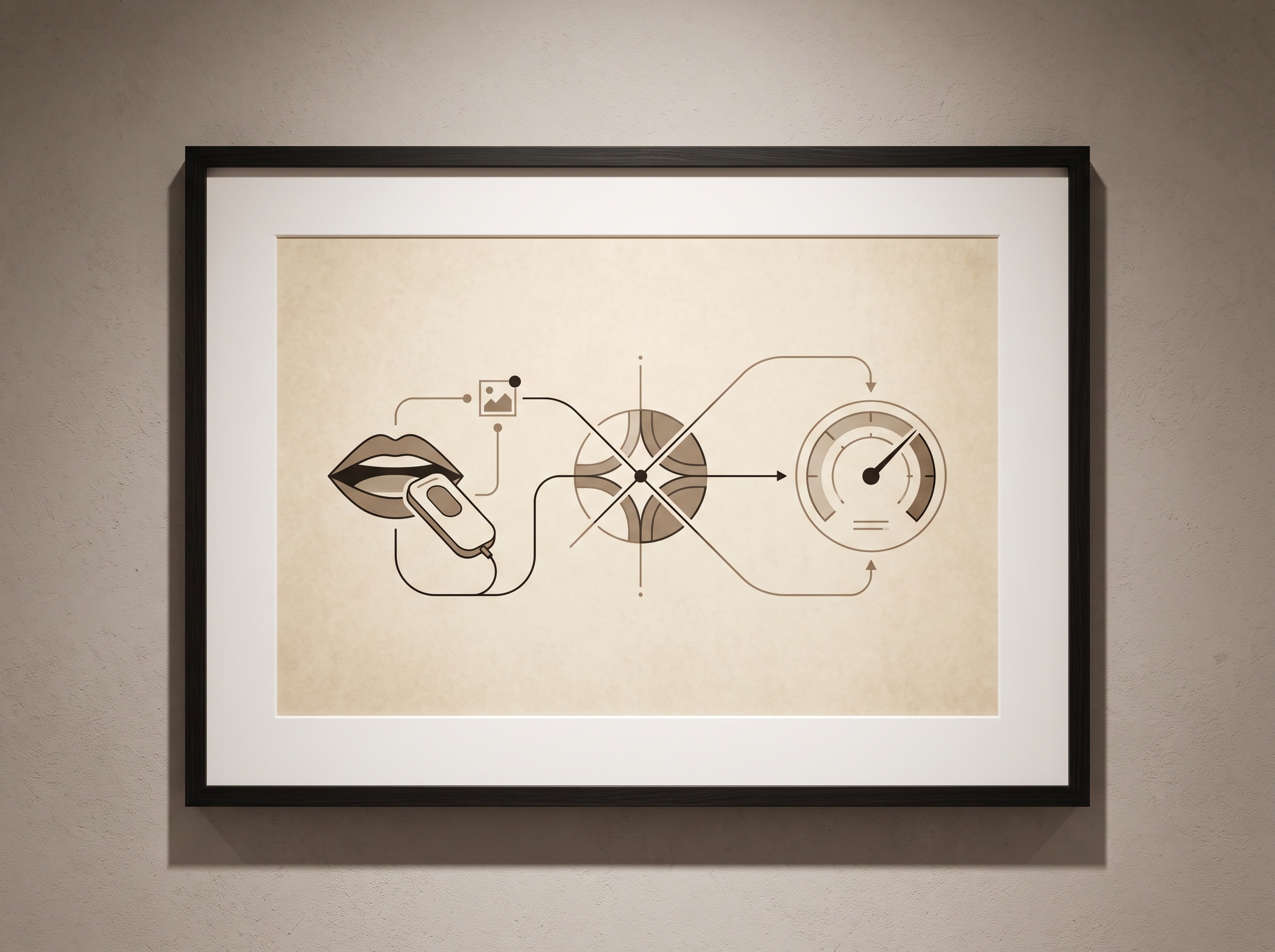
The grant is not starting from a blank page. It builds on an earlier study in which the platform was taken somewhere dentistry rarely goes — into obstetric visits, screening pregnant patients for oral disease during a medical appointment they were already attending. In that work the system detected early-stage decay in 71.7% of participants: not a diagnosis handed down by a dentist in a clinic, but a meaningful signal pulled from imaging captured at the point of care, by people who were not there to do dentistry. That number is the proof of concept the larger award is meant to harden. It says the idea is not merely theoretically appealing but clinically detectable: that a portable capture, read by a model, in a room never designed for radiography, can still find the disease early enough to matter. The pregnancy setting is pointed, too — oral health and pregnancy are linked, and the visit was a door into imaging for a population that often slips through dentistry’s net.
Two machines, two readers
The team plans to build the platform in two forms, and the split is instructive. One is a clinical tool, meant for dental hygienists and other health professionals — a point-of-care instrument that extends a trained reader rather than replacing the clinic. The other is far more radical: a mobile self-screening version, meant for the patient to use on themselves. These are not the same product with different skins. A self-screening tool has to assume the least-controlled capture imaginable, the least-expert hand, and a reader — the person — who cannot interpret what they have made and is trusting the machine entirely. The provenance and trustworthiness of a machine-read oral image become acute when the human is removed from the loop of judgment; the same questions that surround the authenticity and provenance of AI-touched radiographs arrive here from a different direction, because a screening a patient performs and a model alone interprets has no clinician standing between the image and the conclusion.
From a map of disease to a forecast of it
What lifts SMARTeeth++ above ordinary computer-aided detection is its ambition to predict rather than merely find. The platform is described as including a model meant to flag the patients at highest risk of rapid progression — not only whether disease is present today, but whose disease is likely to accelerate. That is a different and harder use of an image. It asks the picture, together with the patient’s history, to carry information about the future: to separate the quiet lesion that will sit unchanged for years from the aggressive one that will become a crisis. A model that can do this turns a screening into triage, directing scarce clinical attention toward the people who will benefit most from it soonest. It also raises the stakes on every input, because a forecast built on a poorly captured image or an incomplete history is a confident error, and a confident error about the future is worse than an honest uncertainty about the present. Any such model is only as good as the images and annotations it learned from — which is exactly why the carefully labeled datasets beneath these systems are not a technicality but the foundation the whole promise rests on.
What it would mean to succeed
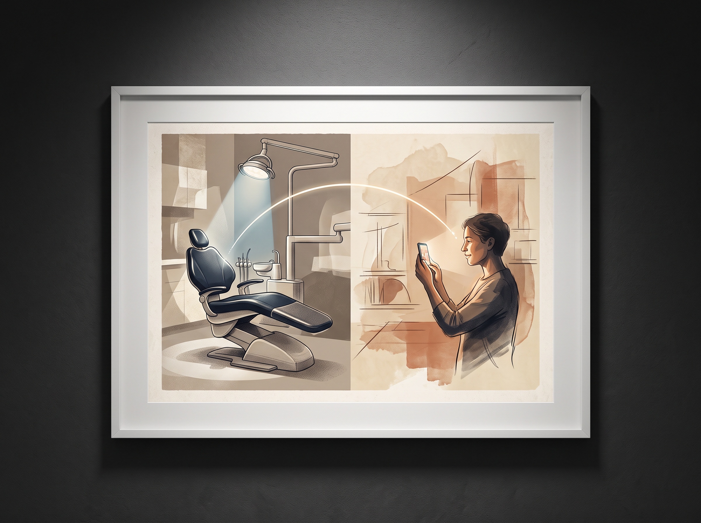
The stated goal of the project is to move oral healthcare from a clinic-based model toward one focused on prevention and outcomes — to catch disease in the many who never make it to the chair, early enough that it can be reversed or arrested rather than drilled and filled. If it works, the quiet consequence is that the dental image stops being something you travel to a special room to obtain and becomes something that can be made almost anywhere, for almost anyone, as a routine act of prevention. That is a genuine democratization of imaging, and it would reach exactly the people traditional dentistry reaches last. The honest caveats remain the ones this kind of project always carries: a 71.7% detection rate is a promising signal, not a finished clinical instrument; portable capture under real-world conditions is unforgiving; and a tool a patient uses on themselves demands a standard of robustness, calibration, and plain trustworthiness that a clinic instrument, watched over by a professional, does not. This award buys five years to find out whether those gaps can be closed.
Future Developments
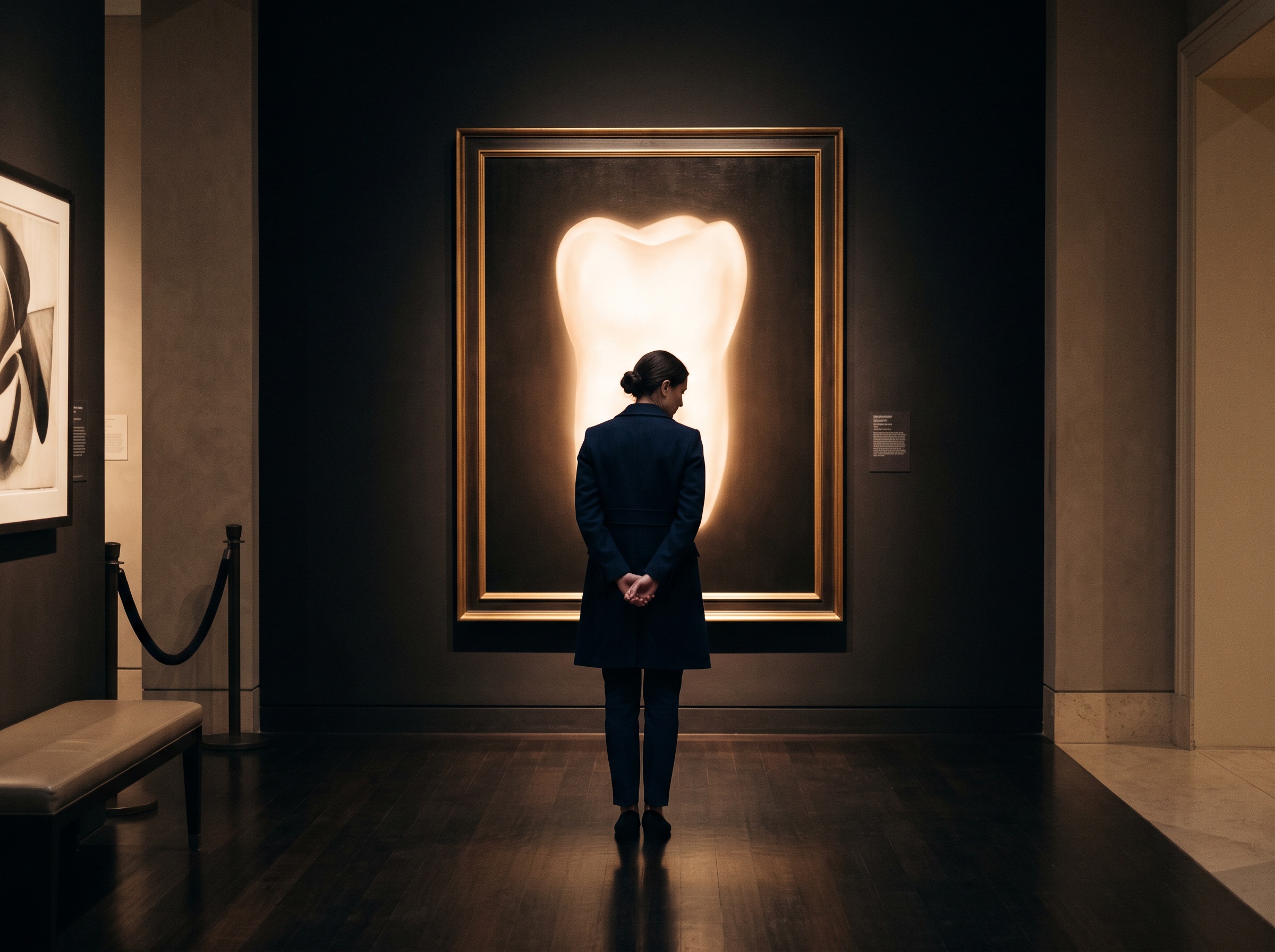
The direction this points is toward imaging as ambient rather than occasional — oral pictures captured often, lightly, and far from any clinic, read by models that grow steadily better at seeing disease in imperfect images and at forecasting which of those findings will matter. The frontier is not a sharper radiograph but a more portable trust: capture devices rugged and simple enough for untrained hands, models honest enough to say when a picture is too poor to read, and risk forecasts transparent enough that a clinician or a patient can see why the machine is worried. For a gallery that frames the dental image as both science and art, there is something fitting in watching the image try to leave the gallery altogether — to be made in the ordinary rooms of ordinary lives rather than in the controlled light of the operatory. The question that follows it out the door is the one worth keeping: what makes an image good enough to be trusted when it is made by anyone, anywhere, and read by no human at all. Answer that well, and imaging stops being something care waits for and becomes something care carries with it.
Sources & further reading:
- New York dental school awarded $3.1M to advance AI-driven oral disease platform
- How AI could help detect tooth decay and gum disease earlier (University of Rochester)
No Comments