


October 17, 2026 When No One Can Tell a Fake: AI-Generated Radiographs and the Coming Question of Image Provenance
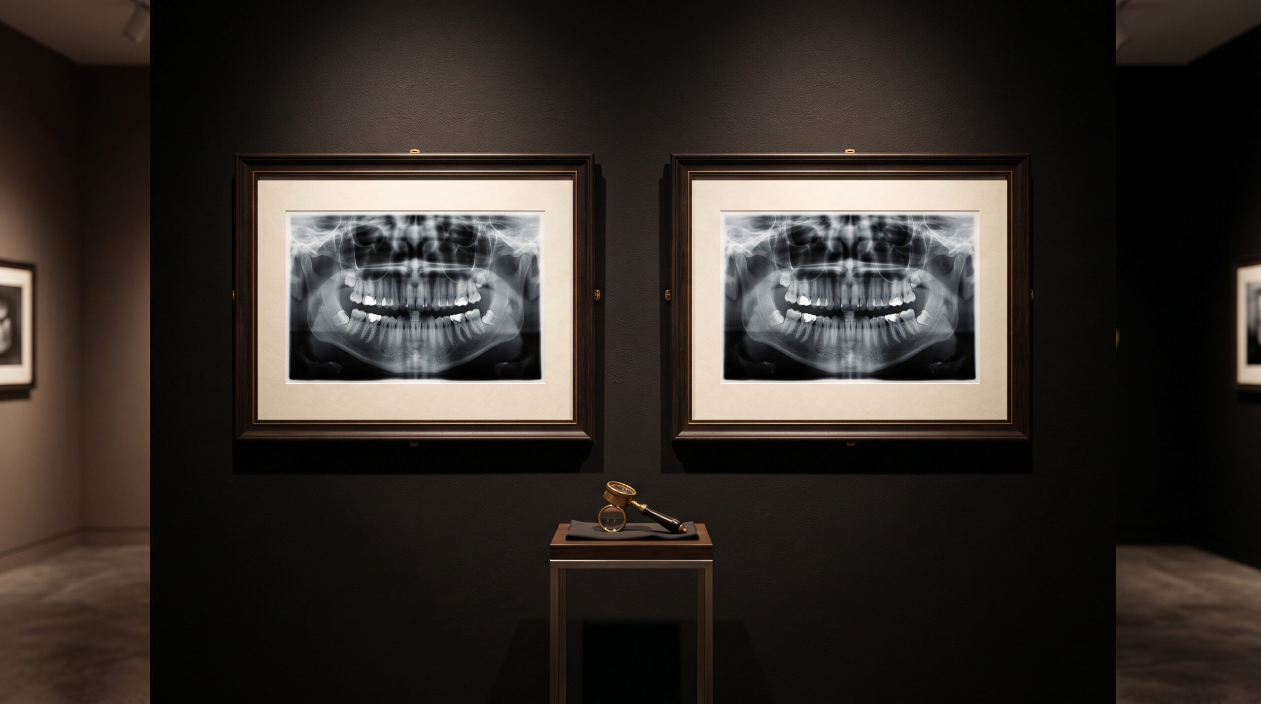
Every art gallery is, underneath its hush and its careful lighting, a machine built to answer a single stubborn question about each picture on its wall: is this real, and can we prove where it came from? The connoisseur’s eye, the raking lamp, the microscope trained on a brushstroke — all of it serves the quieter, less glamorous discipline of provenance, the documented chain of hands and places an object has passed through on its way to the frame. For most of the history of radiography, dentistry has been spared that anxiety. A radiograph was, by its nature, a trace of something physical: photons that had actually passed through a real patient’s jaw and struck a real sensor. The image was evidence because the process that made it could not easily lie. That quiet certainty is now ending.
The study that should unsettle every imaging discipline
In early 2026 a study published in Radiology set out to test something that until recently would have sounded like science fiction. Researchers used generative AI to synthesize chest X-rays that had never passed through any patient, then asked both experienced radiologists and several advanced multimodal AI models to sort the fakes from authentic clinical images. The result, reported by the RSNA, was sobering: neither the humans nor the machines could reliably tell the difference, even when they were explicitly warned that forgeries were mixed into the set. Radiologists and the AI models failed in different ways — the clinicians were better at flagging certain diffusion-model images, the models slightly better at confirming genuine ones — but the headline finding is the one that matters. The trained eye, the instrument we have always trusted to read the image, can no longer be trusted to authenticate it.
The study looked at chest radiographs, not dental ones, and it would be a mistake to pretend the two are identical; a panoramic or periapical image has its own anatomy, its own characteristic noise, its own failure modes. But the generative machinery is indifferent to subject matter. The same diffusion models that can convincingly hallucinate a lung field can be pointed at teeth and bone, and there is no physical principle that makes a dental radiograph harder to counterfeit than a chest film. If anything, the smaller, more repetitive geometry of intraoral imaging may make it easier.
When the image stops being its own evidence
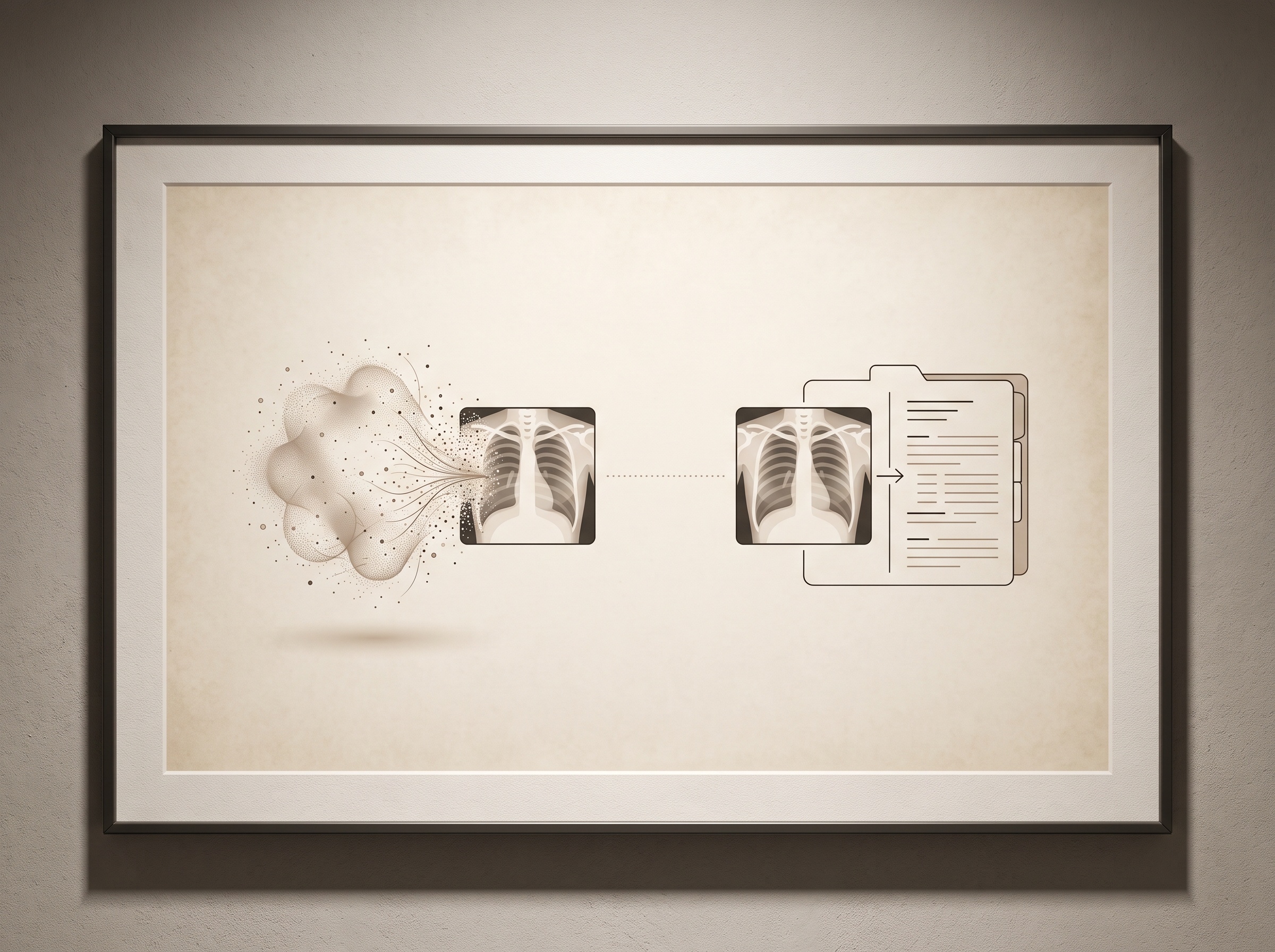
It helps to be precise about what has actually changed, because the temptation is to leap straight to fraud. The deeper shift is epistemic. A radiograph used to authenticate itself: its very existence implied a physical act of exposure, and the only real question was how well that act had been performed. Generative models sever that implication. An image can now be photorealistic without being a photograph — perfectly plausible grayscale anatomy that corresponds to no exposure, no patient, no moment in time. The pixels look like evidence while carrying none of evidence’s provenance.
This is not, for the most part, a story about dentists forging records, though insurance fraud and litigation are the obvious dark corners and they are real. The more pervasive risk is ordinary and procedural. Generative tools are already woven into the imaging pipeline for entirely legitimate reasons — denoising a low-dose capture, reconstructing a sparse scan, even painting back the truncated edge of a cone-beam volume. Each of those is a case where a model has, in some measured degree, invented pixels that the sensor never recorded. The line between “enhanced” and “fabricated” is not a wall; it is a gradient, and we are already standing somewhere on it. Synthetic images are also increasingly used to augment the datasets that train diagnostic AI, which means fabricated radiographs are becoming part of the raw material of the field whether we track them or not. The question is no longer whether a dental image might be partly machine-made. It is whether anyone can say, later, how much.
What the art world already knows
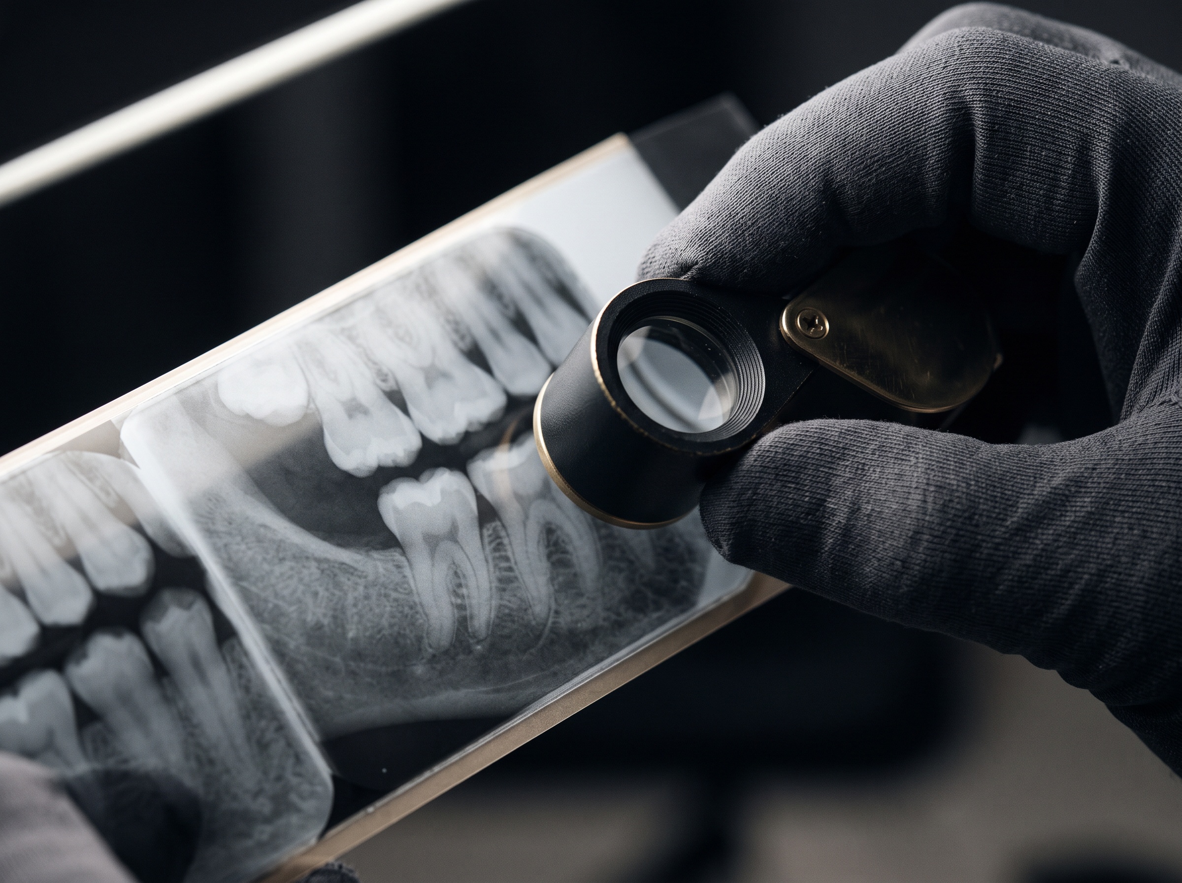
Here the gallery metaphor stops being decorative and becomes a working model. The art world solved — or at least learned to live with — exactly this problem centuries ago, in an era when a skilled forger’s hand could defeat any expert’s eye. Its answer was never to rely on the eye alone. It was to build provenance: an unbroken, documented chain of custody recording where a work came from, who has owned it, and what has been done to it, backed increasingly by material forensics that read the object’s physical substrate for signs of its true age and origin. Authentication became less about whether a picture looked right and more about whether its paper trail and its physical grain told a consistent story.
Dentistry’s radiographs need the same scaffolding, and the good news is that imaging is far better positioned than paint to supply it. A digital image can carry its own history in a way a canvas never could. The DICOM standard already records acquisition metadata — the device, the exposure settings, the timestamp — and a complete audit trail of who opened and altered a study can be kept at the system level. The forensic counterpart to craquelure analysis is active research too: the subtle statistical fingerprints a generator leaves in an image’s noise, the very signals the Radiology study showed are growing too faint for eye or model to catch reliably, but which dedicated detectors and quantitative texture analysis may still surface. Provenance and forensics, working together, are how the gallery has always outlasted the forger.
Signing the image at the moment of capture
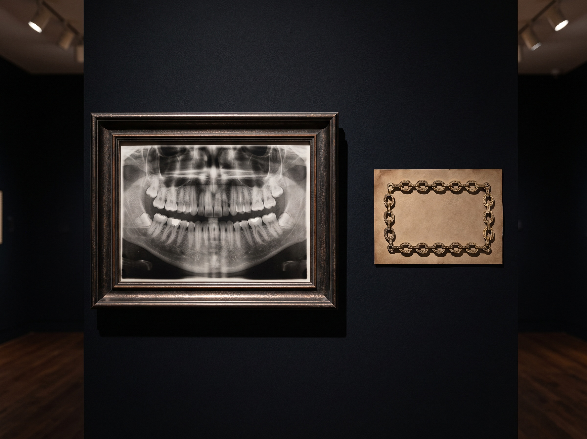
The most promising path borrows an idea that the broader media world is already racing to adopt in response to deepfakes: cryptographic content provenance. The principle is to sign an image at the instant of its birth. Imagine a sensor that, the moment it records an exposure, wraps the pixels in a tamper-evident cryptographic seal — a signature that encodes what device made the image, when, and under what settings, and that breaks visibly if a single pixel is altered downstream. Any later processing, however legitimate, appends to that record rather than erasing it, so the image arrives in the record carrying an honest biography: captured here, denoised there, annotated by this clinician on that date. Standards for exactly this kind of signed, traceable content credential already exist in the consumer-imaging world, and there is no technical reason dental sensors and imaging software cannot adopt them. The authentication question would then no longer rest on whether a human or an AI happens to be fooled by the pixels — a contest we now know we lose — but on whether the image can produce a valid, unbroken signature. An unsigned radiograph would not be presumed fake; it would simply be, like an unprovenanced painting, an object whose origin cannot be vouched for, to be weighed accordingly.
Future Developments
The near term will be a quiet arms race between generators that leave ever-fainter traces and detectors straining to find them, and the Radiology findings suggest the detectors, used alone, are already losing. The durable answer will not come from sharpening the eye — human or artificial — but from changing what we ask of an image in the first place. Expect provenance to migrate from an afterthought buried in DICOM headers to a first-class, cryptographically enforced property of every clinical capture, with signing built into the sensor and chain-of-custody verification built into the viewer, so that opening a study someday means seeing its authenticated biography alongside its anatomy. The galleries learned long ago that beauty and plausibility prove nothing about truth, and that trust has to be manufactured deliberately, through documentation and forensics rather than through confidence in the eye. Dental imaging, having enjoyed a long holiday from that lesson, is about to have to learn it. The images on these walls have always been evidence; the task ahead is to make sure they can still prove it.
Sources & further reading:
No Comments