


September 27, 2026 The Light That Goes Dark: Autofluorescence and the Craft of Imaging Oral Cancer Before It Shows
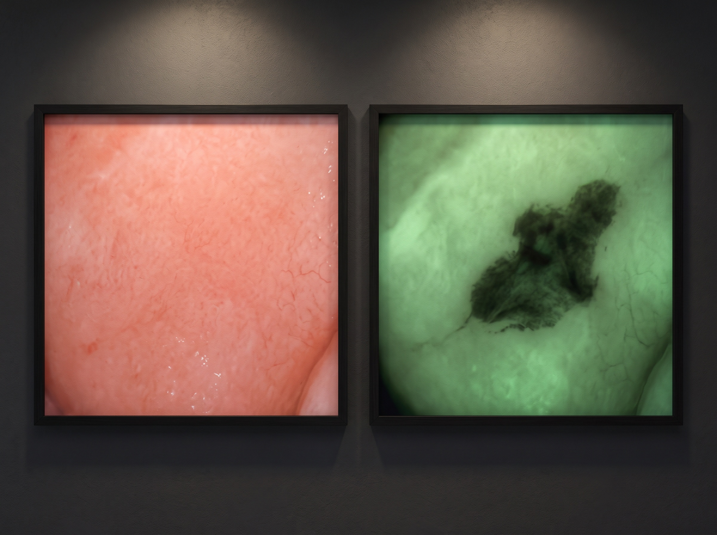
Ask a clinical portrait to show you an early oral malignancy and it will often show you nothing at all. A field of dysplasia, or even an early squamous cell carcinoma, can live in mucosa that still reads as ordinary pink tissue to the eye and the camera — smooth, unremarkable, easy to pass over in a routine soft-tissue exam. This is the blind spot tissue autofluorescence was built for. Instead of photographing reflected light, we ask the tissue to emit its own, then photograph the places where that emitted light goes missing.
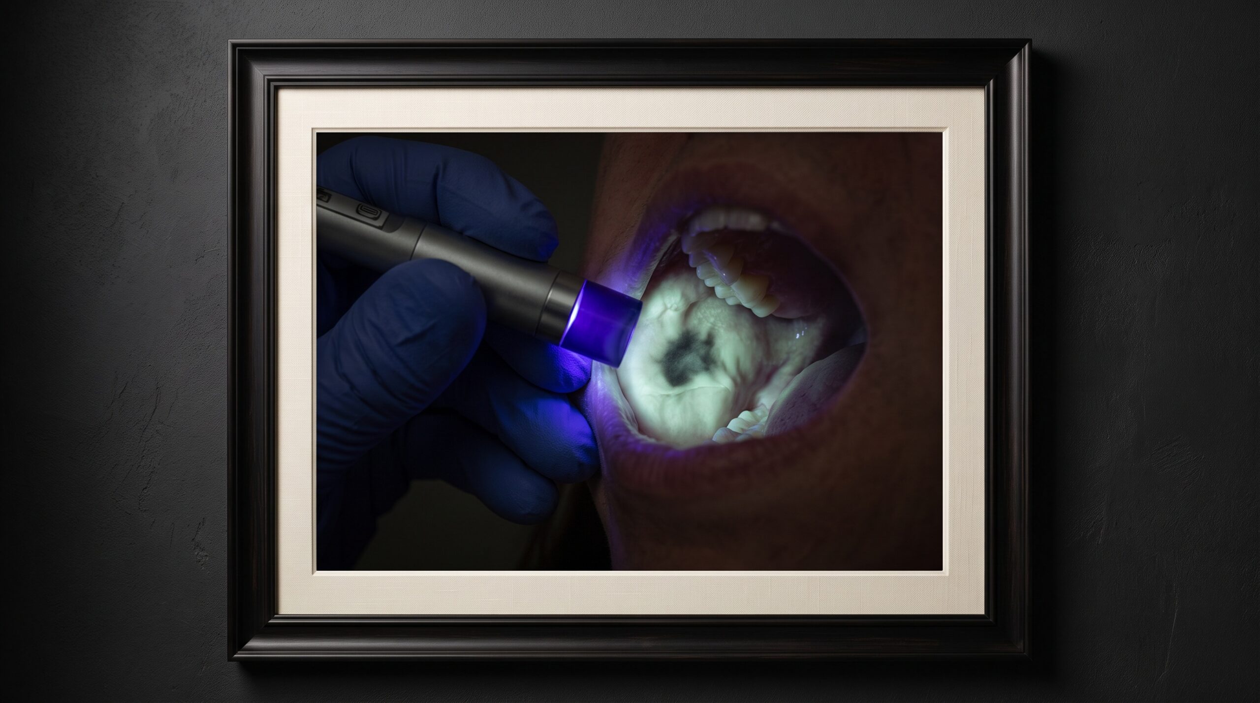
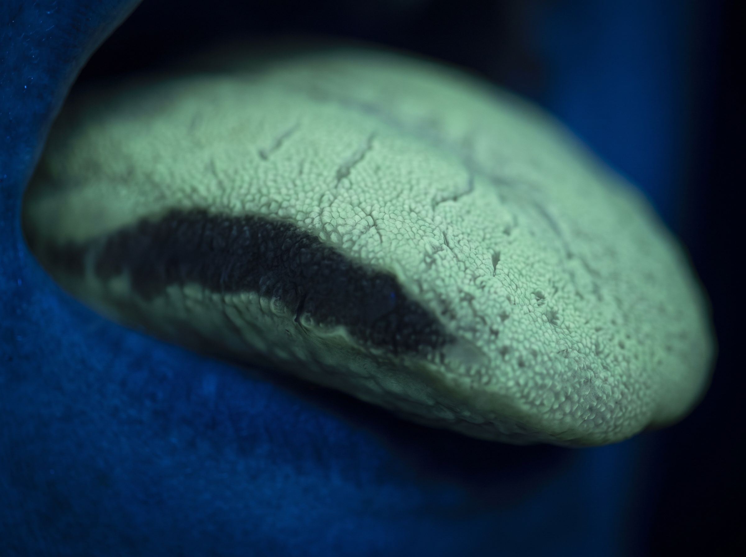
It is one of the more quietly dramatic ideas in oral imaging. Bathed in a narrow band of blue light, healthy mucosa fluoresces — the collagen and other molecules in the tissue absorb the blue and give back a soft, even green. Where the tissue architecture is disturbed by dysplasia or malignancy, that fluorescence collapses, and the region prints as a dark patch against the surrounding glow. The clinician, looking through a filter that blocks the reflected blue and passes only the emitted green, sees a landscape of light with a shadow drawn precisely where the trouble may live.
The light a healthy mucosa gives back
Autofluorescence is intrinsic; nothing is injected or painted on. Certain molecules in the mucosa and the collagen matrix beneath it — the tissue’s natural fluorophores — absorb blue excitation light, typically around 400 to 460 nanometres, and re-emit it at a longer, greener wavelength. In healthy tissue the effect is uniform and bright: a continuous, pale green sheet across the cheek, the floor of the mouth, or the lateral border of the tongue.
What matters diagnostically is not the glow itself but its interruption. When epithelium thickens, when the collagen scaffold below is remodelled by disease, and when blood flow and metabolic activity change in a developing lesion, the tissue absorbs more of the returning green light and emits less of it. The net effect the observer sees is a loss of fluorescence — a dark zone. It is the photographic negative of health: not something new appearing, but something expected going missing.
When the glow goes dark
A loss of fluorescence is a flag, not a verdict. Its value is that it can reveal a suspicious field before the surface has changed enough to catch the eye, and it can show that a visible lesion extends further than its clinical margins suggest — the dark zone often reaches beyond the white or red patch a clinician can see under ordinary light. That extension is genuinely useful information: it can guide where a scalpel or a biopsy punch should go, and it can widen the field a surgeon considers.
But shadows deceive. Inflammation, ulceration, heavy pigmentation, amalgam tattoo, and even a vein close to the surface can all dampen fluorescence and mimic pathology. This is why autofluorescence sits firmly in the category of adjunct — a way to decide where to look harder, not a way to diagnose. The same principle governs every optical modality that reads tissue rather than bone; like the spectral fingerprinting behind hyperspectral imaging of the oral cavity, it narrows the search, and the biopsy still writes the answer.
The instrument and the darkened room
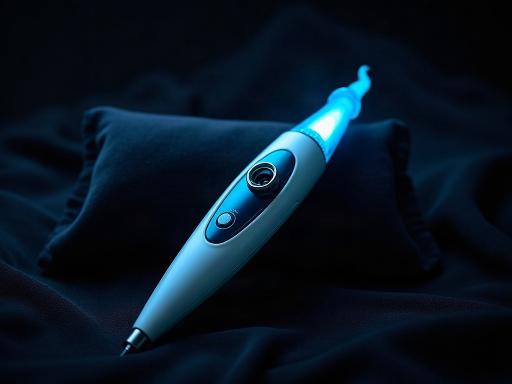
The hardware is elegantly simple. A handheld device emits a tightly controlled band of blue light and provides a viewport — an eyepiece or a camera path — fitted with a filter that rejects the reflected blue and transmits only the longer-wavelength green the tissue emits. Without that filter the whole field would drown in reflected blue and no fluorescence contrast would survive. With it, the emitted green becomes the entire picture.
Photographically, three conditions decide whether the image is worth anything:
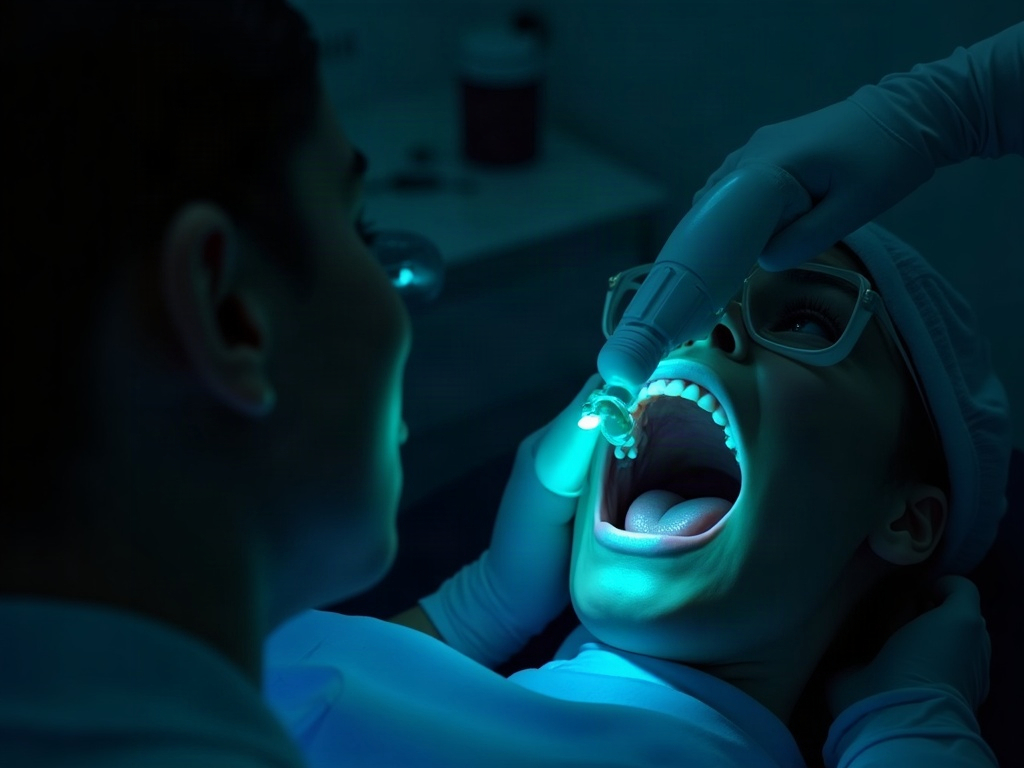
- Darkness. Autofluorescence is a low-light phenomenon. Overhead and operatory lights flood the faint emitted glow and erase the contrast between healthy and suspicious tissue. The room goes dark, exactly as it does for a transillumination capture.
- Geometry. The excitation light and the viewport must sit close to coaxial with the tissue so the whole field is evenly lit; an oblique angle throws false shadow that reads like disease.
- A clean, dry field. Saliva, food debris, and blood scatter light at the surface and add noise. A wiped, retracted field gives the cleanest map of the true tissue fluorescence.
The discipline is the one that governs any light-based diagnostic capture — the same logic that lets transillumination reveal a crack the X-ray cannot: control the light rather than accept it, and let the tissue tell you where it has changed.
Photographing what the eye can barely hold
Documenting autofluorescence is a genuine exposure problem, because the subject is dim, self-luminous, and framed by near-black surroundings. Left on full automatic, a camera meters off the dark field, opens up, and blows the delicate green glow into a flat, contrast-free wash — destroying the very shadow the image exists to show. Manual control is the craft:
- Expose for the glow, not the room. Meter off the fluorescing tissue so the healthy green sits bright but unclipped and the dark zone keeps its edge.
- Small aperture, patient shutter. Depth of field across a curved mucosal surface is unforgiving; stop down for front-to-back sharpness, then let the shutter gather the faint light, bracing the camera against motion.
- Kill the white-light flash. The ring or twin flash that serves a normal clinical portrait is the enemy here — it floods the surface with reflected light and abolishes the fluorescence contrast entirely.
- Fix the white balance deliberately. Let the emitted green read as green and the loss as true shadow, not as a wandering colour cast that a viewer later mistakes for signal.
A well-made autofluorescence image is not only diagnostic evidence; it is a dated, reproducible record. Standardize the excitation, the angle, and the exposure, and a suspicious field photographed today can be honestly compared with the same field in three months — the difference between an impression and a documented change.
Reading the image honestly
Interpretation is where autofluorescence earns respect or squanders it. An even, uninterrupted green suggests healthy architecture. A discrete, well-demarcated dark zone — especially at a high-risk site such as the lateral or ventral tongue, the floor of the mouth, or the soft palate — is the finding that warrants a closer look and, frequently, a scalpel. A diffuse, ill-defined dimming is more often inflammation than malignancy, and the clinician weighs it against the patient’s history, risk factors, and the appearance of the tissue under ordinary light.
The honest framing is this: autofluorescence raises the probability that a region deserves biopsy; it does not lower it to zero when the tissue glows normally. Lesions can and do fluoresce. The image is one witness among several — the white-light exam, palpation, the patient’s symptoms, and ultimately histopathology — and it is at its most powerful when it changes a decision, prompting a biopsy that a purely visual exam would have deferred.
Where autofluorescence fits in the imaging library
Think of autofluorescence not as a rival to any other modality but as a channel tuned to soft tissue and to the earliest disturbances of its architecture — the very changes that radiographs ignore entirely and that a clinical portrait renders as ordinary pink. It is fast, comfortable, radiation-free, and, photographed well, genuinely arresting. Alongside a radiograph’s account of bone and an optical coherence tomogram’s cross-section of enamel and mucosa, an autofluorescence map completes a picture of the mouth as a surface of living, glowing tissue — and marks, in shadow, the places where that glow has begun to fail.
Future developments
The frontier here is measurement and machine reading. Today’s devices largely rely on a clinician’s trained eye to judge whether a glow has dimmed; the next generation is calibrated and quantitative, capturing standardized digital images and assigning a fluorescence value to a region rather than a subjective impression. Multispectral units that combine autofluorescence with reflectance and other wavelengths promise to separate true dysplasia from inflammation and pigment with far greater specificity, cutting the false-positive rate that has long dogged the technique. And as software learns to segment a dark zone, measure its area, and track its change from one visit to the next, autofluorescence is likely to shift from a chairside yes-or-no check into a quantified, longitudinal channel — a way to watch a suspicious field the way we already watch bone. The craft at its heart will not change: darken the room, bathe the tissue in blue, and read the places where the light refuses to come back.
Related Reading
- DIAGNOdent Laser Fluorescence Imaging: Precision Caries Detection Technology for Modern Dental Practice
- Quantitative Light-Induced Fluorescence (QLF) Imaging: Revolutionary Caries Detection Technology in Modern Dentistry
- Red Fluorescence Plaque Detection: Revolutionary 405nm LED Technology for Enhanced Dental Imaging
Sorry, the comment form is closed at this time.