28 Mar Quantitative Light-Induced Fluorescence (QLF) Imaging: Revolutionary Caries Detection Technology in Modern Dentistry
Quantitative Light-Induced Fluorescence (QLF) represents a revolutionary advancement in dental caries detection and monitoring, offering clinicians unprecedented precision in identifying early demineralization processes that traditional visual and radiographic methods often miss. This non-invasive optical imaging technique harnesses the natural fluorescence properties of tooth structure to reveal subtle changes in mineral density, transforming how dental professionals approach caries diagnosis and treatment planning.
The Science Behind QLF Technology
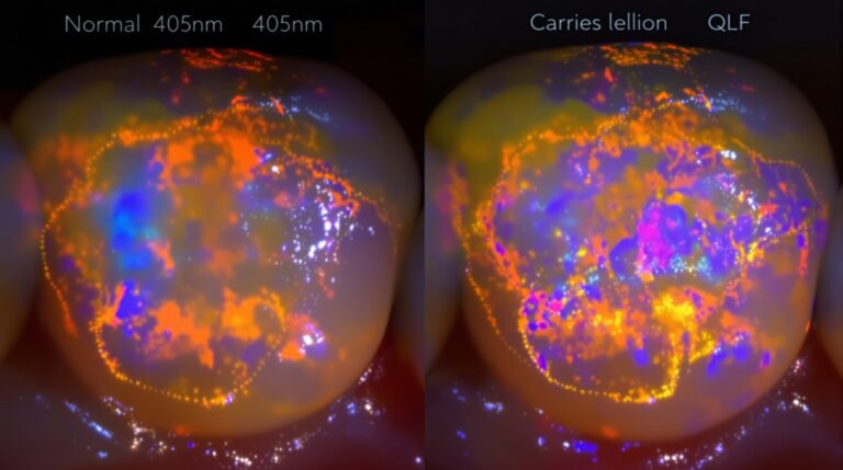
QLF technology operates on the principle that healthy tooth enamel exhibits distinct fluorescence patterns when illuminated with blue light at 405 nanometers. When enamel undergoes demineralization during the caries process, this natural fluorescence is significantly reduced, creating characteristic dark spots or reduced fluorescence areas that correspond to mineral loss. The system captures and quantifies these changes through sophisticated image analysis software, providing both qualitative visual assessment and precise quantitative measurements of lesion severity.
The 405nm wavelength specifically excites fluorophores naturally present in healthy dental hard tissues, primarily porphyrins and other organic compounds embedded within the enamel matrix. As carious lesions develop, these fluorescent compounds are either destroyed or masked by bacterial byproducts and mineral changes, resulting in measurable fluorescence loss that directly correlates with the extent of demineralization.
Clinical Applications and Diagnostic Capabilities
Modern QLF systems excel in detecting occlusal caries, smooth surface lesions, and interdental pathology that may escape detection through conventional methods. The technology proves particularly valuable for monitoring white spot lesions and incipient caries, allowing practitioners to track lesion progression or regression over time with remarkable precision. This capability transforms caries management from a reactive treatment model to a proactive monitoring and prevention approach.
Primary teeth benefit significantly from QLF assessment, as the technology demonstrates exceptional reliability in detecting all types of dental caries in deciduous dentition. The visual feedback provided by QLF imaging helps both clinicians and patients understand the extent of demineralization, facilitating informed treatment decisions and patient education about oral hygiene effectiveness.
Integration with Modern Dental Practice Workflows
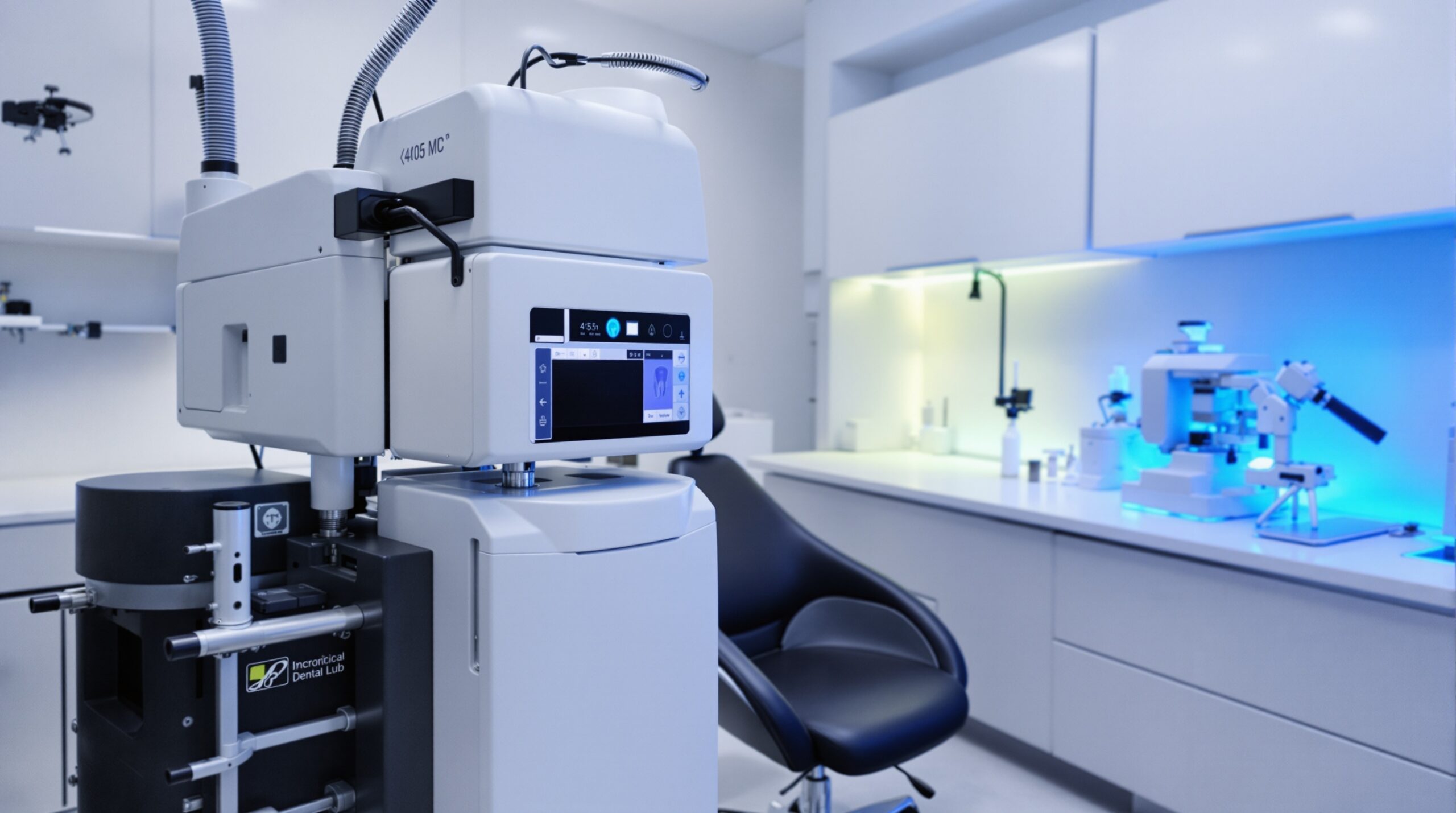
Contemporary QLF systems integrate seamlessly into existing clinical workflows, requiring minimal additional chairside time while providing comprehensive documentation capabilities. The real-time imaging allows for immediate patient education, showing both healthy fluorescence patterns and areas of concern in clear, understandable visual formats. This immediate feedback enhances patient comprehension and treatment acceptance rates significantly.
The quantitative nature of QLF measurements enables standardized assessment protocols across different practitioners and treatment sessions. Fluorescence loss values (ΔF) and red fluorescence measurements (ΔR) provide objective metrics that support evidence-based treatment decisions and facilitate communication between healthcare providers regarding lesion severity and progression rates.
Comparative Advantages Over Traditional Detection Methods
Unlike conventional radiographic techniques that require significant mineral loss before lesions become radiographically apparent, QLF can detect early demineralization at the molecular level. This early detection capability enables intervention during the reversible stages of caries development, potentially preventing the need for restorative treatment through targeted remineralization therapies and enhanced preventive protocols.
Visual examination, while fundamental to dental practice, often fails to identify subtle changes in enamel opacity and early smooth surface lesions. QLF technology enhances clinical observation by revealing subsurface demineralization patterns invisible to the naked eye, particularly in areas with complex anatomical contours or challenging visual access.

Future Developments and Technology Integration
Emerging applications of QLF technology include integration with artificial intelligence algorithms for automated lesion detection and classification. Machine learning models trained on extensive QLF image databases show promising results in standardizing diagnosis and reducing inter-operator variability in lesion assessment. These developments point toward more consistent and objective caries detection protocols across diverse clinical settings.
The combination of QLF with other advanced imaging modalities, such as optical coherence tomography and near-infrared imaging, creates comprehensive diagnostic platforms that provide multidimensional assessment of tooth structure integrity. This multimodal approach enhances diagnostic confidence and enables more personalized treatment planning based on individual patient risk profiles and lesion characteristics.
Implementation Considerations and Best Practices
Successful QLF implementation requires proper calibration protocols and standardized imaging techniques to ensure consistent and reproducible results. Factors such as plaque removal, tooth drying, and consistent positioning significantly influence measurement accuracy and must be carefully controlled during clinical assessments.
Training protocols for clinical staff should emphasize both technical proficiency and interpretation skills, as the technology’s diagnostic value depends heavily on proper image acquisition and analysis. Regular calibration checks and quality assurance procedures maintain system accuracy and ensure reliable longitudinal monitoring capabilities.
As dental practices increasingly adopt precision medicine approaches, QLF technology provides the quantitative foundation necessary for individualized caries risk assessment and tailored prevention strategies. The technology’s ability to monitor treatment outcomes objectively supports evidence-based practice models and enhances the overall quality of patient care in contemporary dental settings.
Related Reading
- DIAGNOdent Laser Fluorescence Imaging: Precision Caries Detection Technology for Modern Dental Practice
- Near-Infrared Transillumination (NIRT): Advanced Dental Imaging for Early Caries Detection
- Polarized Light Microscopy in Enamel Analysis: Advanced Birefringence Techniques for Caries Detection and Remineralization Studies
Sorry, the comment form is closed at this time.