01 May Metal Artifact Reduction Techniques in Dental Imaging: Advanced Strategies for Enhanced Diagnostic Quality
Metal artifacts in dental imaging present significant diagnostic challenges, creating streaking patterns and distortions that can obscure critical anatomical structures. Modern dental practices increasingly rely on sophisticated artifact reduction techniques to maintain image quality while preserving essential diagnostic information.
Understanding Metal Artifact Formation
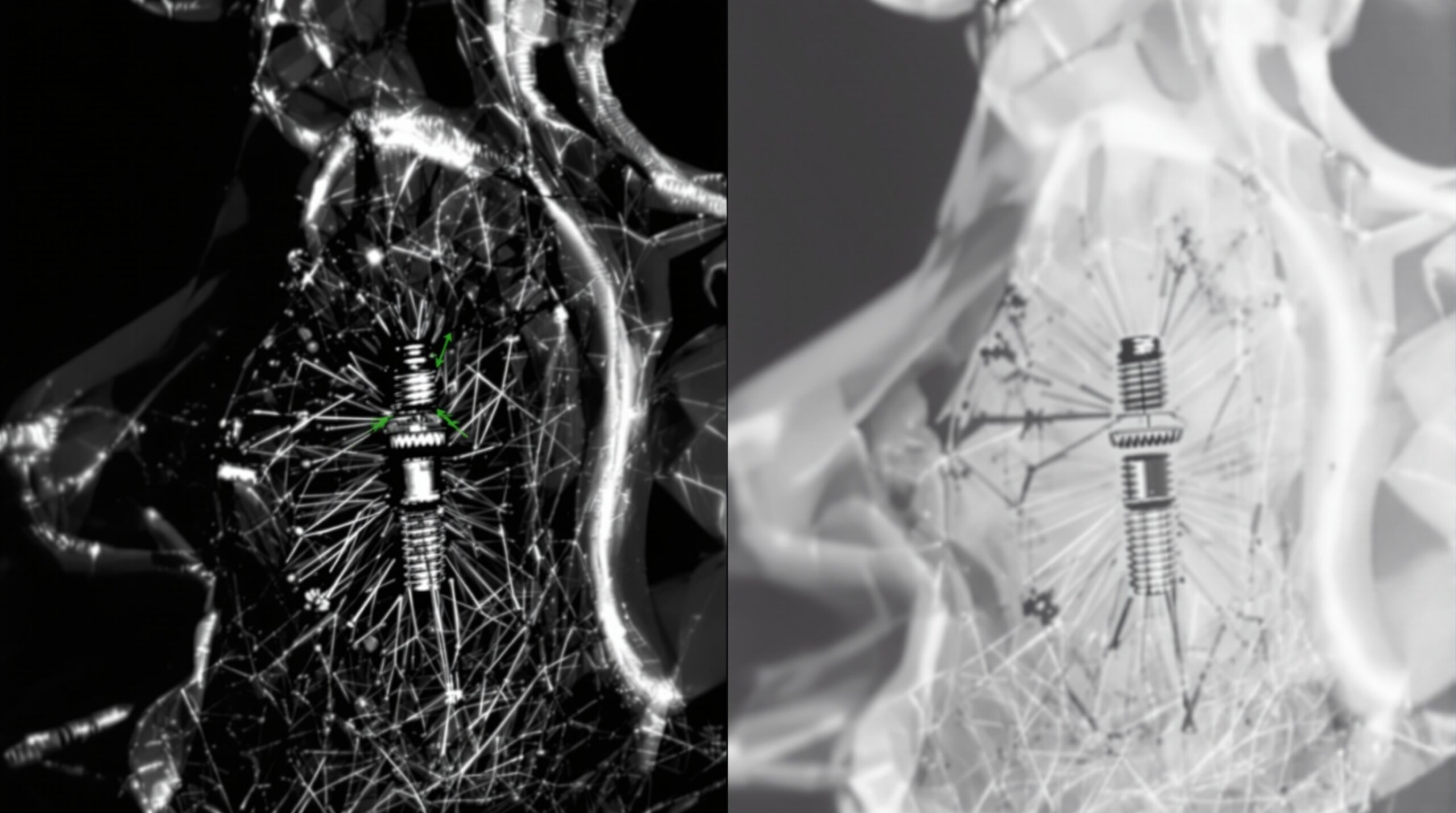
Metal artifacts occur when X-ray beams encounter high-density materials such as dental implants, crowns, or orthodontic appliances. These materials absorb or scatter radiation differently than surrounding tissues, creating characteristic streak artifacts that radiate outward from the metal object. The severity of artifacts depends on factors including metal composition, object size, beam energy, and reconstruction algorithms.
Hardware-Based Reduction Strategies
Modern CBCT systems incorporate several hardware-based approaches to minimize metal artifacts. Dual-energy imaging allows differentiation between metal and soft tissue by utilizing two different X-ray energy spectra. Advanced filtration systems can reduce beam hardening effects, while optimized tube voltage settings help balance contrast and artifact reduction.
Geometric positioning plays a crucial role in artifact management. Strategic patient positioning can minimize the path length through metal objects, reducing artifact severity. Some systems offer specialized scanning protocols designed specifically for metal artifact reduction, incorporating modified reconstruction parameters and extended acquisition times.
Software-Based Correction Algorithms
Image processing techniques represent the most versatile approach to metal artifact reduction. Linear interpolation methods replace corrupted projection data with estimated values based on adjacent projections. More sophisticated algorithms employ iterative reconstruction techniques that model the metal objects and compensate for their effects during image reconstruction.
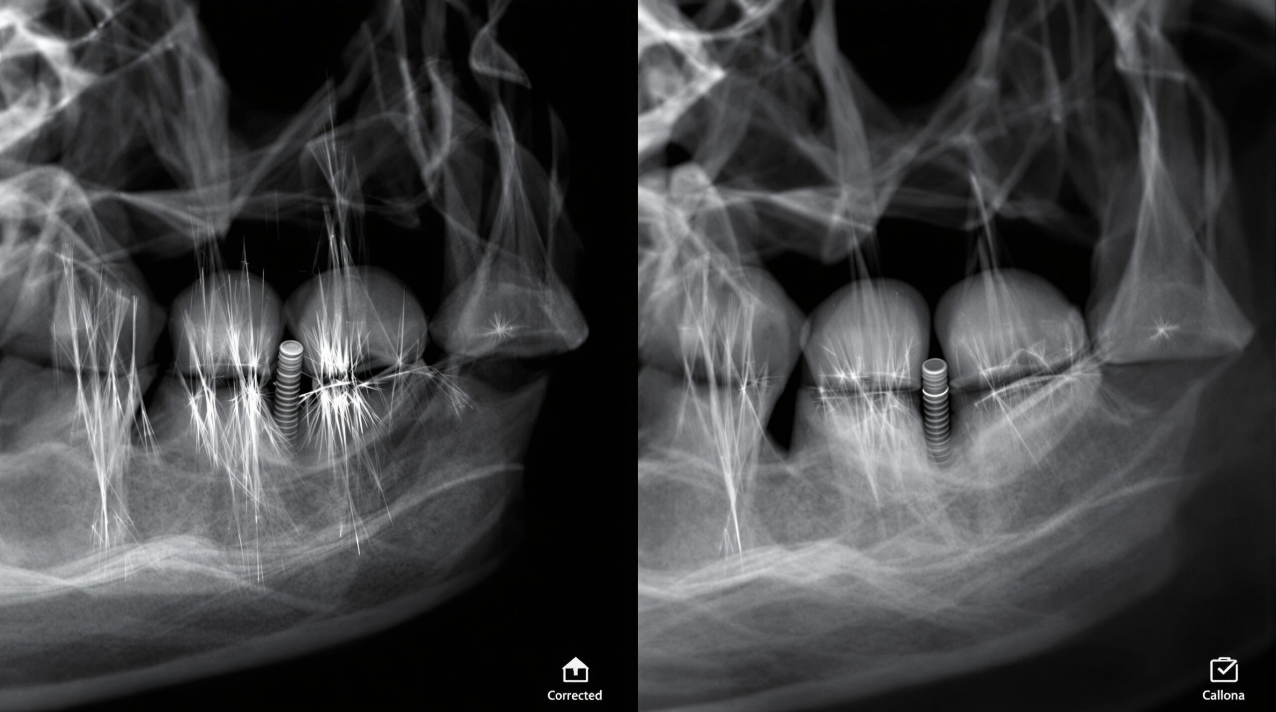
Sinogram completion methods identify and correct affected projection data in the raw acquisition space before reconstruction. These techniques can effectively reduce streaking while preserving edge sharpness and anatomical detail. Machine learning algorithms increasingly supplement traditional methods, offering adaptive artifact reduction based on image content analysis.
Clinical Implementation Guidelines
Successful metal artifact reduction requires systematic approach combining multiple strategies. Pre-scan evaluation should assess metal object locations and compositions to optimize acquisition parameters. During acquisition, proper patient positioning and immobilization minimize motion artifacts that compound metal-related distortions.
Post-processing workflows should incorporate artifact reduction as standard practice rather than corrective measures. Quality assurance protocols ensure consistent artifact reduction performance across different metal types and clinical scenarios. Staff training emphasizes recognition of artifact patterns and appropriate selection of correction techniques.
Quality Assessment and Validation
Effective artifact reduction maintains diagnostic quality while minimizing computational overhead. Validation requires phantom studies using various metal compositions and geometries to establish optimal parameters. Clinical validation ensures artifact reduction doesn’t introduce new artifacts or compromise diagnostic accuracy.
Quantitative metrics including noise reduction, contrast preservation, and geometric accuracy provide objective assessment of reduction techniques. Regular calibration ensures consistent performance across different imaging conditions and metal configurations.
Future Developments
Emerging technologies promise enhanced metal artifact reduction capabilities. Photon-counting detectors offer improved spectral discrimination, enabling better material characterization and artifact suppression. Advanced reconstruction algorithms incorporate prior knowledge about metal objects and surrounding anatomy.
Artificial intelligence applications continue expanding, offering real-time artifact detection and adaptive correction strategies. Integration with treatment planning systems enables artifact reduction optimization based on specific clinical requirements and diagnostic objectives.
Related Reading
- Radiopaque Contrast Agents in Dental Imaging: Advanced Material Science for Enhanced Diagnostic Visibility
- Quality Assurance Phantom Testing: Essential Calibration Protocols for Digital Dental Imaging Systems
- Close-Range Photogrammetry in Dentistry: Revolutionary 3D Imaging Technology for Precision Diagnostics
Sorry, the comment form is closed at this time.