22 Mar Advanced Panoramic Radiography Techniques: 2026 ADA Guidelines and Digital Innovations
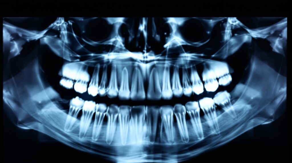
Panoramic radiography continues to evolve as a cornerstone of dental imaging, offering comprehensive visualization of the maxillofacial structures in a single exposure. The American Dental Association’s updated 2026 guidelines emphasize evidence-based practices that maximize diagnostic value while minimizing patient radiation exposure.
Current ADA Recommendations for Panoramic Imaging
The latest ADA guidelines establish panoramic radiographs as the preferred initial imaging modality for monitoring tooth eruption before orthodontic treatment begins. This recommendation reflects extensive clinical research demonstrating superior visualization of root alignment patterns and developmental anomalies compared to traditional intraoral series.
Key applications include comprehensive evaluation of impacted teeth, assessment of temporomandibular joint morphology, and detection of pathological conditions affecting the entire maxillofacial complex. The guidelines stress that panoramic imaging should follow ALARA principles while providing maximum diagnostic information.
Digital Advances in Panoramic Technology
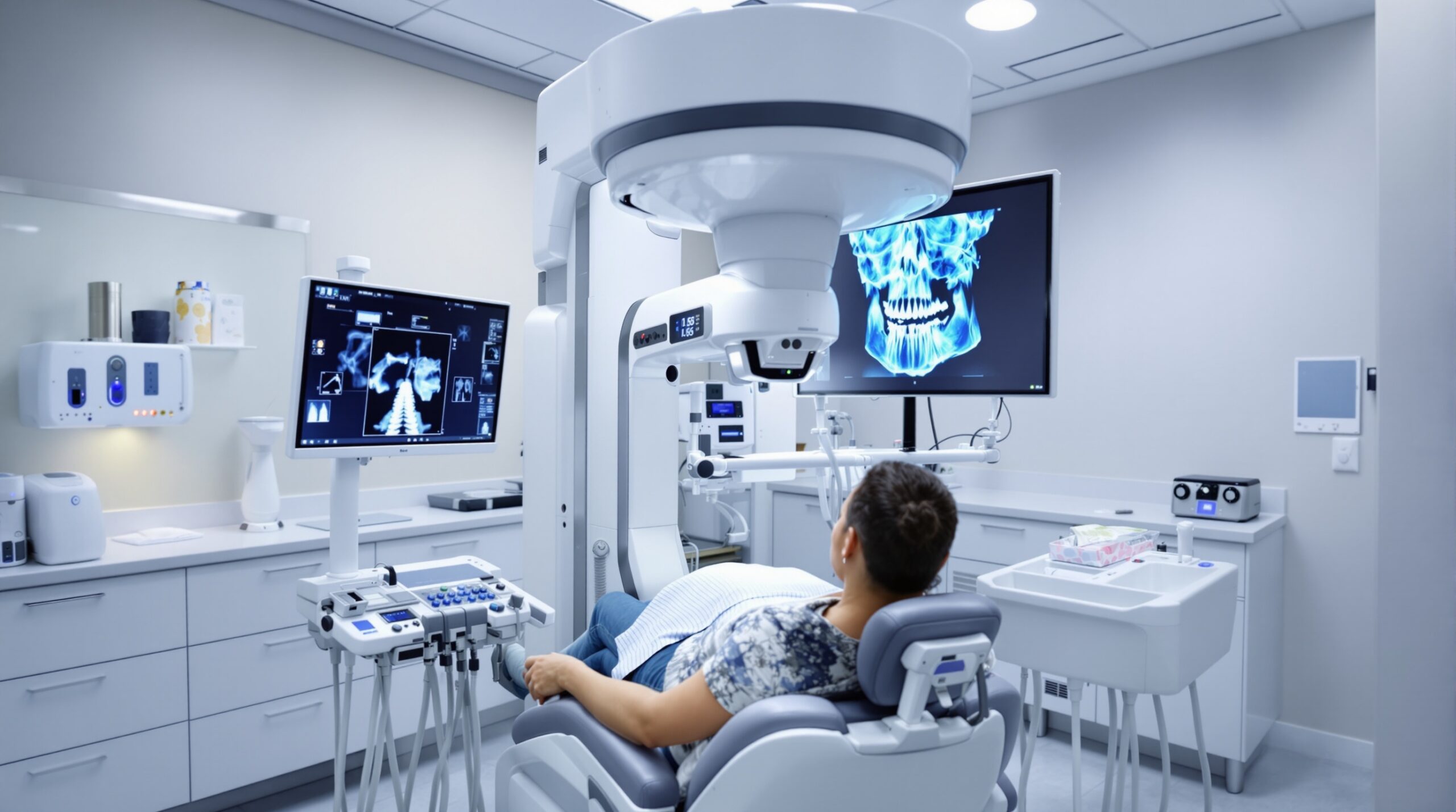
Modern digital panoramic systems incorporate automatic exposure control mechanisms that adapt X-ray beam energy to individual patient anatomy. This technology ensures optimal image quality while maintaining minimal radiation exposure levels. Advanced sensors and processing algorithms enhance image sharpness and contrast resolution significantly.
Enhanced Image Processing Capabilities
Contemporary panoramic units feature sophisticated software that corrects common imaging artifacts, including ghost images and magnification distortions. Real-time image preview allows immediate quality assessment, reducing the need for retakes and further limiting patient radiation exposure.
Clinical Applications and Best Practices
Panoramic radiography excels in orthodontic treatment planning, providing essential information about root parallelism, crown angulation, and potential complications. The imaging technique offers superior visualization of the mandibular third molar region compared to intraoral radiographs.
Patient Positioning and Technique Optimization
Precise patient positioning remains critical for diagnostic quality panoramic images. Modern units incorporate laser positioning guides and chin supports that ensure reproducible patient alignment. Proper technique reduces common errors including anterior teeth narrowing and cervical spine superimposition.
Integration with Digital Workflows
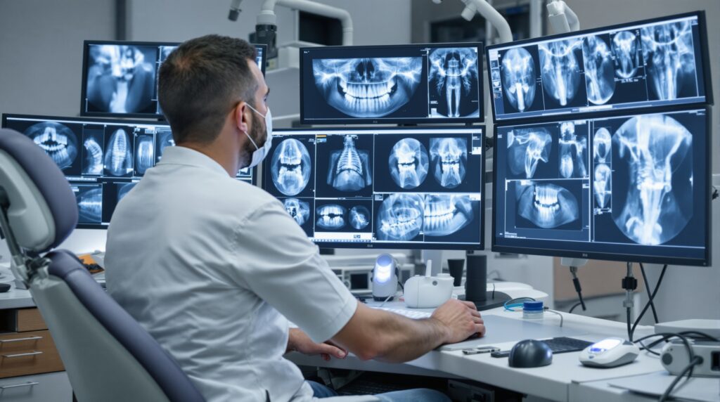
Advanced panoramic systems seamlessly integrate with practice management software and digital imaging networks. Instant image availability facilitates immediate consultation and treatment planning discussions with patients. Cloud-based storage solutions ensure secure access to imaging records from multiple locations.
Future Developments in Panoramic Imaging
Emerging technologies including artificial intelligence-assisted diagnosis and enhanced low-dose imaging protocols promise further improvements in panoramic radiography capabilities. Research continues into advanced reconstruction algorithms that may approach CBCT-level detail while maintaining the simplicity and cost-effectiveness of traditional panoramic imaging.
The evolution of panoramic radiography demonstrates the dental profession’s commitment to evidence-based imaging practices that prioritize patient safety while maximizing diagnostic effectiveness. These advancements ensure panoramic radiography remains an essential component of contemporary dental practice.
Sorry, the comment form is closed at this time.