12 Apr AI-Powered Image Segmentation in Dental Imaging: Revolutionizing Diagnostic Accuracy and Clinical Workflow
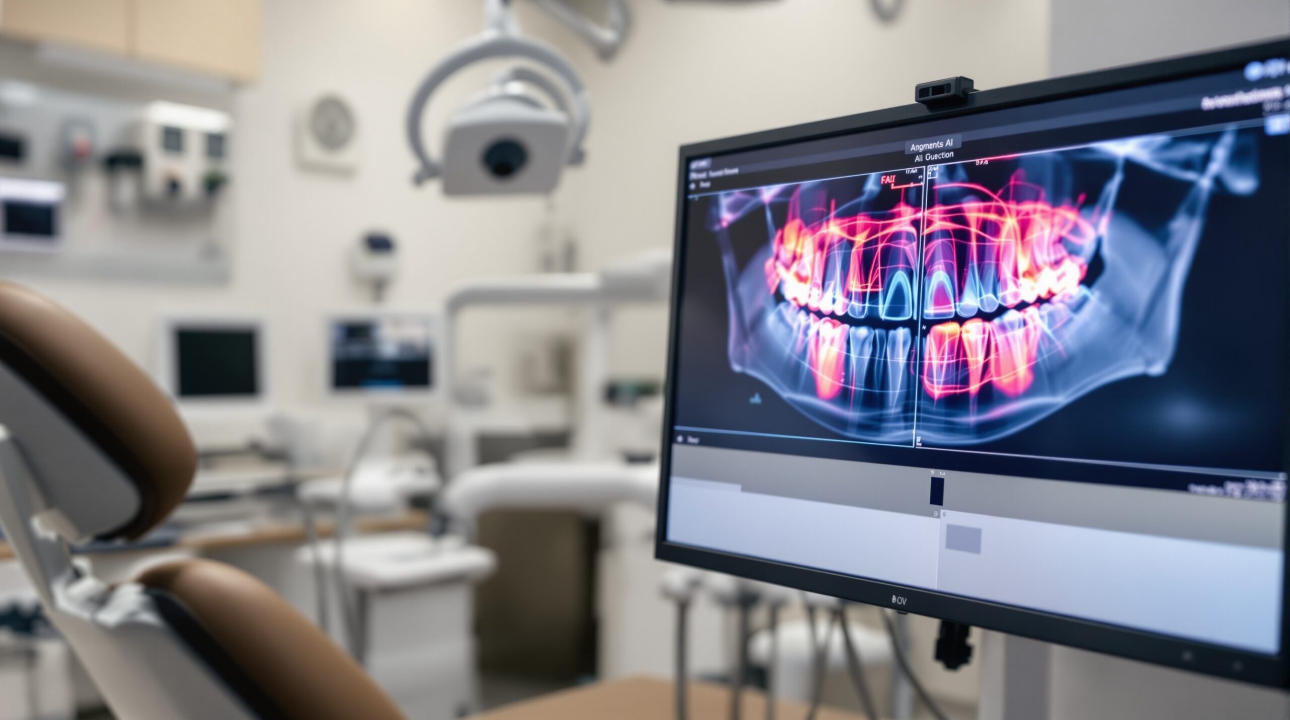
Artificial intelligence has transformed dental imaging through advanced segmentation technologies that automatically identify and highlight anatomical structures in radiographic images. AI-powered image segmentation represents a paradigm shift from manual interpretation to automated analysis, enhancing diagnostic accuracy while streamlining clinical workflows.
Understanding AI Image Segmentation Technology
AI image segmentation utilizes sophisticated algorithms to automatically partition dental images into distinct regions of interest. These systems employ deep learning models trained on vast datasets of radiographic images to recognize and delineate anatomical structures including teeth, bone, periodontal ligaments, and pathological conditions.
The technology operates through computer vision techniques that analyze pixel patterns, contrast variations, and spatial relationships within digital radiographs. Machine learning algorithms continuously refine their accuracy through exposure to diverse clinical cases, resulting in increasingly precise segmentation capabilities.
CBCT Volumetric Segmentation Advances
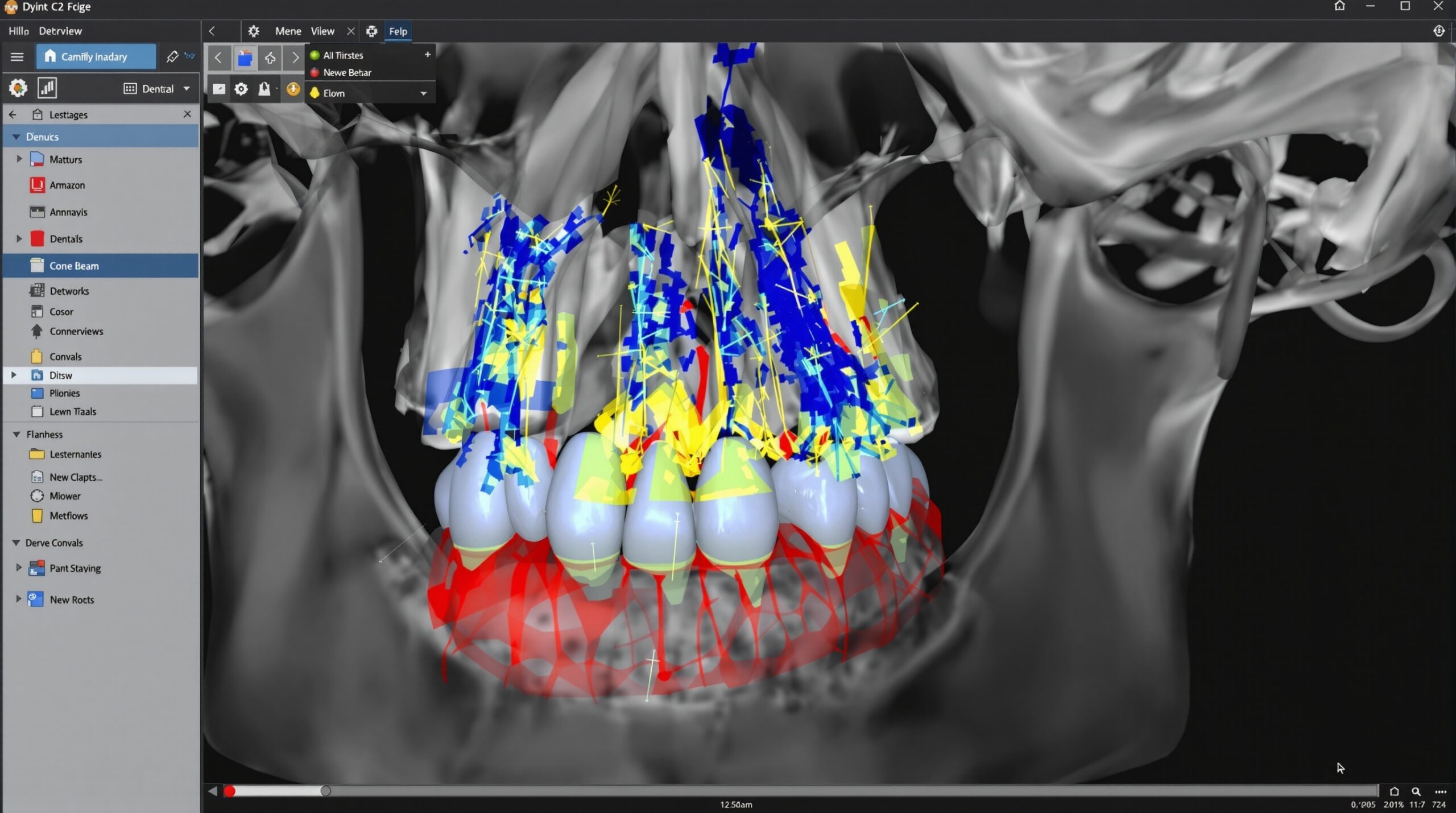
Cone Beam Computed Tomography (CBCT) imaging benefits significantly from AI segmentation technology. Three-dimensional volumetric data provides comprehensive anatomical visualization, with AI algorithms capable of automatically segmenting complex structures including root canal systems, maxillary sinuses, and mandibular nerves.
Advanced segmentation protocols can differentiate between cortical and cancellous bone, identify impacted teeth orientations, and precisely map anatomical landmarks for surgical planning. This technology reduces interpretation time while improving diagnostic consistency across different practitioners.
Clinical Applications and Diagnostic Enhancement
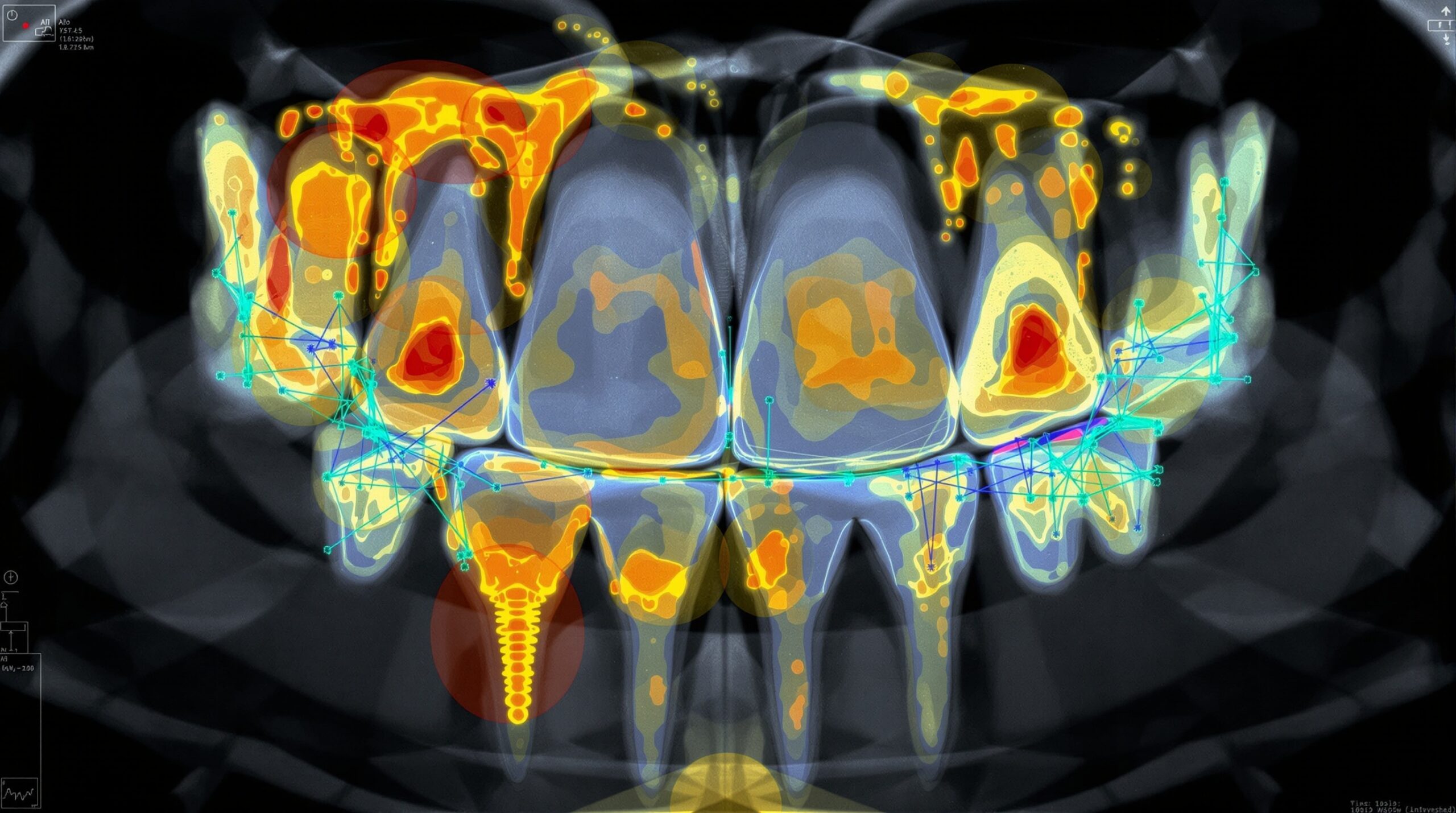
AI segmentation technology enhances multiple aspects of dental diagnosis. Caries detection algorithms can automatically identify and quantify decay progression, while periodontal assessment tools measure bone loss patterns and pocket depths with sub-millimeter accuracy.
Endodontic applications include automated root canal identification, apex localization, and treatment planning optimization. Orthodontic practitioners benefit from automated cephalometric landmark detection and growth pattern analysis through longitudinal image comparison.
Workflow Integration and Practice Benefits
Integration of AI segmentation into existing practice workflows requires minimal infrastructure changes while delivering substantial efficiency gains. Automated image analysis reduces radiographic interpretation time from minutes to seconds, allowing practitioners to focus on treatment planning rather than manual measurements.
Quality assurance protocols benefit from AI consistency, eliminating inter-observer variability in radiographic interpretation. Standardized segmentation criteria ensure uniform diagnostic approaches across different practitioners within group practice environments.
FDA-Approved Solutions and Implementation
Several AI segmentation platforms have received FDA clearance for clinical use, including solutions from Diagnocat, VideaAI, and CranioCatch. These systems integrate seamlessly with existing PACS infrastructure through DICOM compatibility and cloud-based processing capabilities.
Implementation typically involves software installation, staff training protocols, and gradual workflow integration. Most platforms offer subscription-based pricing models with per-image processing fees, making advanced AI technology accessible to practices of all sizes.
Future Developments and Clinical Impact
Emerging AI segmentation technologies focus on real-time analysis during image acquisition, predictive modeling for treatment outcomes, and integration with treatment planning software. Machine learning algorithms continue advancing through federated learning approaches that improve accuracy while maintaining patient privacy.
The integration of AI segmentation with augmented reality systems promises enhanced surgical guidance, while automated report generation capabilities will further streamline documentation requirements. These advances position AI-powered image segmentation as an essential component of modern digital dentistry practices.
Sorry, the comment form is closed at this time.