28 Apr Bitewing Radiography Positioning Techniques: Mastering Patient Comfort and Diagnostic Accuracy
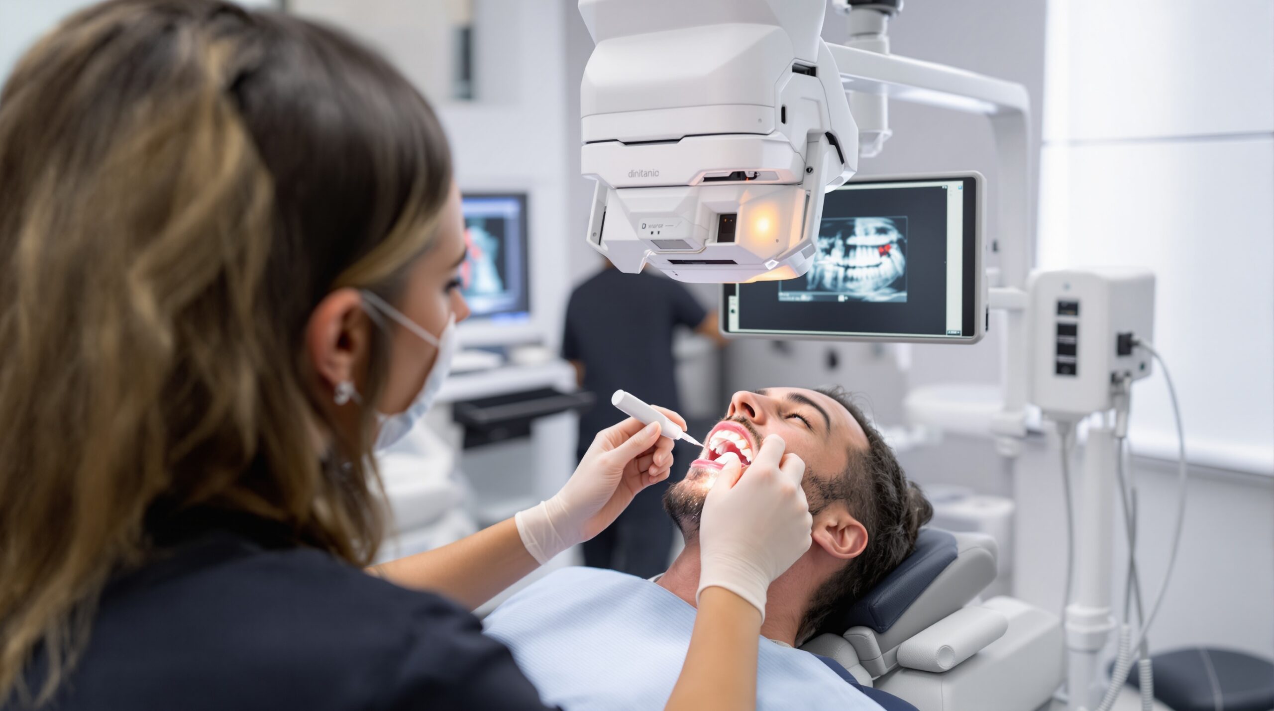
Bitewing radiography remains one of the most frequently performed dental imaging procedures, essential for detecting interproximal caries, evaluating bone levels, and monitoring periodontal health. However, achieving optimal diagnostic quality while ensuring patient comfort requires mastering specific positioning techniques and understanding the nuances of sensor placement.
Fundamentals of Bitewing Positioning
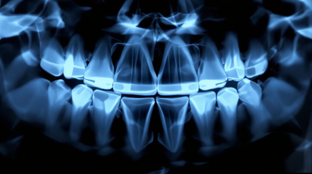
Proper bitewing technique relies on precise sensor placement to capture the crowns of both maxillary and mandibular teeth, along with the supporting alveolar bone. The goal is to visualize the contact areas clearly while maintaining proper geometric relationships between the X-ray beam, sensor, and teeth.
The horizontal bitewing technique utilizes a landscape orientation of the digital sensor, positioned lingually to the teeth with the bite tab extending through the occlusal plane. This orientation maximizes the coverage area and provides optimal visualization of multiple teeth in a single exposure.
Step-by-Step Positioning Protocol
Patient Preparation
Begin by adjusting the dental chair to a comfortable height for both operator and patient. Position the patient’s head in a natural, upright posture with the occlusal plane parallel to the floor. Remove any eyeglasses, earrings, or removable appliances that might interfere with the imaging process.
Sensor Placement Technique
Place the digital sensor in its protective sleeve, ensuring the bite tab is properly centered. Position the sensor holder with the bite tab extending perpendicular to the long axis of the sensor. The sensor should be placed as far lingually as possible while maintaining patient comfort.
For premolar bitewings, center the sensor over the contact area between the first and second premolars. The anterior edge of the sensor should extend to include the distal surface of the canine. For molar bitewings, center the sensor over the first and second molar contact area, ensuring the third molar area is included when erupted.
Patient Instruction and Comfort
Instruct the patient to close slowly and bite gently on the bite tab. Avoid having patients “bite hard” as this can cause discomfort and movement artifacts. The patient should maintain a relaxed closure throughout the exposure.
Optimizing Patient Comfort
Patient comfort significantly impacts the quality of bitewing radiographs. Uncomfortable patients may move during exposure, resulting in motion blur or requiring retakes that increase radiation exposure.
Ergonomic Considerations
Digital sensors can feel sharp or uncomfortable due to their rectangular shape and electronic components. Consider using sensor cushioning products or bite blocks designed to reduce discomfort. Some practices utilize foam padding or specialized sensor holders that distribute pressure more evenly.
Communication Strategies
Clear communication helps reduce patient anxiety and ensures cooperation. Explain each step of the procedure beforehand and provide an estimated duration for each exposure. Let patients know they can signal if they experience discomfort, and be prepared to adjust positioning if needed.
For patients with strong gag reflexes, consider pre-treating with topical anesthetic, using smaller sensors when available, or employing desensitization techniques such as controlled breathing exercises.
Common Positioning Errors and Solutions
Overlapping Contact Areas
Overlapped contacts occur when the horizontal angulation is incorrect. Ensure the X-ray beam is perpendicular to the interproximal surfaces and directed through the contact areas. Slight adjustments in tube head positioning can eliminate this common error.
Cone Cutting
Cone cutting results from improper alignment between the PID (Position Indicating Device) and the sensor. Always verify that the PID completely covers the sensor area and that the central ray is directed toward the center of the sensor.
Distortion Issues
Excessive vertical angulation can cause foreshortening or elongation of tooth images. Maintain a vertical angulation of approximately +10 degrees for maxillary teeth and -15 degrees for mandibular teeth in most cases, adjusting as needed for individual patient anatomy.
Advanced Positioning Techniques
Horizontal Offset Method
For difficult anatomical situations, the horizontal offset technique can improve sensor placement. This involves positioning the sensor slightly anterior or posterior to the ideal location while adjusting the horizontal angulation to maintain proper geometric relationships.
Modified Bite Tab Techniques
In patients with limited mouth opening or anatomical restrictions, modified bite tab positioning may be necessary. This can include angling the bite tab or using alternative sensor holding devices designed for challenging cases.
Quality Assurance Considerations
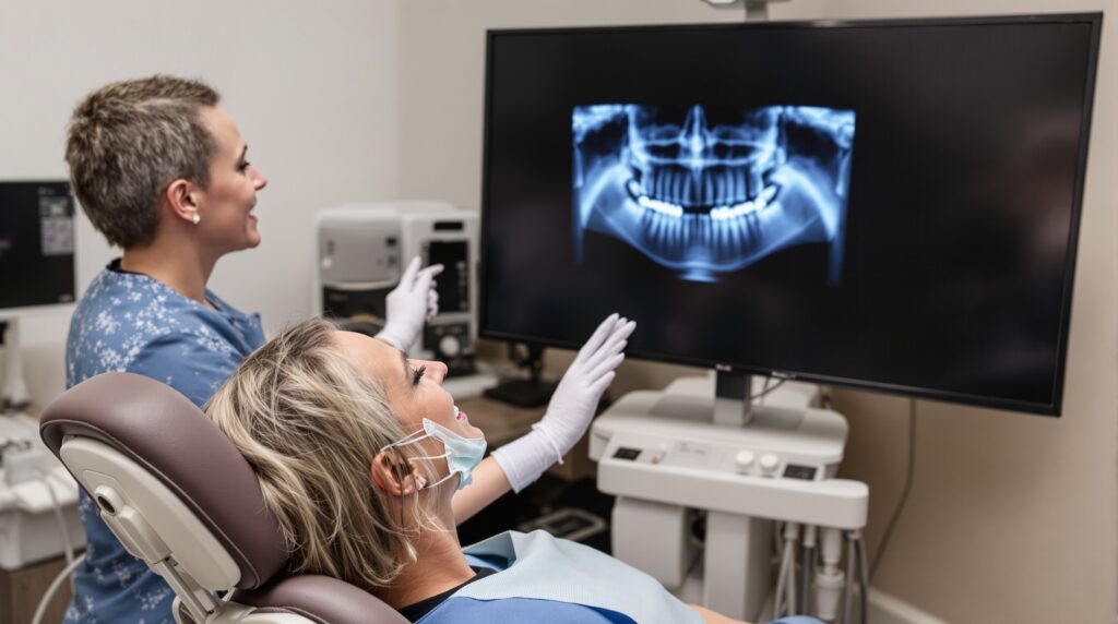
Implement a systematic approach to evaluate each bitewing image for diagnostic adequacy. Check for proper coverage of anatomical structures, absence of positioning errors, optimal contrast and density, and overall image sharpness.
Document any modifications to standard techniques in patient records, particularly for patients who require special accommodations. This ensures consistency in future imaging sessions and helps maintain diagnostic quality over time.
Conclusion
Mastering bitewing radiography positioning techniques requires understanding both the technical aspects of sensor placement and the human factors that influence patient comfort. By combining proper geometric principles with patient-centered care approaches, dental professionals can consistently produce high-quality diagnostic images while minimizing patient discomfort and radiation exposure.
Regular training updates and technique refinements help maintain proficiency in bitewing radiography, ensuring optimal diagnostic outcomes for comprehensive dental care. The investment in proper positioning technique pays dividends in improved diagnostic accuracy and enhanced patient experience.
Sorry, the comment form is closed at this time.