08 Apr Digital Subtraction Radiography: Advanced Change Detection Technology for Enhanced Dental Diagnostics
Digital Subtraction Radiography (DSR) represents a revolutionary advancement in dental imaging technology, offering unparalleled precision in detecting and monitoring subtle changes in oral and maxillofacial structures. This sophisticated technique has transformed how dental professionals approach diagnostic imaging by eliminating anatomical noise and highlighting even the smallest pathological changes that might otherwise go undetected.
By comparing sequential radiographs and mathematically subtracting unchanging structures, DSR creates enhanced images where only the areas of change are visible, dramatically improving diagnostic accuracy for conditions such as periodontal disease, caries progression, and post-treatment monitoring.
The Science Behind Digital Subtraction Radiography
DSR operates on the principle of temporal subtraction, where two standardized radiographs taken at different times undergo digital processing to remove static anatomical structures. The result is a subtraction image that displays only the areas where changes have occurred between the two imaging sessions.
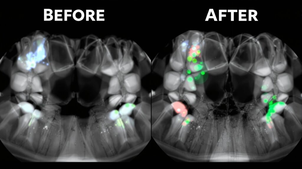
The process involves precise geometric alignment of sequential images, followed by pixel-by-pixel mathematical subtraction. Areas of bone loss appear as darker regions in the subtraction image, while areas of bone formation or repair appear brighter, creating a stark visual contrast that makes subtle changes immediately apparent to the clinician.
Clinical Applications and Diagnostic Benefits
DSR has proven invaluable in numerous clinical scenarios where conventional radiography may fall short in detecting early pathological changes:
- Periodontal Disease Monitoring: Detection of alveolar bone changes as small as 1-5% per unit volume, enabling early intervention
- Caries Progression Analysis: Enhanced visualization of demineralization patterns and treatment efficacy
- Implant Assessment: Precise monitoring of peri-implant bone levels and early detection of complications
- Endodontic Outcomes: Evaluation of periapical healing and bone repair following root canal therapy
- Orthodontic Treatment: Assessment of root resorption and alveolar bone changes during tooth movement
Research has demonstrated that DSR can detect crestal bone height changes as small as 0.78mm, significantly more sensitive than conventional radiographic interpretation, which typically requires 30-50% mineral loss before changes become radiographically apparent.
Technical Requirements and Implementation
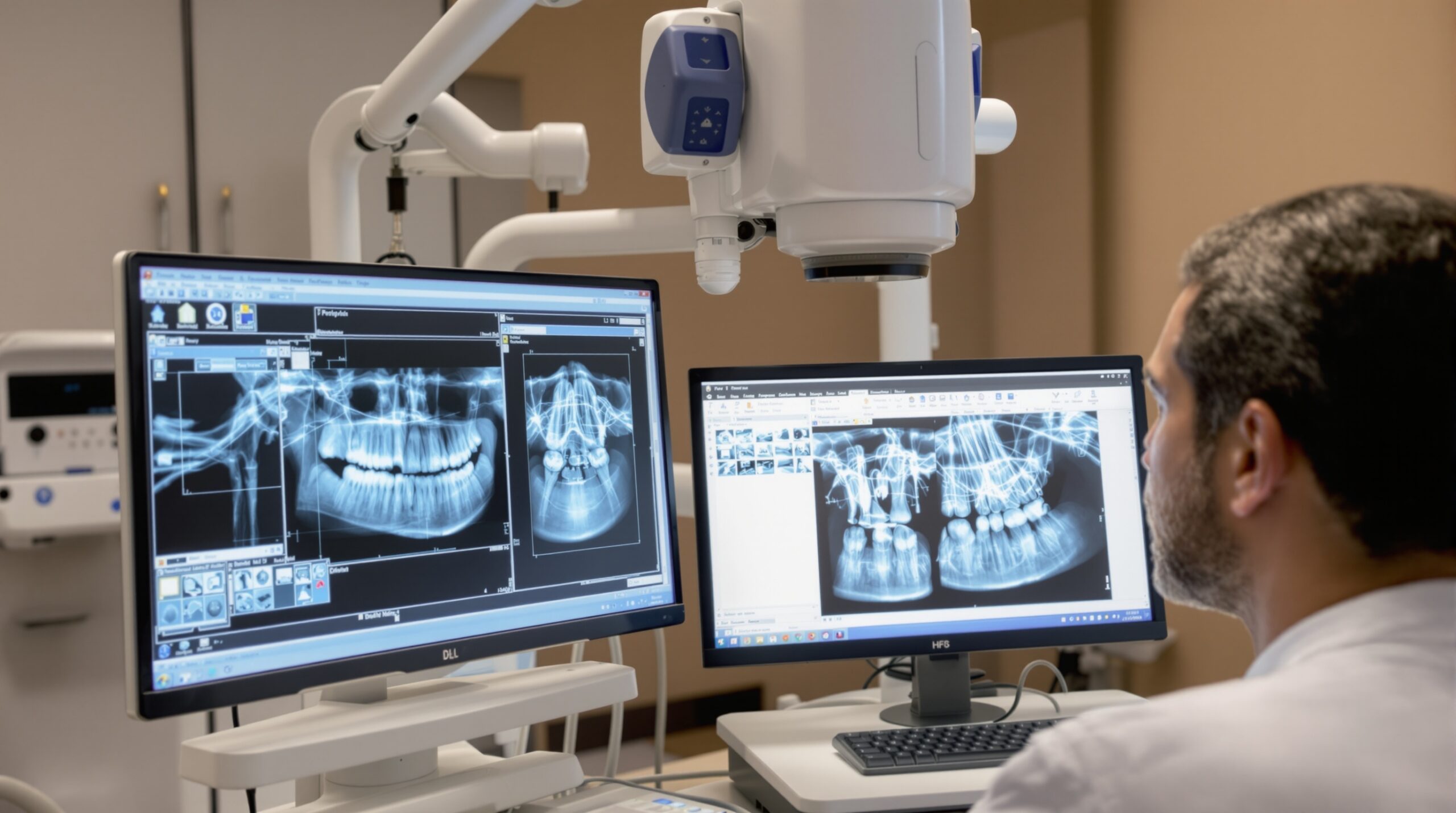
Successful implementation of DSR requires stringent standardization protocols to ensure optimal results. The imaging geometry must remain consistent between sequential radiographs, necessitating the use of positioning devices and bite blocks for intraoral radiography or precise head positioning systems for extraoral imaging.
Modern digital imaging systems have greatly facilitated DSR implementation, offering:
- Immediate image acquisition and processing
- Automated alignment algorithms for geometric correction
- Enhanced contrast manipulation capabilities
- Integration with practice management systems for longitudinal tracking
- Standardized exposure parameters for consistent image quality
Advantages Over Conventional Radiography
DSR offers several distinct advantages that make it an invaluable diagnostic tool in modern dental practice:
Enhanced Sensitivity
The removal of anatomical noise allows for detection of changes that would be masked by the complex trabecular patterns in conventional radiographs. This enhanced sensitivity enables earlier intervention and improved treatment outcomes.
Objective Analysis
DSR provides quantifiable measurements of change, reducing subjectivity in radiographic interpretation and improving consistency among different observers.
Improved Patient Communication
The stark visual contrast in subtraction images makes it easier to demonstrate disease progression or treatment success to patients, enhancing treatment acceptance and compliance.
Limitations and Considerations
While DSR represents a significant advancement in dental imaging, several factors must be considered for successful implementation:
Patient movement between exposures can create artifacts that compromise image quality and diagnostic accuracy. Additionally, changes in soft tissue positioning or radiographic projection angles can introduce errors in the subtraction process.
The technique requires additional time for image processing and analysis, and the learning curve for proper interpretation may be steep for practitioners unfamiliar with subtraction imaging principles.
Future Developments and Integration
Emerging technologies are enhancing DSR capabilities through artificial intelligence and machine learning algorithms. These developments promise automated change detection, predictive modeling for disease progression, and integration with other advanced imaging modalities such as CBCT and intraoral scanning.
The integration of DSR with practice management systems allows for comprehensive longitudinal patient monitoring, creating valuable databases for research and evidence-based treatment planning.
Conclusion
Digital Subtraction Radiography represents a paradigm shift in dental diagnostic imaging, offering unprecedented sensitivity for detecting subtle pathological changes in oral and maxillofacial structures. Its ability to remove anatomical noise and highlight areas of change makes it an invaluable tool for early disease detection, treatment monitoring, and improved patient outcomes.
As dental practices continue to embrace digital technologies, DSR stands as a testament to the potential of advanced imaging techniques to enhance diagnostic accuracy and clinical decision-making. The continued development of automated processing algorithms and artificial intelligence integration promises to make this powerful diagnostic tool even more accessible and effective in routine clinical practice.
Sorry, the comment form is closed at this time.