


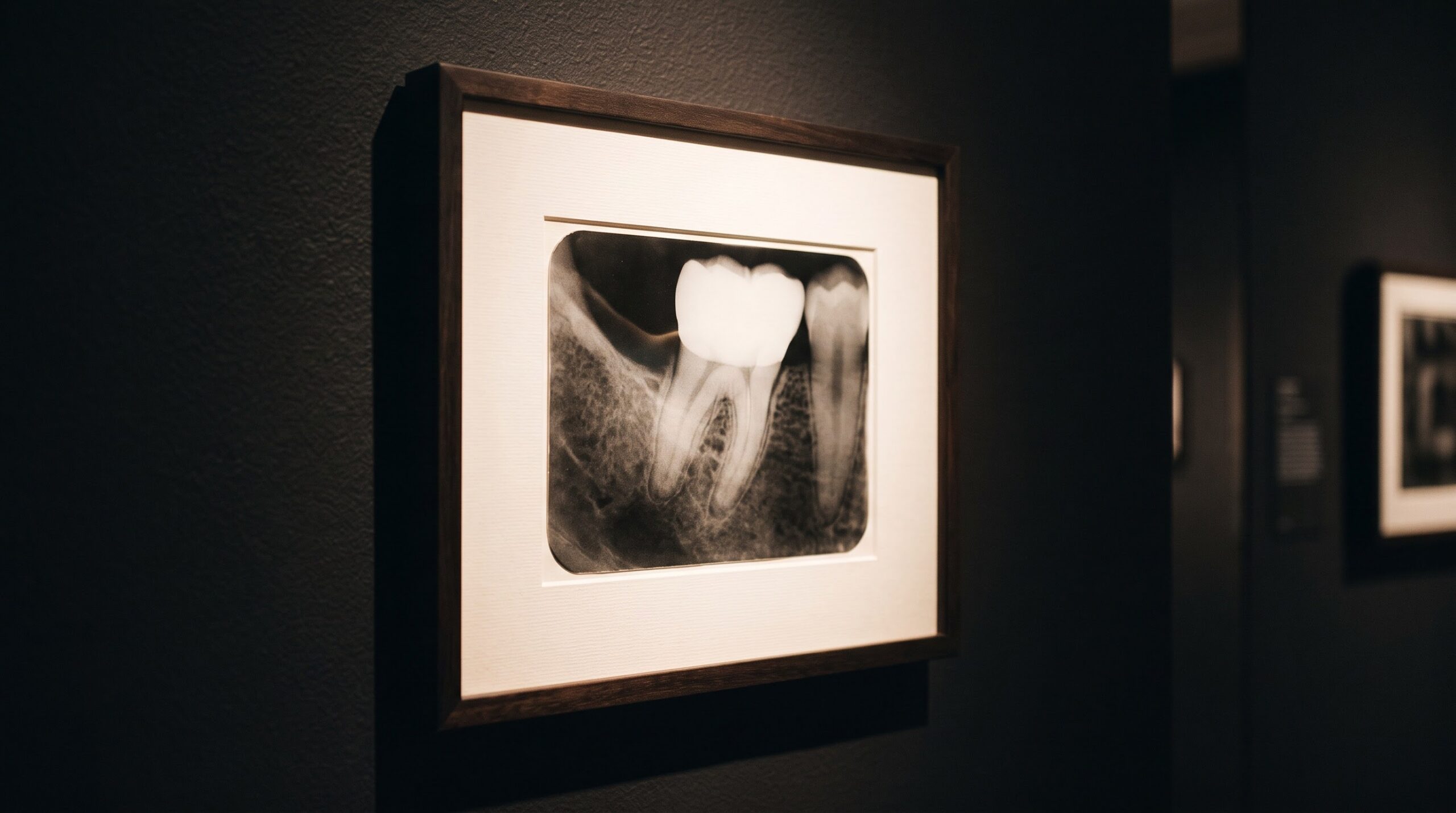
November 19, 2026 The Mach Band Illusion: When the Eye Invents a Shadow the X-Ray Never Recorded
A radiograph is usually trusted as a faithful witness: whatever gray it shows, we assume, is a gray the x-ray actually measured. Most of the time that trust is earned. But some of the most convincing marks on a dental film were never recorded by the beam at all. They are drawn, in real time, by the viewer’s own visual system — fabrications so precise that they settle exactly where disease would sit and persuade an experienced eye to see a cavity in empty enamel. The most elegant of these forgeries has a name borrowed from nineteenth-century physics: the Mach band.
Ernst Mach, better known for the speed that bears his name, noticed in the 1860s that where two regions of different but even brightness meet, the human eye refuses to accept the honest transition. It exaggerates. On the lighter side of the boundary it perceives an extra-bright stripe; on the darker side, an extra-dark one. Neither stripe exists in the light arriving at the eye. They are inventions of perception — and on a radiograph, that darker invented stripe reads as a radiolucency, the single most loaded word in image interpretation.
An illusion built into the retina
The Mach band is not a failure of attention or a trick of a tired reader; it is wired into the hardware of sight. Photoreceptors in the retina do not report light in isolation. Each one is suppressed by the activity of its neighbors, a mechanism called lateral inhibition that normally serves us beautifully by sharpening the edges of the world. At a sharp luminance boundary, however, that inhibition becomes lopsided: receptors just inside the bright region are inhibited less than their neighbors and fire harder, while those just inside the dark region are inhibited more and fire less. The result is an overshoot in both directions — a bright band and a dark band straddling an edge that is, in truth, a clean step.
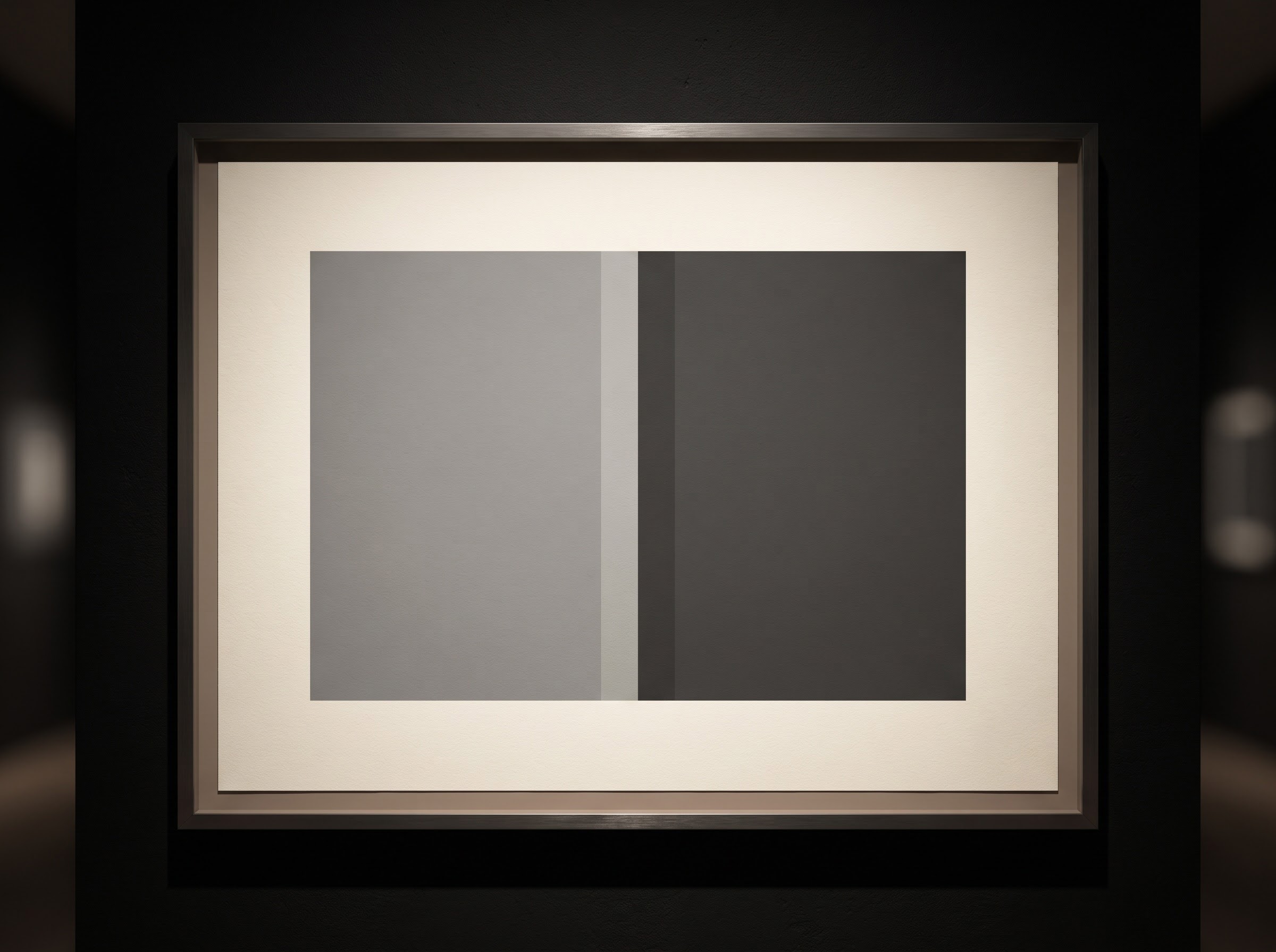
Strip the phenomenon down to two plain tonal blocks and it becomes undeniable: the boundary glows on one side and deepens on the other, yet a light meter sweeping across the image records nothing but two flat values and a single step between them. This is the crucial fact to carry to the viewing screen. The band is a perceptual event, not a photographic one. It lives in the visual cortex, never in the pixel.
Where it hides on a dental film
Dentistry is unusually rich in the sharp density boundaries that breed Mach bands, because teeth are a study in abrupt contrast: dense enamel against softer dentin, a radiopaque restoration against native tooth, the bright lamina dura against the dark periodontal space, bone against the air of a sinus. Wherever a bright structure abuts a darker one, the eye is primed to lay down a false line. The most notorious site is the cementoenamel junction, where the bright crown gives way to the darker root. A thin dark Mach band drapes itself across the neck of the tooth — exactly the location and appearance of cervical caries or root-surface demineralization.
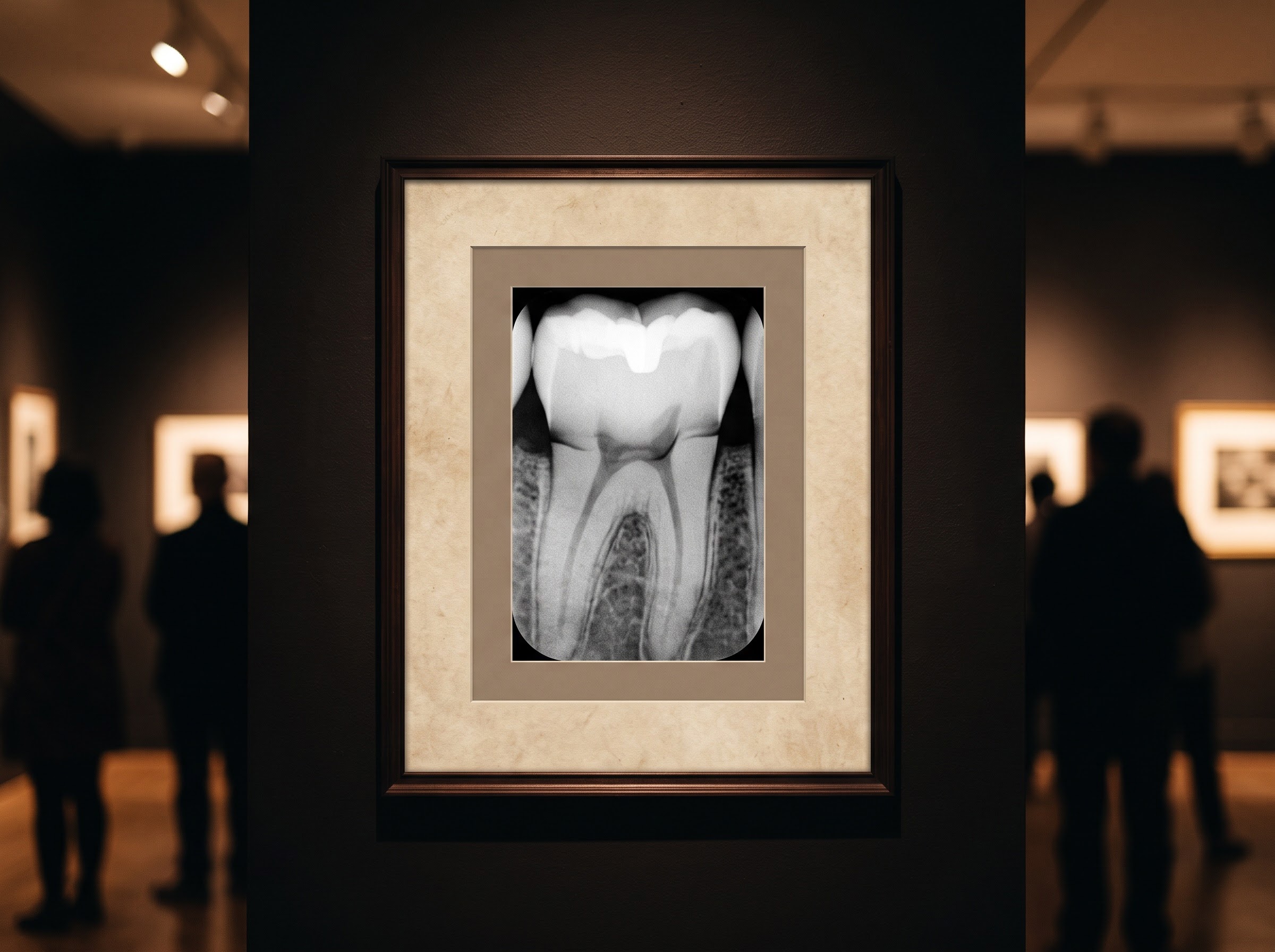
The illusion is just as happy to impersonate other pathologies. A dark band along the margin of an amalgam or crown can read as recurrent decay or an open margin; a bright band beside a radiolucent lesion can appear to sharpen or enlarge its border; a band at the alveolar crest can suggest early bone loss. This is the same genre of diagnostic trap we examined in the anatomy that pretends to be disease — except here the mimic is not a shadow of real structure but a shadow of pure perception, which makes it harder to dismiss because moving the film does not move it.
The test that unmasks it
Because the Mach band depends entirely on the neighboring luminance edge, it carries its own exposure. Cover the brighter region beside the suspicious line — with a card, a thumb, or a digital mask — and remove the contrast that feeds the illusion. A true radiolucency stays put; a Mach band vanishes the instant its neighbor is hidden. This simple maneuver is the most reliable bedside discriminator there is, and it costs nothing but a moment’s discipline.
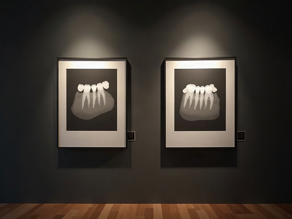
Digital imaging adds a second, decisive tool. Because the band is perceptual, the recorded density values beneath it are unchanged; sampling the pixel brightness across the suspect line will show a smooth step, not the true dip that demineralization produces. The same software powers that can be turned against us — aggressive sharpening and contrast that manufacture edges, a hazard we traced in the radiolucent illusion that burns out the neck of the tooth — can here be turned in our favor, letting us interrogate the actual data rather than the impression it leaves. The eye proposes; the density value disposes.
Reading as a discipline, not a reflex
None of this means the eye is a poor instrument. Lateral inhibition is why we can see form at all, and the same mechanism that invents the Mach band lets a clinician register a genuine lesion in a fraction of a second. The point is not to distrust perception but to know its signature failures and build small habits around them: treat a thin dark line that sits perfectly along a density boundary with suspicion, mask before you commit, sample before you diagnose, and correlate with the clinical picture rather than convicting a tooth on a shadow. An image is not only something to look at; it is something to read with the patient as a shared text, and a false line misread aloud becomes a treatment that should never have begun.
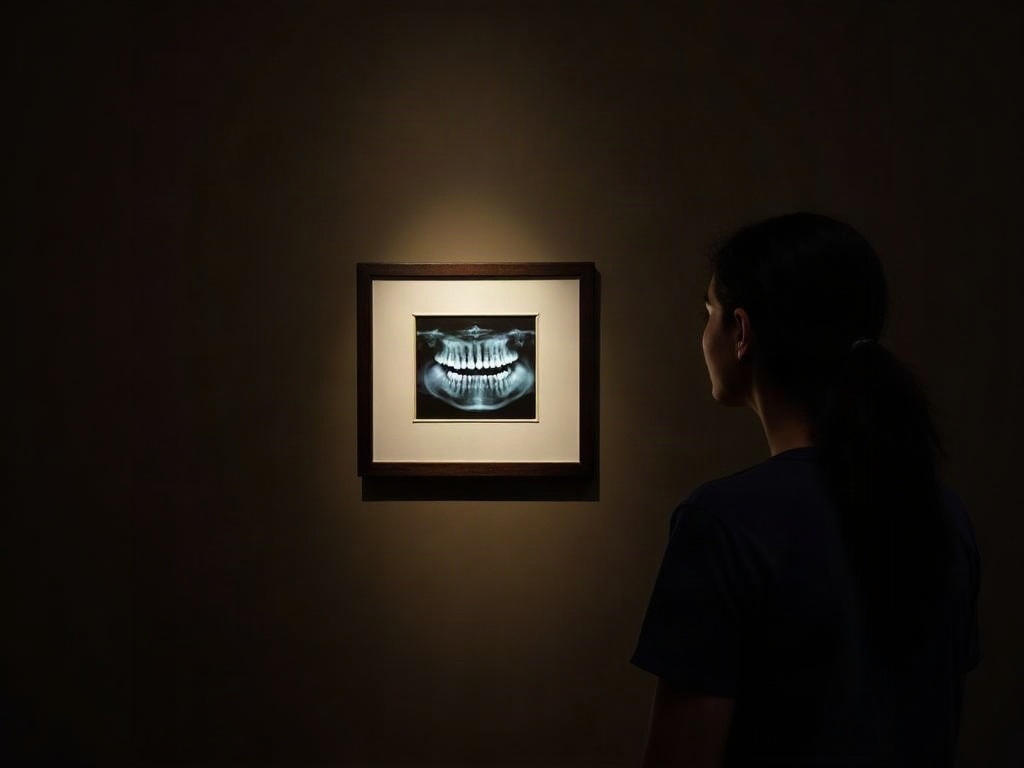
The Mach band is, in the end, a reminder of where the image truly lives. A radiograph is not finished when the sensor captures it; it is completed — and sometimes quietly edited — by the nervous system that views it. The craft of interpretation is as much about understanding that final, invisible stage of rendering as it is about mastering exposure and geometry.
Future Developments
As diagnostic software matures, the honest response to perceptual illusions is to make the underlying data easy to consult. Density-profile tools, one-click local masking, and perception-aware display calibration can turn the Mach band from a hidden trap into a transparent one — a line the reader can test in a second rather than a shadow they must simply resist. There is a deeper prospect, too: machine-vision systems do not possess a retina and feel no lateral inhibition, so they are immune to this particular illusion even as they fall prey to others of their own. The most interesting imaging of the coming years will pair the two readers deliberately — the human eye, with its brilliant, fallible edge-sharpening, and the algorithm that measures without perceiving — each used to check the other’s characteristic blind spot. The gallery has always asked us to look carefully. The next discipline is to look carefully and then confirm what we saw, because the finest forgery in the room may be the one our own eyes signed.
Sources & further reading:
No Comments