01 Apr Stationary Intraoral Tomosynthesis: Bridging 2D and 3D Dental Imaging with Revolutionary Layer Visualization
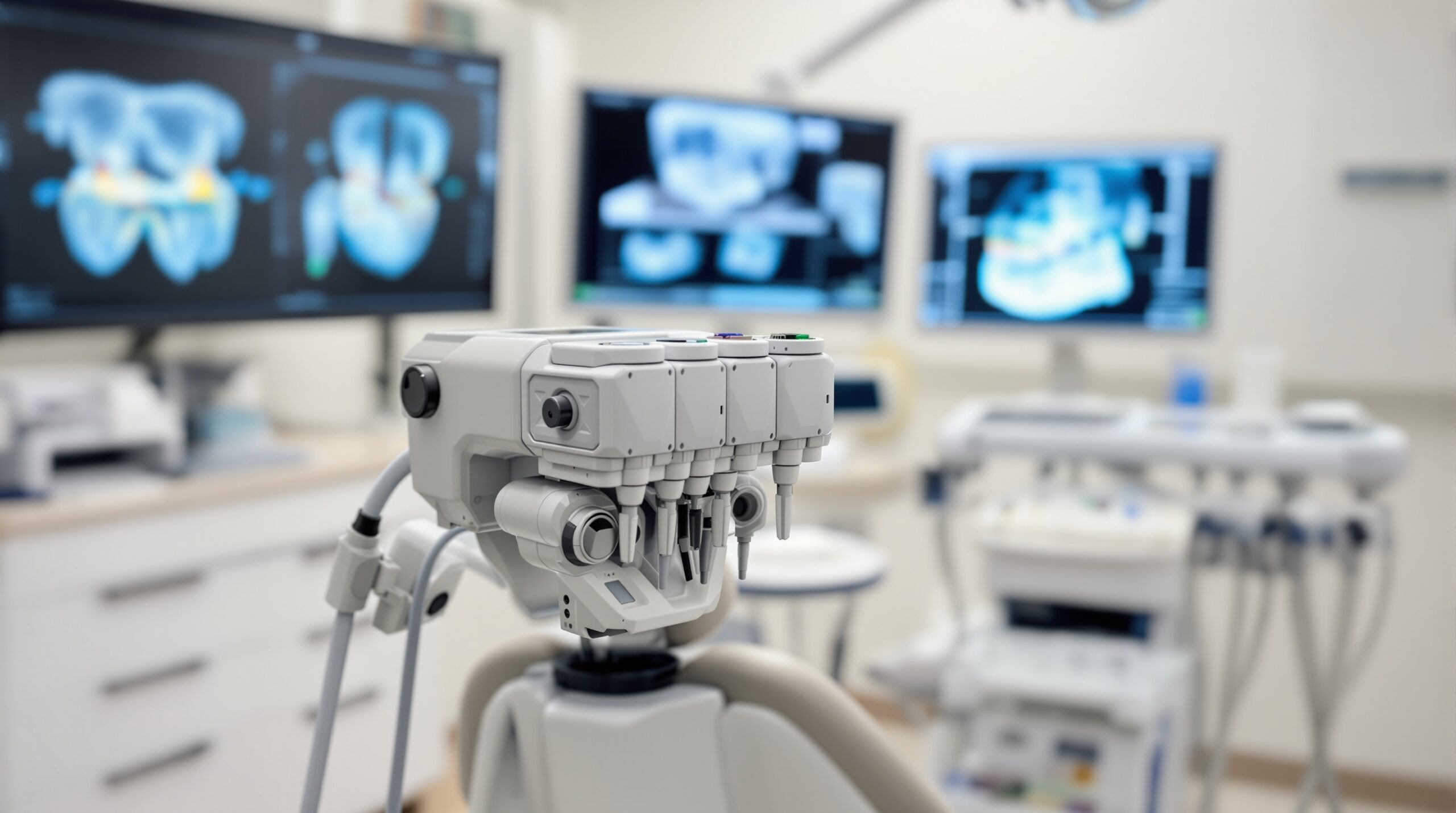
Stationary Intraoral Tomosynthesis (sIOT) represents a revolutionary advancement in dental imaging technology, offering clinicians an innovative solution that bridges the gap between traditional 2D radiography and full 3D cone-beam computed tomography (CBCT). This cutting-edge imaging modality provides enhanced diagnostic capabilities while maintaining radiation doses comparable to conventional intraoral radiographs.
Understanding Tomosynthesis Technology
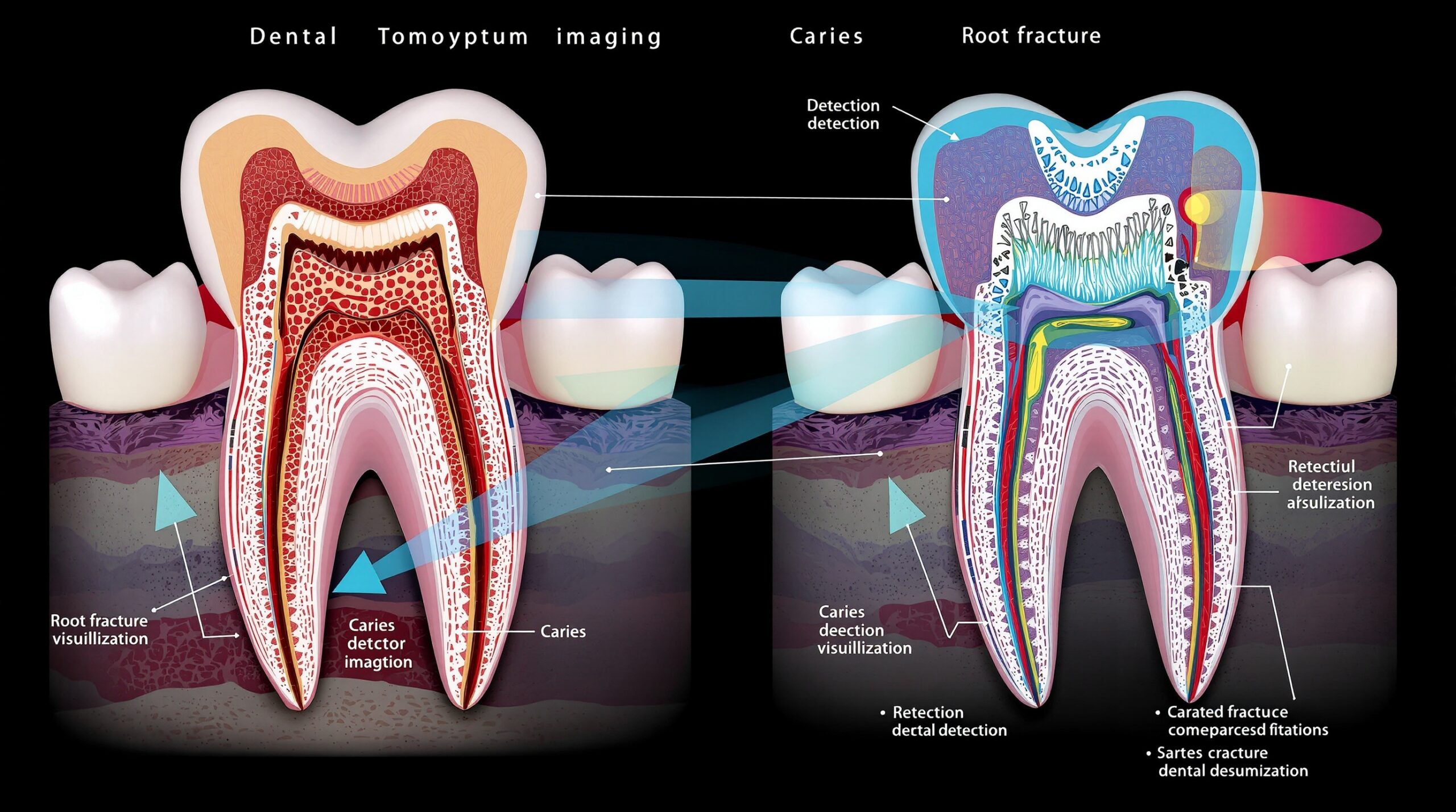
Digital tomosynthesis adapts principles from mammography to dental applications, utilizing a stationary multisource X-ray system that captures multiple projection images from different angles. Unlike conventional radiography that produces a single flat image, tomosynthesis generates a series of cross-sectional slices through the area of interest, effectively eliminating the structural overlap that often obscures critical diagnostic information.
Clinical Applications and Advantages
Stationary intraoral tomosynthesis excels in several key clinical scenarios where traditional 2D imaging faces limitations:
- Vertical Root Fracture Detection: The layer-by-layer visualization eliminates confusing overlapping structures that often mask hairline fractures in conventional radiographs
- Caries Adjacent to Restorations (CAR): Enhanced contrast and reduced artifact interference provide superior detection of decay near existing fillings or crowns
- Periodontal Assessment: Clearer visualization of bone levels and defects without the masking effects of overlying structures
- Endodontic Evaluation: Improved assessment of root canal morphology and periapical pathology
- Implant Planning: Better visualization of available bone volume and adjacent anatomical structures
Technology Advantages Over Traditional Modalities
The stationary intraoral tomosynthesis system offers several compelling advantages over both conventional radiography and CBCT imaging:
Compared to 2D Radiography:
- Eliminates anatomical structure overlap
- Provides depth information through multiple image layers
- Reduces the need for multiple radiographic projections
- Maintains familiar intraoral positioning techniques
Compared to CBCT:
- Significantly lower radiation dose (comparable to bitewing radiographs)
- Reduced metal artifacts due to limited-angle acquisition
- Higher spatial resolution for detailed structural analysis
- More cost-effective implementation
- Faster image acquisition and processing
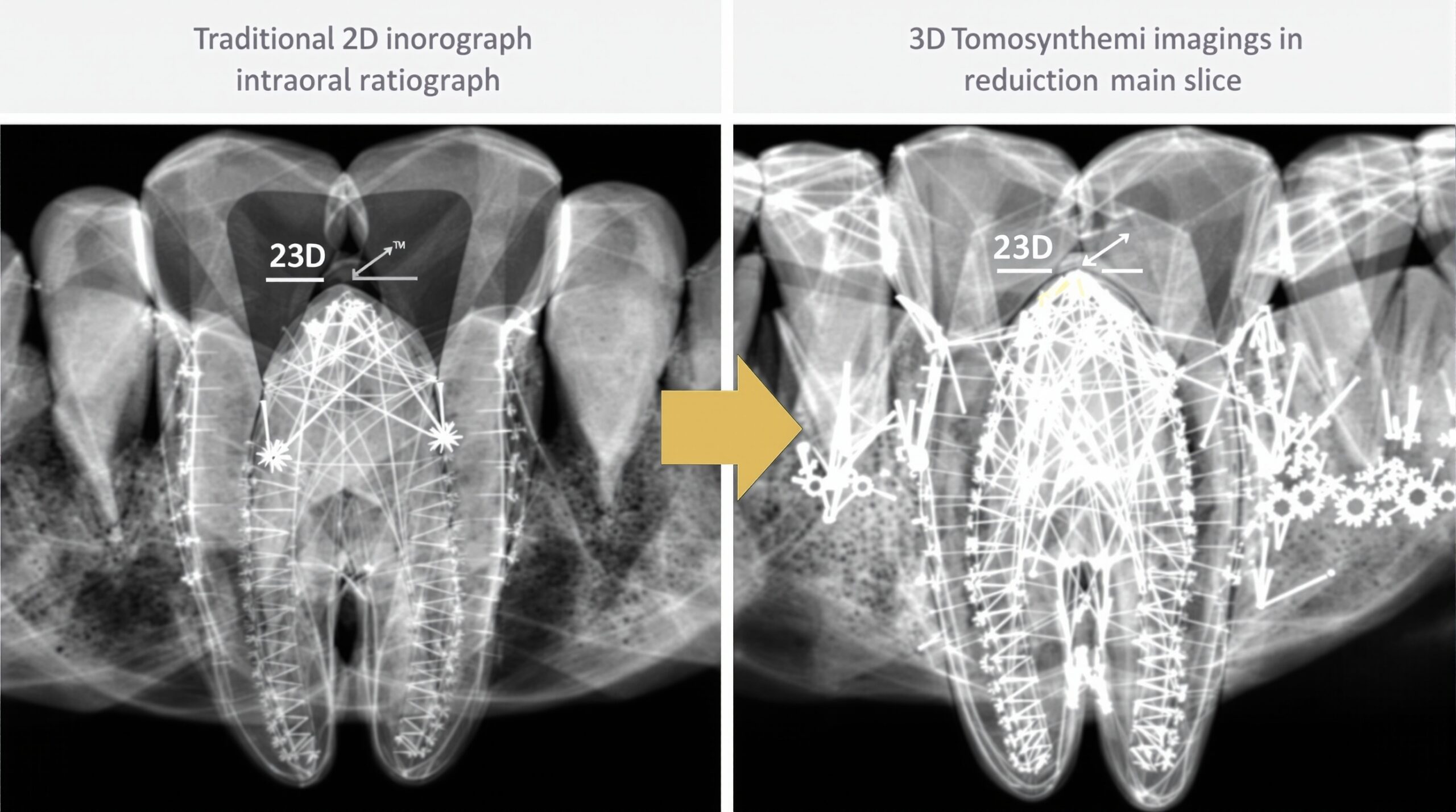
Synthetic Radiography Enhancement
One of the most innovative aspects of modern tomosynthesis systems is the integration of synthetic radiography capabilities. This technology combines information from multiple tomosynthesis slices to generate synthetic 2D images that enhance familiar diagnostic patterns while providing the depth information of 3D imaging.
Research has demonstrated that synthetic radiography can significantly improve diagnostic accuracy for challenging cases such as vertical root fractures and caries adjacent to restorations, where traditional radiography often produces inconclusive results.
Clinical Integration and Workflow
The implementation of stationary intraoral tomosynthesis into existing dental practices requires minimal workflow modifications. The familiar intraoral positioning techniques allow dental professionals to leverage their existing radiographic skills while gaining access to enhanced diagnostic capabilities.
Key workflow advantages include:
- Rapid image acquisition (typically 3-5 seconds)
- Immediate image review and analysis
- Integration with existing practice management systems
- Minimal additional training requirements for dental staff
- Patient-friendly positioning similar to conventional intraoral radiography
Future Implications and Research Directions
As digital tomosynthesis technology continues to evolve, several areas of development show particular promise for dental applications:
- Artificial Intelligence Integration: Machine learning algorithms are being developed to automatically detect pathology across tomosynthesis slice data
- Dose Optimization: Ongoing research focuses on further reducing radiation exposure while maintaining diagnostic quality
- Enhanced Resolution: Advanced detector technologies promise even higher spatial resolution for microscopic detail visualization
- Expanded Clinical Applications: Research continues into additional diagnostic scenarios where tomosynthesis may provide superior information
Limitations and Considerations
While stationary intraoral tomosynthesis offers significant advantages, clinicians should be aware of certain limitations:
- Limited field of view compared to panoramic or CBCT imaging
- Learning curve required for optimal image interpretation
- Higher initial equipment investment compared to conventional systems
- Ongoing need for validation studies in diverse clinical populations
Conclusion
Stationary intraoral tomosynthesis represents a paradigm shift in dental imaging, offering clinicians a powerful diagnostic tool that combines the accessibility of conventional radiography with enhanced three-dimensional visualization capabilities. As this technology continues to mature and gain clinical validation, it promises to become an essential component of comprehensive dental diagnosis, particularly for challenging cases where traditional imaging methods provide limited diagnostic confidence.
The integration of tomosynthesis into dental practice workflows represents not just a technological advancement, but a fundamental improvement in diagnostic precision that can lead to better treatment outcomes and enhanced patient care. For dental professionals seeking to optimize their diagnostic capabilities while maintaining reasonable radiation exposure levels, stationary intraoral tomosynthesis offers a compelling solution that bridges the gap between 2D and 3D imaging modalities.
No Comments