17 Mar CBCT Technology Revolutionizes Dental Diagnosis: A Complete Guide to Cone Beam Computed Tomography
Cone Beam Computed Tomography (CBCT) has fundamentally transformed dental imaging, offering unprecedented three-dimensional visualization of oral and maxillofacial structures. This advanced imaging technology provides dental professionals with detailed anatomical information that was previously impossible to obtain through traditional radiographic methods.
Understanding CBCT Technology
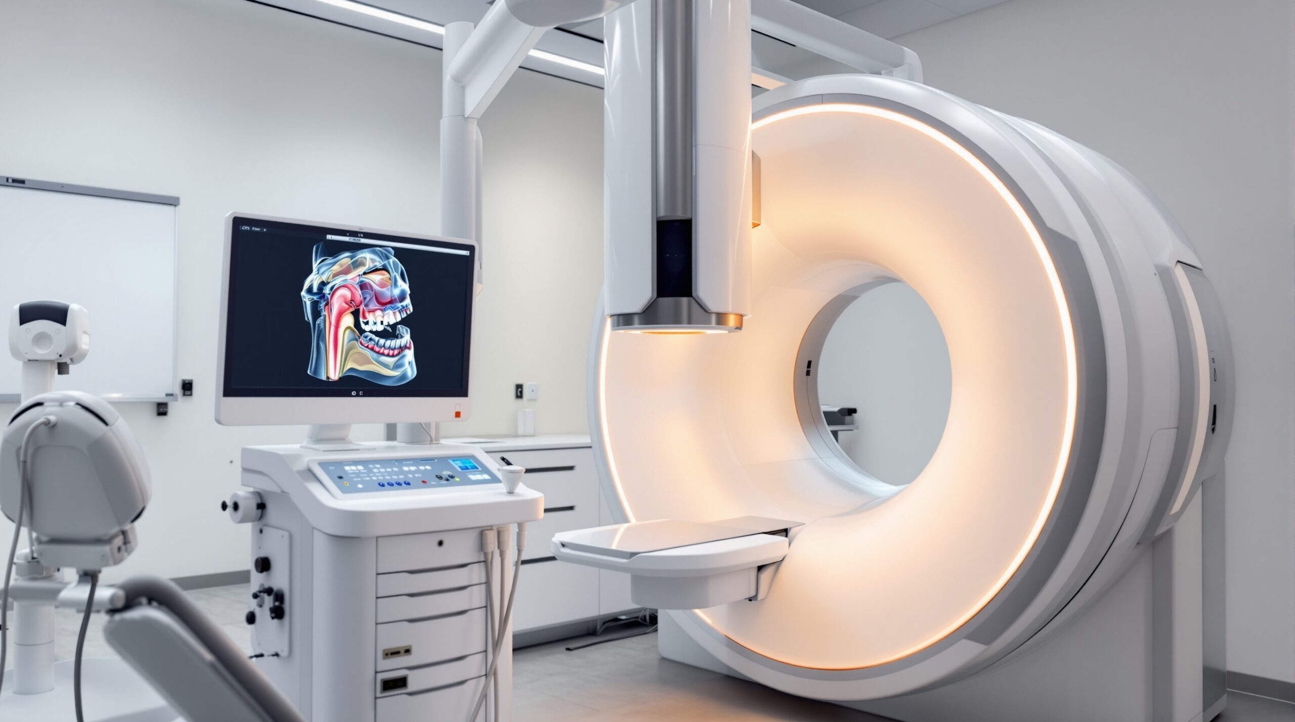
CBCT uses a cone-shaped X-ray beam that rotates around the patient’s head, capturing up to 600 distinct images in a single rotation. Unlike traditional CT scans that use a fan-shaped beam, the cone beam configuration allows for comprehensive 3D dataset acquisition while significantly reducing radiation exposure compared to conventional medical CT scans.
Clinical Applications and Diagnostic Benefits
The versatility of CBCT technology extends across numerous dental specialties, each benefiting from the enhanced diagnostic capabilities:
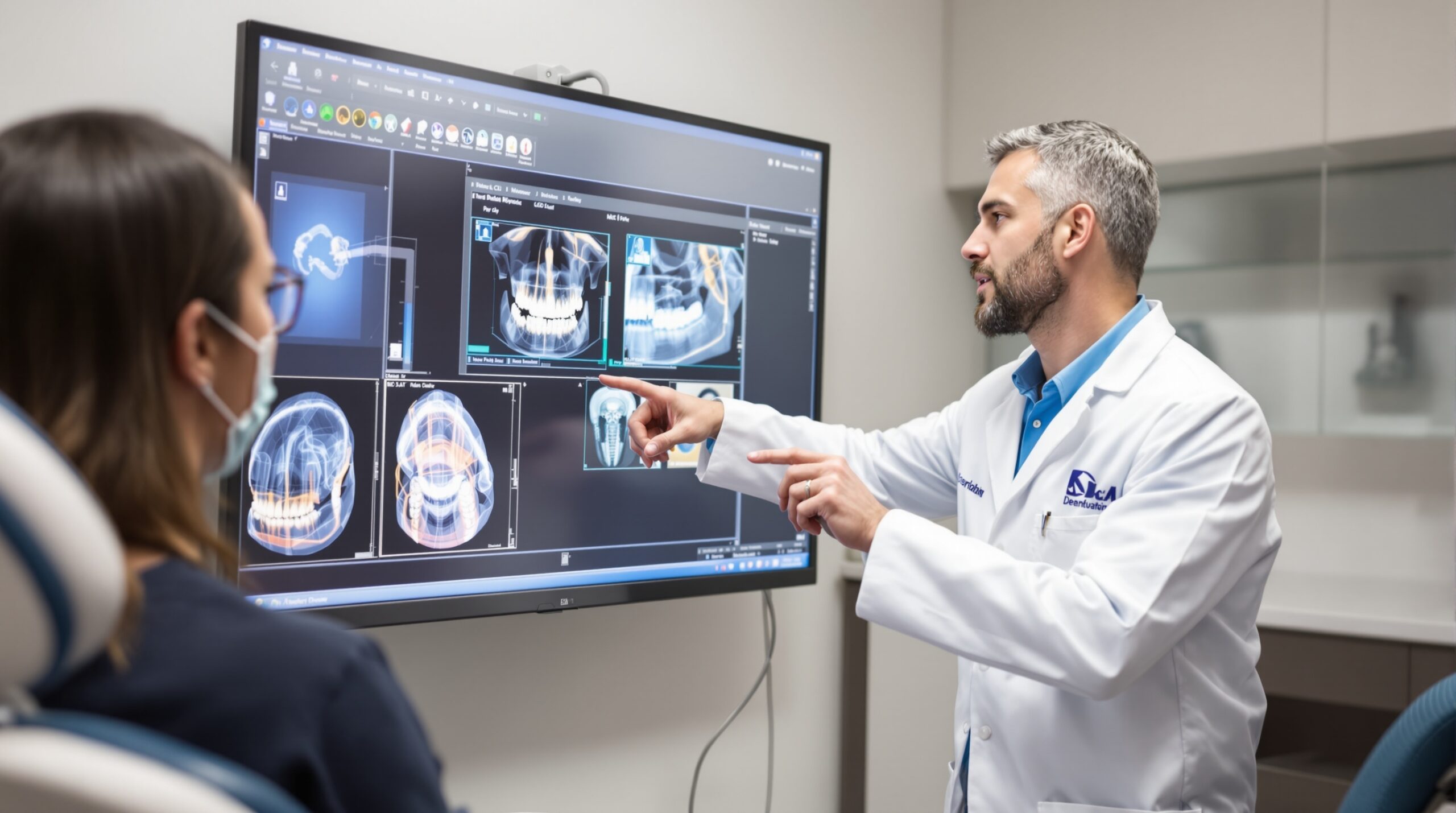
Implant Planning and Placement
CBCT provides precise measurements of bone density, height, and width, enabling optimal implant placement while avoiding vital structures such as the inferior alveolar nerve and maxillary sinuses. The 3D visualization allows practitioners to plan implant trajectories with millimeter accuracy.
Endodontic Applications
Root canal morphology becomes clearly visible through CBCT imaging, revealing complex canal systems, calcifications, and periapical pathology that may be missed on conventional radiographs. This enhanced visualization significantly improves treatment success rates.
Orthodontic Analysis
Comprehensive evaluation of tooth positions, root angulations, and skeletal relationships provides orthodontists with critical information for treatment planning, especially in cases involving impacted teeth or complex malocclusions.
Radiation Safety and Considerations
While CBCT does involve ionizing radiation, the exposure levels are substantially lower than medical CT scans. The effective radiation dose typically ranges from 11-674 μSv, depending on the field of view and imaging protocol. This represents approximately 1-12% of the radiation exposure from a medical CT scan of the head.
Technical Specifications and Image Quality
Modern CBCT systems offer various field of view options, from small localized regions (4cm x 4cm) to full head scans (23cm x 26cm). Voxel sizes typically range from 0.08mm to 0.4mm, providing excellent spatial resolution for detailed anatomical visualization.
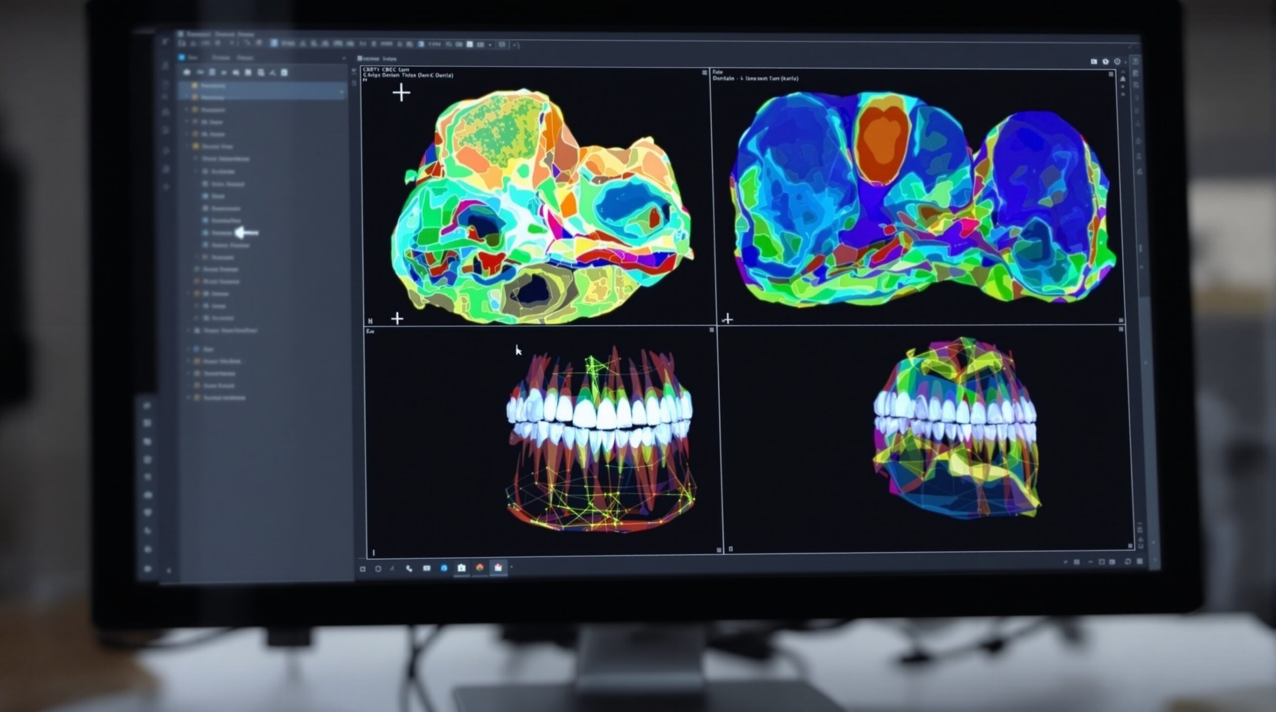
Image Reconstruction and Analysis
Advanced reconstruction algorithms convert the raw projection data into detailed 3D volumes that can be viewed in multiple planes:
- Axial views: Horizontal cross-sections through anatomical structures
- Coronal views: Vertical cross-sections from front to back
- Sagittal views: Vertical cross-sections from side to side
- 3D renderings: Volume-rendered images for comprehensive visualization
Future Developments and Integration
The integration of artificial intelligence and machine learning algorithms is enhancing CBCT interpretation capabilities. Automated detection of anatomical landmarks, pathology identification, and treatment planning assistance are becoming increasingly sophisticated, improving diagnostic accuracy while reducing interpretation time.
Digital Workflow Integration
CBCT data seamlessly integrates with CAD/CAM systems, surgical guide fabrication, and digital treatment planning software, creating comprehensive digital workflows that enhance precision and predictability in dental procedures.
Conclusion
CBCT technology represents a paradigm shift in dental imaging, providing three-dimensional visualization capabilities that enhance diagnostic accuracy, treatment planning precision, and patient outcomes. As the technology continues to evolve with improved image quality and reduced radiation exposure, CBCT is becoming an indispensable tool in modern dental practice.
The investment in CBCT technology, while significant, offers substantial returns through improved diagnostic capabilities, enhanced treatment planning, and increased patient satisfaction. Dental practices considering CBCT implementation should evaluate their specific clinical needs and patient demographics to determine the most appropriate system specifications and imaging protocols.
Sorry, the comment form is closed at this time.