


September 25, 2026 Against the Void: The Black Contrastor and the Craft of Isolating a Tooth
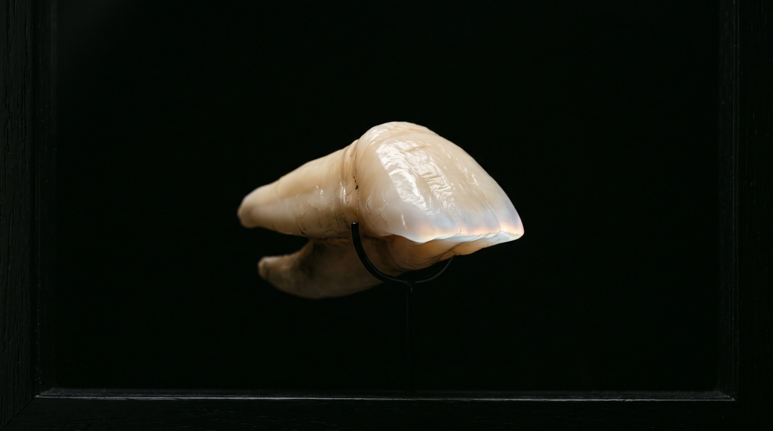
Look at any well-made anterior dental photograph and the first thing you notice is what is missing. There is no glistening lip, no shadowed vestibule, no pink tongue crowding the edge of the frame. There is only the tooth, suspended in an even, velvet darkness, its incisal edge catching light like the rim of a glass. That darkness is not an accident of exposure. It is an instrument—a thin black plate called a contrastor—and learning to use it well is one of the quiet arts of clinical imaging.
We tend to treat the background of a photograph as the part we don’t have to think about. In the mouth, the opposite is true. The background is the loudest thing in the room, and until you silence it, the tooth cannot really be seen.
Why the mouth fights the photographer
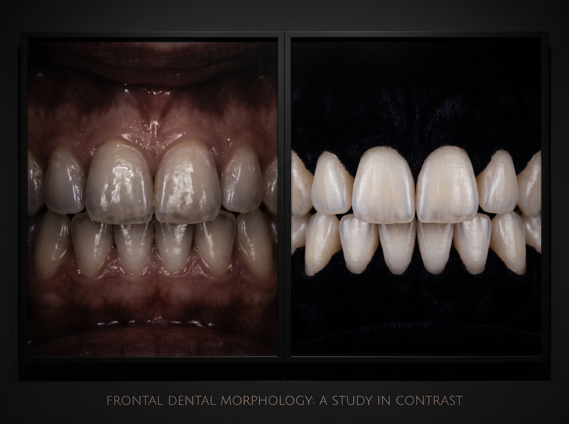
An intraoral scene is a hostile studio. Every surface is wet, and wet surfaces throw specular highlights back at the lens. The mucosa is saturated red, a color that competes with the warm dentin tones you are trying to record. The palate and opposing arch sit just millimeters behind the teeth, close enough to fall inside the depth of field and clutter the frame with anatomy that has nothing to do with the subject. Worst of all, a bright or reflective background bounces light through a translucent tooth from behind, washing out precisely the internal character—the halos, the mamelons, the subtle gradient from cervical to incisal—that a shade-matching or aesthetic photograph exists to capture.
The result, without intervention, is a picture that is technically of a tooth but tells you almost nothing about it. The eye, given a busy field, refuses to settle.
What a contrastor actually is
A contrastor is disarmingly simple: a flat, matte-black plate—typically anodized aluminum or a coated polymer—on a handle, sized and shaped to sit behind the teeth being photographed. Anterior contrastors are curved to follow the arch; occlusal versions are broader and flatter. The matte finish is the entire point. A glossy black surface would still reflect the flash and defeat the purpose; a properly anodized one absorbs the light that reaches it and returns almost nothing to the sensor.
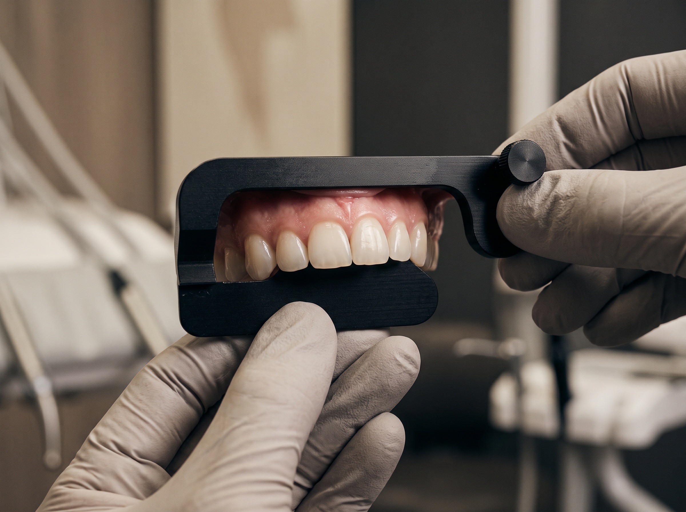
Placed correctly, the plate does two things at once. It hides everything behind the teeth—lips, tongue, opposing arch, the operatory itself—and it stops light from passing through the tooth from behind. In a single move, the mouth disappears and the tooth is left standing alone.
The physics of a tooth against black
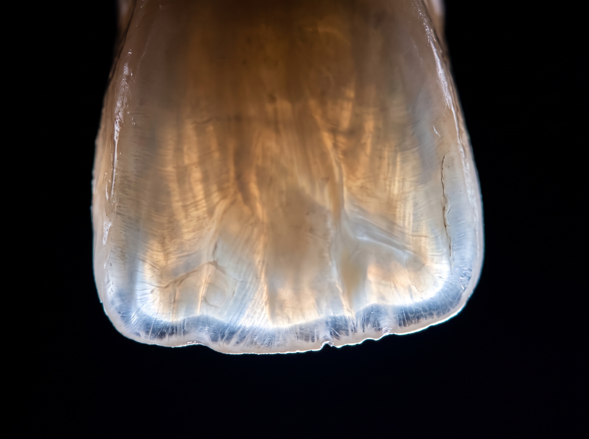
The reason a black background is so revealing has less to do with photography than with the optical nature of enamel. Enamel is not opaque; it is a translucent, light-scattering ceramic. When light strikes a tooth, some reflects off the surface, but a great deal enters the enamel, scatters among its prismatic rods, and re-emerges—this subsurface scattering is what gives natural teeth their depth and their faint inner glow.
Against a bright background, that delicate re-emitted light is overwhelmed. Against black, it becomes the whole show. The incisal third, where enamel is thickest and least backed by dentin, lights up with a translucent halo. Internal features—white flecks, craze lines, the soft opalescent blue that healthy incisal enamel can throw—register as they never would over a busy field. The contrastor doesn’t add anything to the tooth. It simply removes everything that was hiding what was already there.
The craft of placement and light
Using a contrastor well is a matter of millimeters. Held too far back, it lets the vestibule or opposing teeth creep into the frame; held too close, it touches the incisal edges and either casts a hard line or shows up as a smudge of contact. The aim is a small, even gap behind the teeth—close enough to fill the background completely, far enough that the plate never intrudes into focus. The operator’s other hand, a mirror, or an assistant retracts the lip while the plate does its work behind.
Lighting then has to be reconsidered. Because the background returns no light, exposure has to be set for the tooth itself, not averaged across a scene that is now mostly black; most cameras, left on automatic, will try to “lift” the darkness and blow out the highlights. Manual exposure, a controlled flash—ring or twin-point—and a deliberately underexposed field are what let the tooth sit bright and clean on true black rather than muddy grey. Cross-polarizing filters can be layered on top to tame the surface specular highlights further, but even without them, the contrastor alone transforms the image.
Where black is the wrong choice
The technique has limits, and part of the craft is knowing them. Black isolation is made for the anterior segment and for documenting individual teeth, restorations, and translucency. It is the wrong tool for photographs that need to show the tooth in relation to its surroundings—gingival architecture, soft-tissue color, the emergence profile of a crown, the harmony of a smile against the lips and face. Strip those away and you have discarded exactly the context that some clinical questions depend on.
A contrastor is also a poor companion in the posterior, where access, saliva, and the geometry of the arch make placement awkward and the payoff small. The discipline is not to isolate everything, but to isolate deliberately: to reach for black when the subject is the tooth as an object of light, and to leave it in the tray when the subject is the tooth as part of a living mouth.
The portrait beneath the record
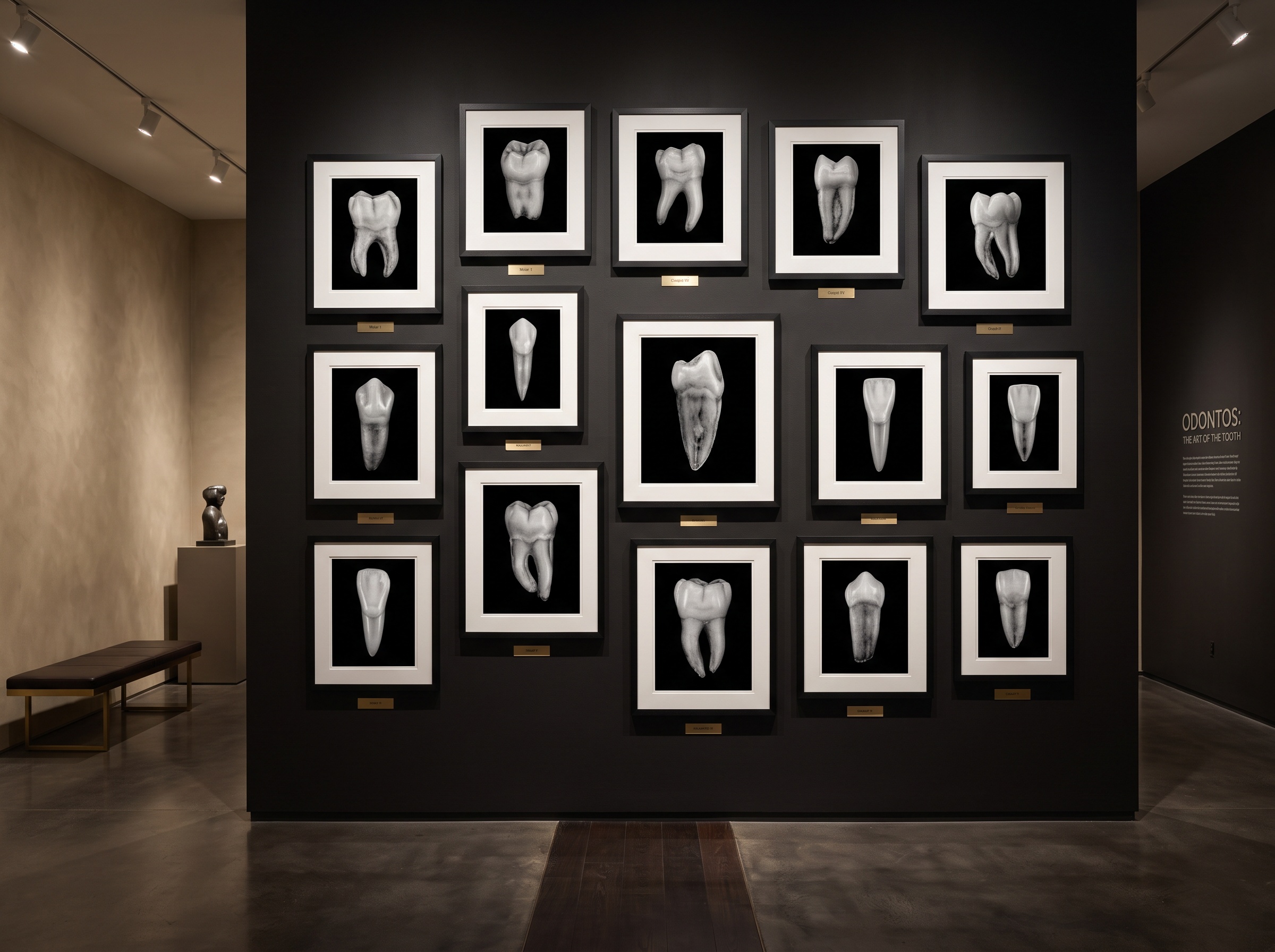
There is a reason the best clinical libraries look, at a glance, like art collections. A row of teeth photographed against black reads as a series of portraits: each one composed, each one lit, each one lifted out of the noise of the mouth and given room to be examined. That is not vanity. A ceramist reading such an image can see the layering they need to reproduce; a clinician reviewing a case years later can see what a tooth truly looked like, not what the operatory happened to reflect that day. The black background is, in the most literal sense, a way of taking the tooth seriously enough to look at it properly.
Future Developments
The contrastor is a nineteenth-century idea—negative space, borrowed from portrait studios—pressed into twenty-first-century service, and it is quietly being absorbed into software. Intraoral scanners and clinical cameras are beginning to segment the tooth from its background computationally, masking the mucosa and rendering a synthetic black field after the fact; AI shade-matching tools increasingly expect that isolated, evenly lit input and struggle without it. As those pipelines mature, the physical plate may become optional for documentation even as the aesthetic it created—the tooth suspended in darkness, read as translucent light—becomes the default way we picture a single tooth. What began as a trick for tidying the frame is turning into the grammar of dental imaging itself: proof, once again, that the craft of seeing a tooth clearly is inseparable from the craft of deciding what to leave out.
Sources & further reading:
- Smile Lite MDP and contrastors in clinical dental photography
- Dental photography technique and imaging craft
Sorry, the comment form is closed at this time.