


February 4, 2026 The Weight in the Hand: Handheld and Wall-Mounted X-Ray, and What the Dose Really Costs
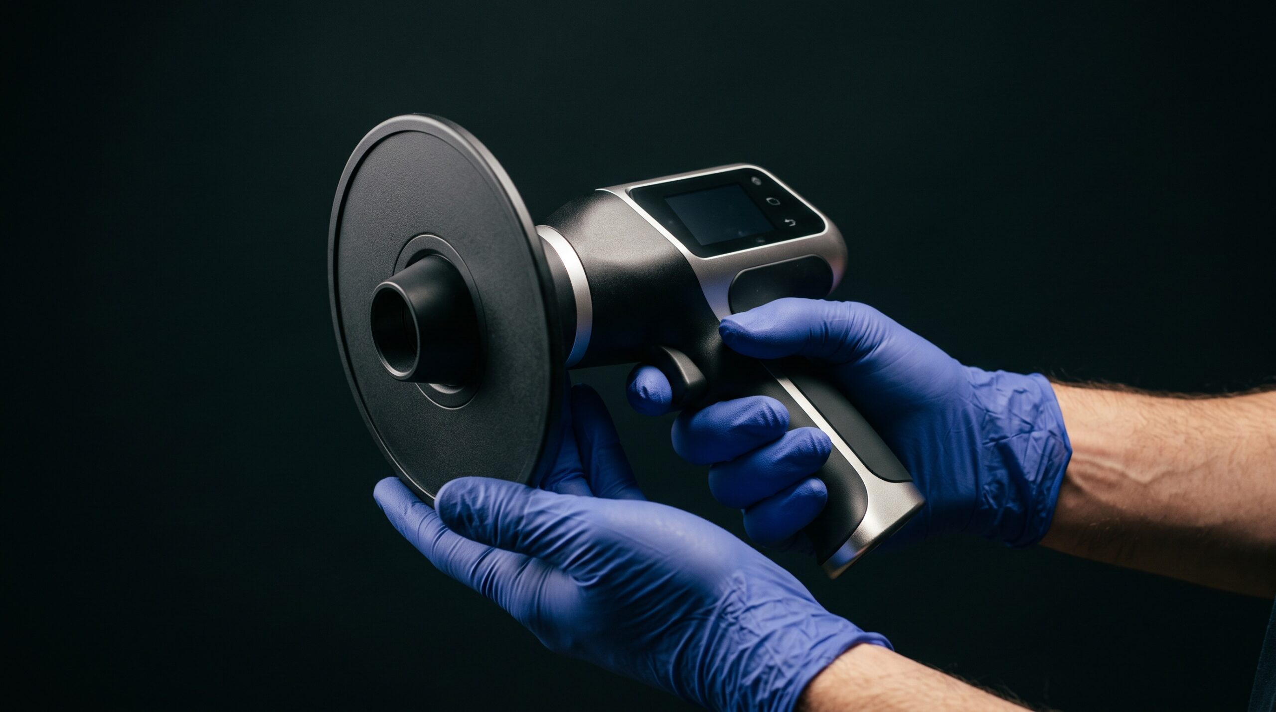
There is a quiet elegance to the handheld intraoral X-ray unit. Where the wall-mounted tube anchors imaging to a single room, tethered to its articulated scissor arm, the handheld device folds the whole apparatus into a shape that fits in two hands and travels to the patient — to the bedbound, the wheelchair-using, the anxious child who will not walk to the operatory, the clinic set up for a morning in a place that has no operatory at all. It is one of those rare instruments that expands what dentistry can reach. And it does so by placing the source of an ionizing beam a few inches from the fingers of the person aiming it. That single fact is the whole story, and a recent phantom-based study has now put a number on it.
The Same Portrait, Made Two Ways
Start with what does not change. A periapical radiograph made with a well-designed handheld unit is, to the diagnosing eye, indistinguishable from one made on a wall-mounted tube. The comparative dosimetry study — measuring a handheld MaxRay Cocoon against a conventional wall-mounted system on a phantom — found the diagnostic image quality and the patient-equivalent dose broadly comparable between the two. The molar looks like the molar. The canal traces its path, the lamina dura holds its line, the caries declares itself or does not. Whatever anxiety surrounds the handheld device, it is not an anxiety about the picture. The image is honest. The difference lives entirely in the geometry of who else the radiation touches.
Where the Dose Goes
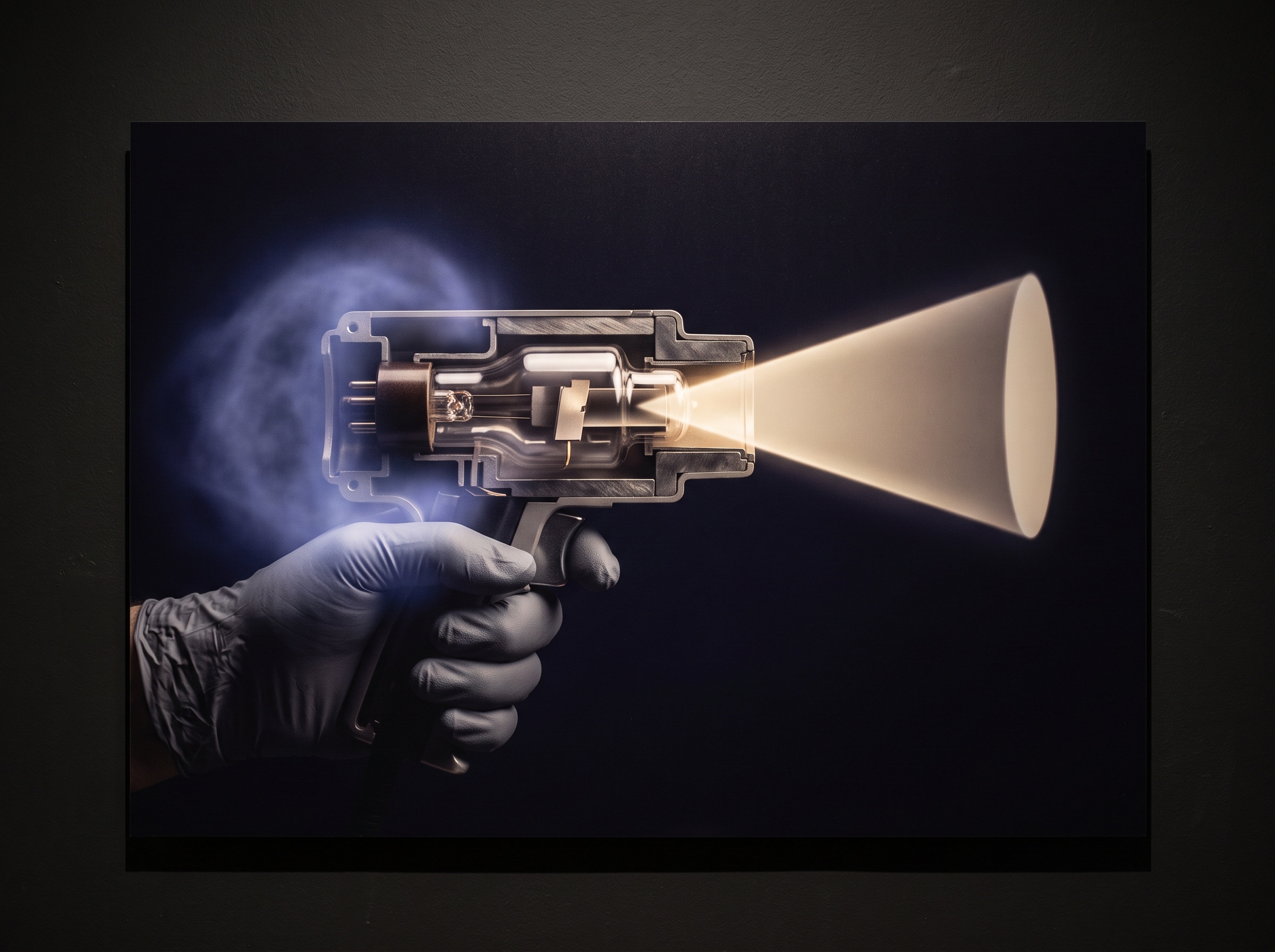
The wall-mounted tube is aimed and abandoned. The operator presses the exposure switch from behind a barrier or at the regulation distance, and in that moment no part of them is near the beam. The handheld unit inverts this completely: the operator holds the tube head throughout the exposure, and their hand rests within the zone of maximum backscattered radiation, immediately around the exit port. The study’s most striking result followed directly from that geometry. The operator finger dose was 5.6 times higher with the handheld device than with the wall-mounted system — not because the handheld is a cruder or dirtier machine, but because the hand is simply there, inside the bloom of scatter that every intraoral exposure throws backward off the patient and the collimator.
It is worth being precise about what that multiple means and does not mean. It is a ratio of two small numbers. A single exposure delivers a trivial dose to the finger by any measure; the handheld’s 5.6-fold figure is still, per shot, modest. The concern is cumulative and occupational: the clinician who takes dozens of exposures a day, hundreds a week, thousands a year, holding the device each time. Over a career, a 5.6-fold difference in extremity dose is not a rounding error. It is the reason the handheld demands a discipline the wall-mounted tube never asked for.
The Backscatter Shield: Small Disc, Large Effect
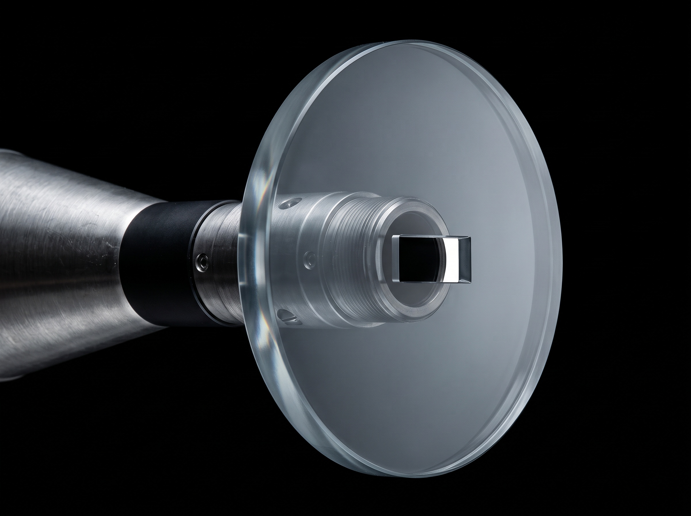
Here the story turns hopeful, because the physics that creates the problem also points straight at the solution. Backscatter is a near-field phenomenon, densest right at the cone tip, and it can be intercepted right there. A separate line of research measured exactly this. Fitting a circular backscatter shield at the cone-tip reduced occupational exposure to around 40% of its unshielded value. A rectangular collimator — which tightens the primary beam to the shape of the sensor and so simply makes less scatter in the first place — reduced it to about 13%. Used together, the shield and the collimator brought occupational exposure down to roughly 7.7% of the bare-device figure. That is more than a tenfold reduction, achieved with two pieces of hardware that add almost nothing to the weight in the hand.
Set those numbers beside the 5.6-fold penalty and the picture resolves. A handheld unit stripped of its shielding, used casually, genuinely does expose its operator far more than a wall-mounted tube. A handheld unit with an integrated backscatter shield and a rectangular collimator, used by someone who respects the geometry, closes most of that gap. The device is not inherently unsafe; it is conditionally safe, and the conditions are cheap, well understood, and easy to meet.
The Craft of Holding It Well
What a wall-mounted tube achieved through architecture, the handheld achieves through technique. The shield must actually be fitted — not left in a drawer because it adds a moment to setup. The rectangular collimator should be the default, both for dose and for the crisper, less-fogged image a tighter beam yields. The operator’s free hand belongs nowhere near the field; the patient, never held by the person taking the exposure. A stable two-handed grip and a braced stance keep the tube steady, which protects both the dose geometry and the sharpness of the image, since a handheld unit shares with every camera the truth that motion is the enemy of resolution. And personal dosimetry — a ring badge, a body badge — turns an abstract 5.6-fold multiple into a measured, personal number that a clinician can actually watch over time.
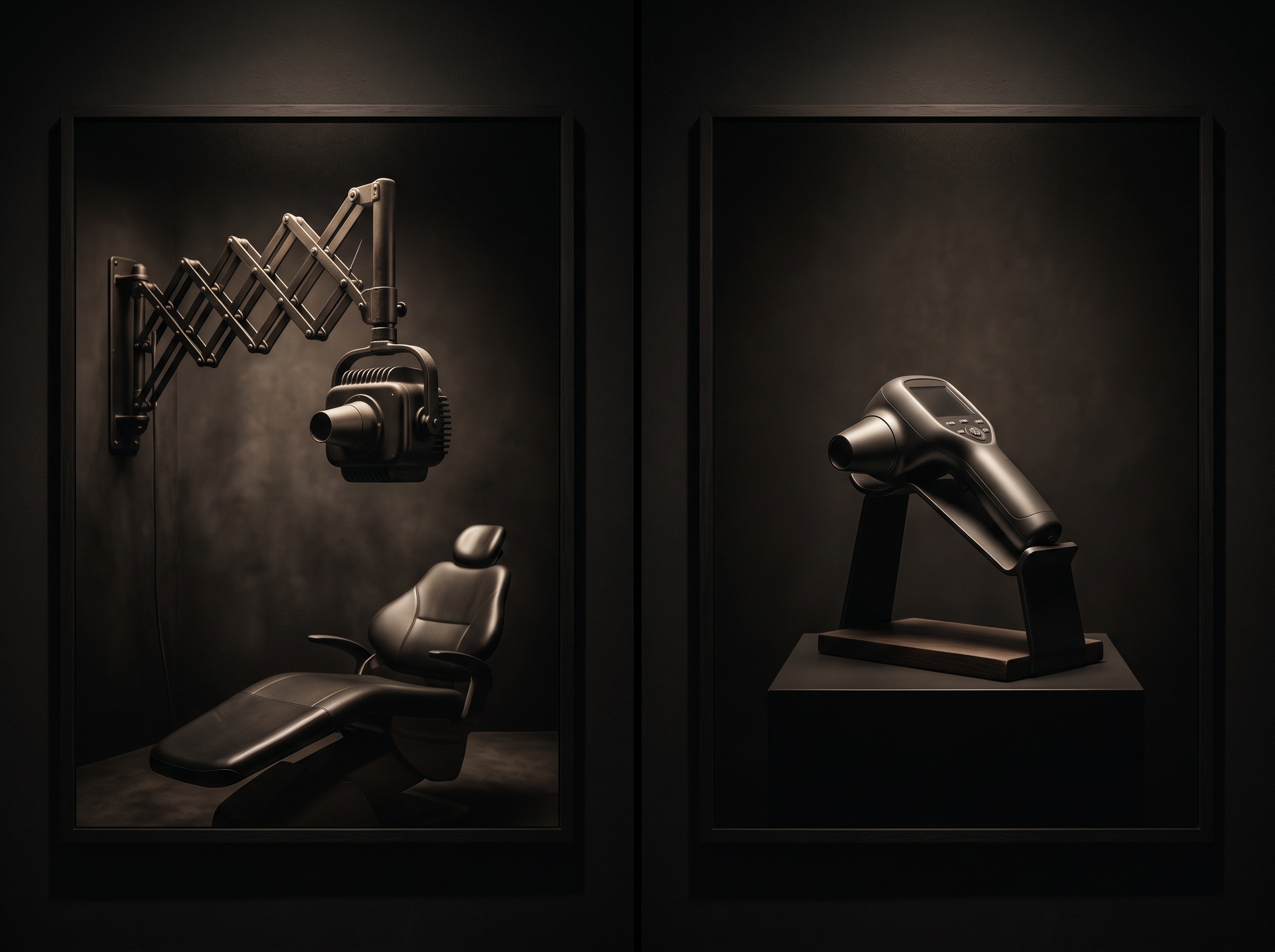
None of this is burdensome. It is simply the etiquette the instrument requires in exchange for its freedom, and it is the difference between a handheld unit that quietly accumulates dose in a career’s worth of fingers and one that is, in practice, as safe to stand behind as the tube bolted to the wall.
Future Developments
The trajectory of the handheld is toward making its discipline automatic rather than optional. Integrated, non-removable backscatter shields and permanently rectangular collimation are increasingly standard rather than accessories, engineering the safe geometry into the device so the operator cannot forget it. Lighter batteries and lower-dose, higher-sensitivity sensors shorten every exposure, and shorter exposures mean less scatter for every hand in the room. One can imagine the next refinements plainly: interlocks that will not fire without the shield seated, position sensors that warn when the free hand drifts into the field, on-board dose logging that tallies each operator’s exposure the way a modern scanner already tallies its own. The handheld unit earned its place by carrying imaging to patients the wall could never reach; its future is to carry the safety of the wall along with it — so that the elegant convenience of holding the source in your hand costs the hand that holds it nothing at all.
Sources & further reading:
- Comparative Dosimetry of Handheld Versus Wall-Mounted Intraoral X-ray Systems: A Phantom-Based Study (Cureus, 2025)
- Stray radiation dose reduction using a backscatter shield and a rectangular collimator for a handheld intraoral X-ray unit (Radiation Protection Dosimetry, 2023)
Related Reading
- Can a Handheld Radiograph Match the Wall-Mount? The Physics of Image Quality in Portable Dental X-Ray
- The X-Ray That Fits in a Bag: NASA’s Portable Imaging Trials and the Craft of Radiography Far From Any Hospital
- The Risk of Not Looking: Weighing a Cone-Beam Scan’s Radiation Against What It Reveals
Sorry, the comment form is closed at this time.