


April 27, 2026 Photoacoustic Imaging in Dentistry: Revolutionary Non-Invasive Periodontal Health Monitoring and Diagnostic Technology
Photoacoustic imaging represents a groundbreaking advancement in dental diagnostic technology, offering clinicians unprecedented capabilities for non-invasive periodontal health assessment. This revolutionary imaging modality combines optical and acoustic technologies to provide high-resolution visualization of dental tissues, periodontal structures, and gingival health parameters with exceptional precision and accuracy.
Understanding Photoacoustic Imaging Technology
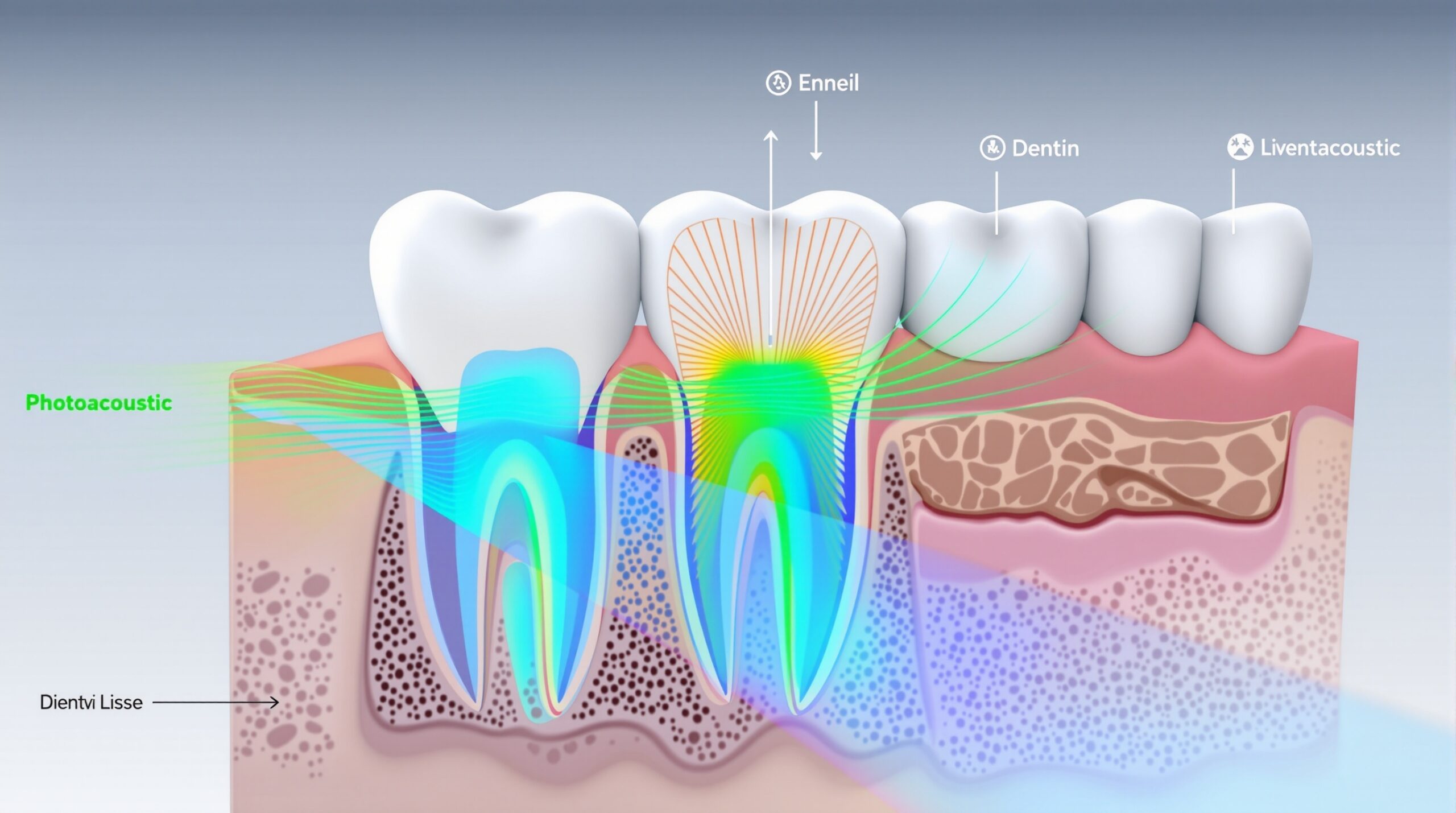
Photoacoustic imaging (PAI) operates on the photoacoustic effect, where pulsed laser light generates acoustic waves in tissues. When laser pulses hit dental structures, they create localized heating that produces ultrasonic signals. These acoustic waves are captured by sensitive detectors and processed to create detailed cross-sectional images of oral tissues.
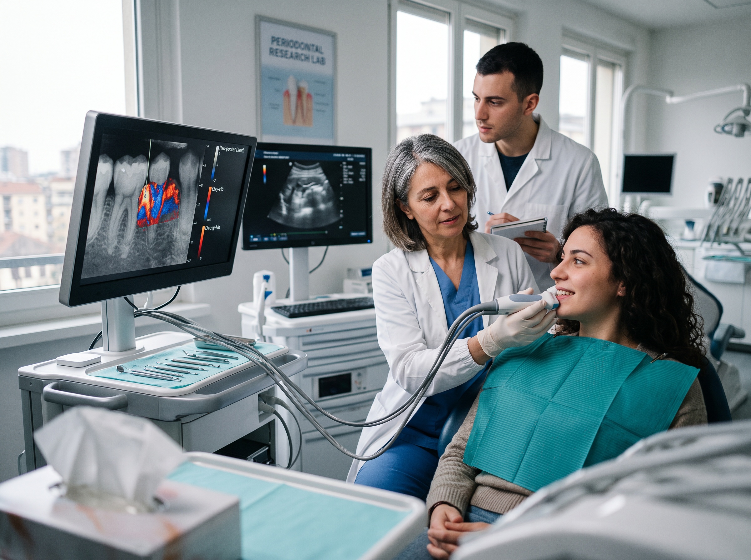
The technology offers several significant advantages over traditional imaging methods:
- Non-invasive visualization of periodontal structures
- Real-time imaging capabilities
- Superior soft tissue contrast
- Radiation-free operation
- Enhanced diagnostic precision
Clinical Applications in Periodontal Assessment
Recent clinical studies have demonstrated photoacoustic imaging’s remarkable effectiveness in periodontal health monitoring. The first human study revealed the technology’s ability to accurately measure periodontal pocket depths with greater precision than traditional periodontal probes.
Key clinical applications include:
- Periodontal pocket measurement: Precise depth assessment with submillimeter accuracy
- Gingival thickness evaluation: Non-invasive soft tissue measurement
- Inflammatory tissue detection: Enhanced visualization of periodontal inflammation
- Treatment monitoring: Real-time assessment of therapeutic outcomes
Advantages Over Traditional Methods
Photoacoustic imaging provides several advantages compared to conventional periodontal assessment techniques. Traditional periodontal probing can be uncomfortable for patients and may not provide comprehensive coverage of all tooth surfaces. PAI offers complete circumferential assessment of periodontal structures without patient discomfort.
The technology’s high spatial resolution enables detection of subtle tissue changes that might be missed by conventional examination methods. This enhanced sensitivity allows for earlier intervention and more effective treatment planning.
Technical Specifications and Performance
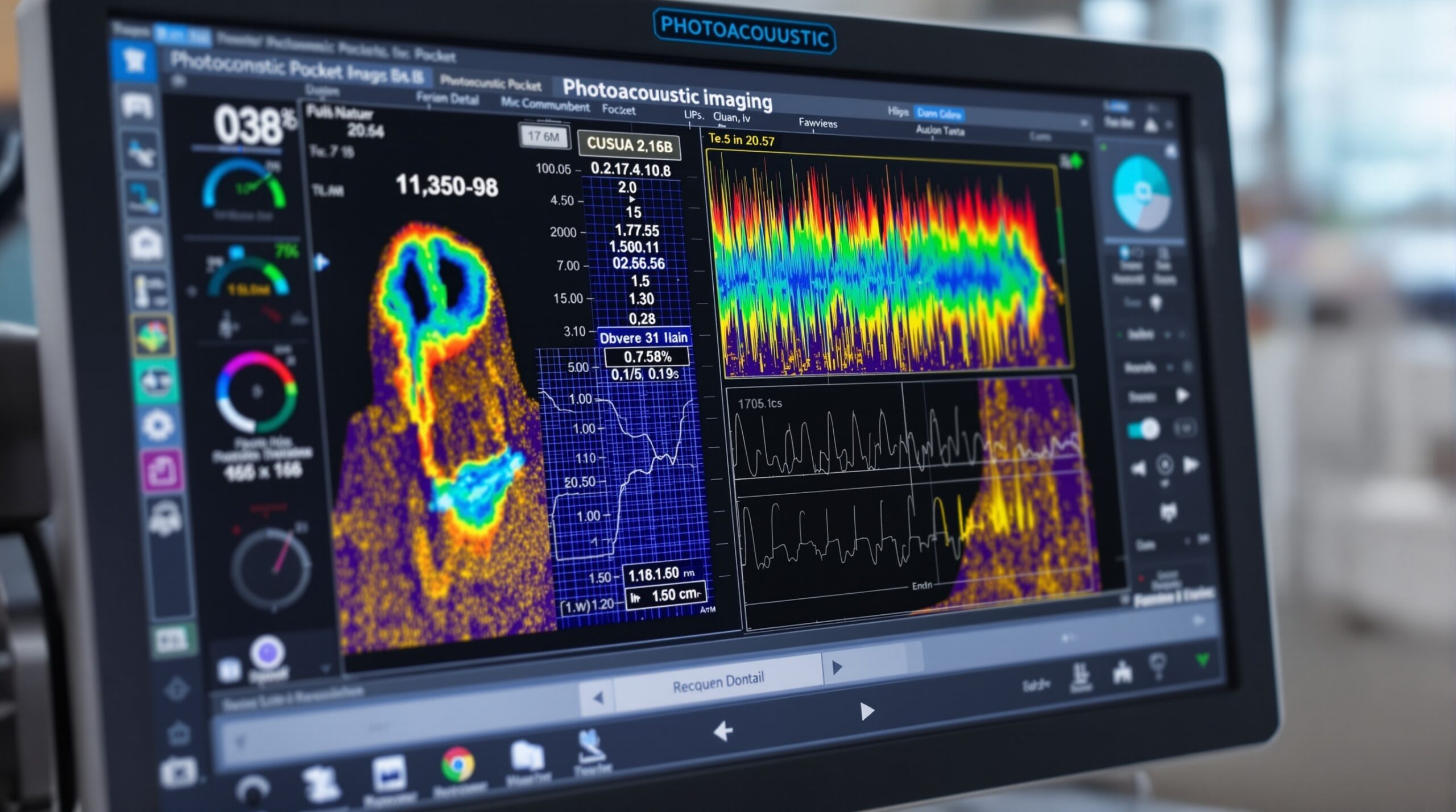
Modern photoacoustic microscopy systems utilize 40 MHz ultrasound frequencies to achieve optimal spatial resolution for periodontal anatomy visualization. This frequency range provides excellent penetration depth while maintaining sufficient resolution to distinguish between tooth, gum, gingival margin, and gingival thickness measurements.
The technology demonstrates exceptional measurement precision, with accuracy levels of 0.01 mm or better in clinical studies. This level of precision surpasses traditional periodontal probing methods and enables detection of minute changes in periodontal health status over time.
Contrast Agent Enhancement
Some photoacoustic imaging protocols utilize food-grade contrast agents to enhance visualization of specific structures. These safe, biocompatible agents improve image contrast and enable better differentiation between various tissue types within the periodontal environment.
Clinical Integration and Workflow
Integrating photoacoustic imaging into clinical practice requires minimal workflow modifications. The non-invasive nature of the technology makes it suitable for routine periodontal assessments, follow-up examinations, and treatment monitoring protocols.
The imaging process is rapid, typically requiring only minutes to complete comprehensive periodontal assessment. Real-time visualization capabilities allow clinicians to immediately review results with patients, enhancing patient education and treatment acceptance.
Patient Benefits
Patients experience numerous benefits from photoacoustic imaging technology:
- Comfortable, pain-free examination
- No radiation exposure
- Immediate results availability
- Enhanced treatment planning accuracy
- Objective monitoring of treatment progress
Future Developments and Research
Ongoing research continues to expand photoacoustic imaging applications in dentistry. Current investigations focus on automated data collection algorithms, enhanced image processing capabilities, and integration with artificial intelligence systems for improved diagnostic accuracy.
Future developments may include portable photoacoustic imaging systems, expanded applications for caries detection, and integration with other advanced imaging modalities for comprehensive oral health assessment.
Research Directions
Contemporary research emphasizes developing photoacoustic imaging protocols for specific clinical scenarios, including periodontal disease progression monitoring, treatment outcome assessment, and early detection of pathological changes in periodontal tissues.
The technology’s potential for integration with digital dental workflows and electronic health records systems represents another significant area of development, promising enhanced clinical efficiency and improved patient care coordination.
Conclusion
Photoacoustic imaging represents a transformative advancement in dental diagnostic technology, offering unprecedented capabilities for non-invasive periodontal health assessment. The technology’s exceptional precision, patient comfort, and real-time imaging capabilities position it as a valuable addition to modern dental practice.
As research continues to refine the technology and expand its applications, photoacoustic imaging is poised to become an essential tool for comprehensive periodontal care, early disease detection, and enhanced treatment monitoring in contemporary dental practice.
Related Reading
- Close-Range Photogrammetry in Dentistry: Revolutionary 3D Imaging Technology for Precision Diagnostics
- Optical Coherence Tomography in Dentistry: Revolutionary Non-Invasive Imaging for Advanced Dental Diagnostics
- DIAGNOdent Laser Fluorescence Imaging: Precision Caries Detection Technology for Modern Dental Practice
Sorry, the comment form is closed at this time.