


November 10, 2026 The Anatomy That Pretends to Be Disease: Reading Radiographic Mimics on the Dental X-Ray
There is a particular kind of unease that visits every clinician who reads radiographs: the dark shape near a root apex that was not there in memory, the faint line crossing a tooth that might be a fracture, the pale shadow floating over the ramus. The reflex is to name it, and the temptation is to name it as disease. Yet some of the most convincing pathology on a dental film is not pathology at all. It is normal anatomy, caught at an unfamiliar angle, or an artifact of the machine’s geometry, pretending for a moment to be something it is not. Learning to recognise these radiographic mimics is one of the quietest and most important disciplines in imaging, because the cost of misreading them is paid by the patient in unnecessary treatment or in missed disease.
The False Positive Is a Reading Error, Not an Imaging One
It is worth stating plainly: a mimic is not a flaw in the radiograph. The image is doing exactly what it should, faithfully recording the superimposed shadows of three-dimensional anatomy onto a two-dimensional plane. The error, when it happens, lives entirely in interpretation. This is a liberating idea, because it means the remedy is not a better machine or a higher dose but a better-trained eye and a disciplined method. The reader who treats every radiolucency as guilty until proven innocent will overdiagnose; the one who dismisses everything as anatomy will eventually miss a lesion that mattered. The craft sits between those two failures, and it is built from a working knowledge of where normal structures live and how they are supposed to look.
Foramina and Canals: Holes That Look Like Lesions
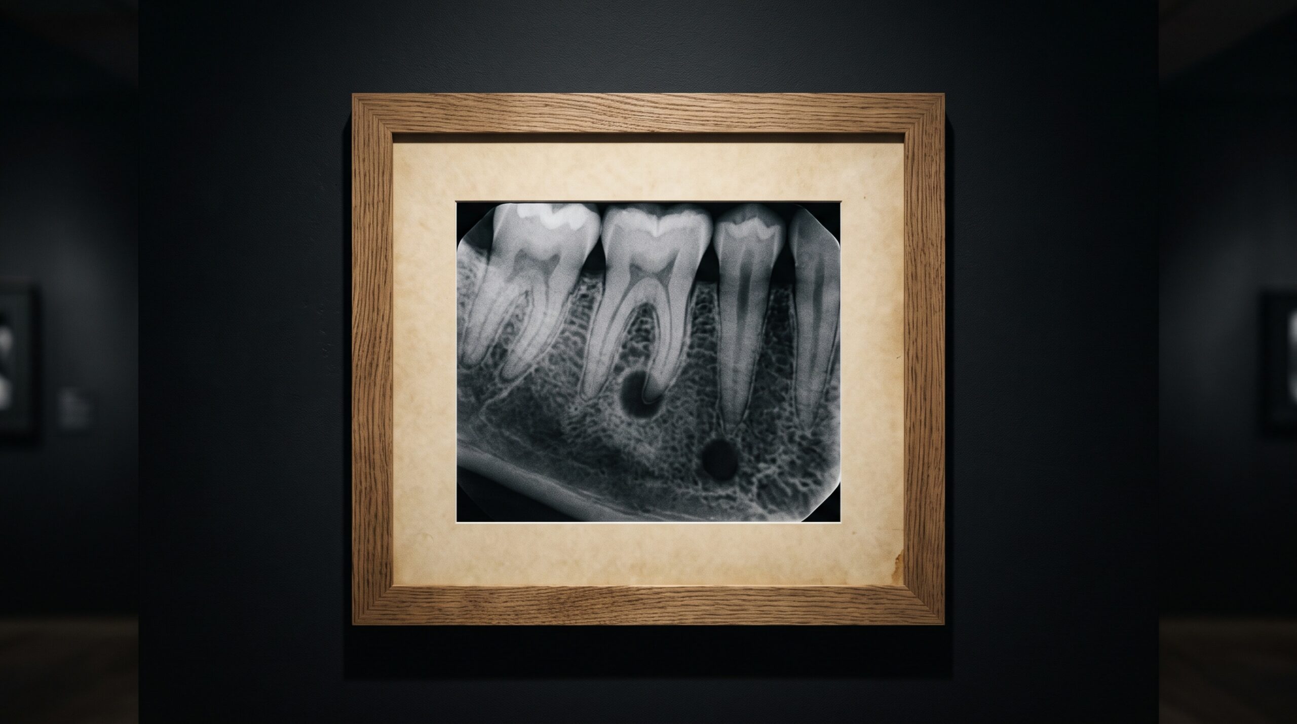
The classic mimic is the mental foramen, the small opening in the mandible through which the mental nerve emerges, typically near the apices of the lower premolars. On a periapical film it can project directly over or beside a root tip as a round, well-defined radiolucency, and it has sent more than one healthy premolar toward unnecessary endodontic treatment. The incisive foramen does the same trick between the maxillary central incisors, where a normally generous opening can be mistaken for a cyst. Nutrient canals, the fine channels that carry vessels through bone, appear as faint linear radiolucencies that an anxious eye reads as fractures or bone loss. In each case the giveaway is context: a tooth with an intact lamina dura and an uninterrupted periodontal ligament space around a vital, asymptomatic root is almost never harbouring the lesion the shadow suggests.
The Overlapping Shadows of the Panoramic
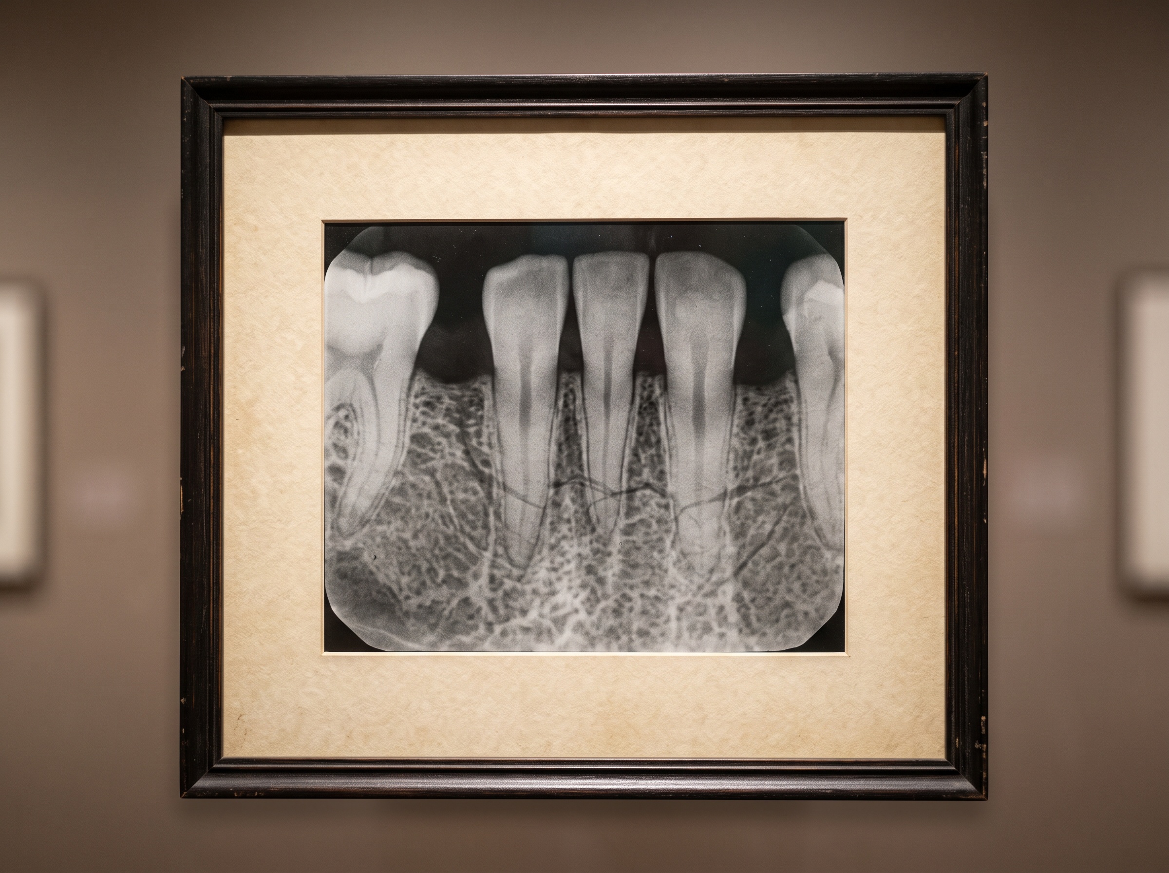
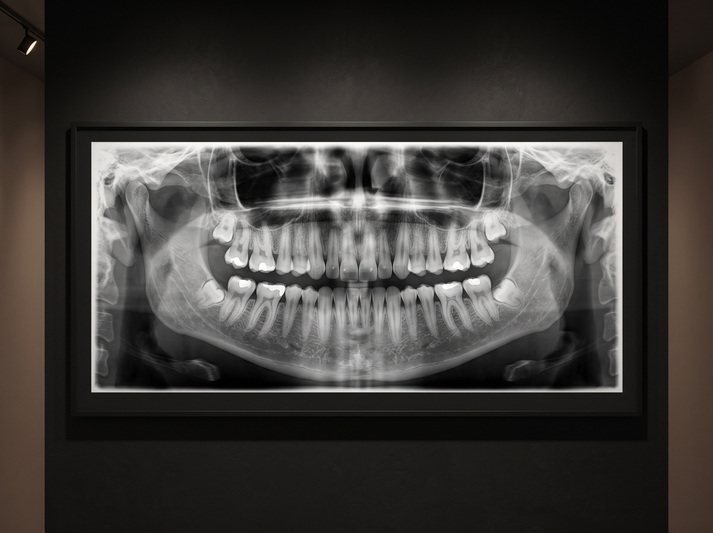
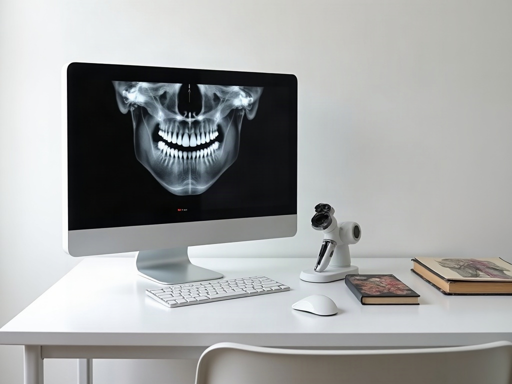
Panoramic imaging multiplies the opportunities for mimicry, because its very design projects distant structures onto the plane of interest. The maxillary sinus is the great offender of the upper arch: a large, dark, air-filled space whose floor dips between the molar roots, and whose normal septa and recesses can suggest cysts or bony lesions where none exist. Soft-tissue shadows, the dorsum of the tongue, the nasal cartilage, the ear and even the cervical spine, all cast their own grey veils across the film. Most striking of all are the ghost images, the reversed and magnified phantoms thrown onto the opposite side when a dense structure passes through the beam twice. A reader who does not know the machine’s geometry can spend a long moment puzzling over a lesion that is simply the mirror of a real object elsewhere. We explored these phantoms in detail in our piece on reading and preventing the panoramic ghost image, and they remain the purest example of anatomy and geometry conspiring to deceive.
Radiolucencies That Belong There
Beyond discrete openings, the jaws are full of normal low-density regions that a cautious reader must learn to welcome rather than fear. The submandibular fossa, a depression on the lingual surface of the mandible below the molars, appears as a diffuse radiolucency that can mimic a lesion but has no defined cortical border of disease. Marrow spaces vary enormously between patients, and a generous, trabeculated pattern in one person is simply their normal bone. The pulp chamber and canals, the furcation of multi-rooted teeth, and the periodontal ligament space itself are all radiolucent by design. The reader’s task is not to treat darkness as a symptom but to ask whether this particular darkness has the character of disease: an expansile outline, a corticated or ragged border, displacement of adjacent structures, or a correlation with clinical findings. Absent those features, a radiolucency in a known anatomical location is most often the anatomy announcing itself.
When the Artifact Imitates the Body
A smaller but real category of mimics comes not from the patient but from the imaging chain. A bend or crease in a film or sensor, a fingerprint, a double exposure, static discharge, or an over-aggressive sharpening filter can each introduce lines, halos and density changes that look biological. Digital processing deserves particular caution, because contrast enhancement and edge sharpening can manufacture the appearance of a lesion margin where the raw data holds none, a hazard that rewards a sceptical eye on any heavily adjusted image. The defence is the same humility that governs all good reading: when a finding appears, ask whether it moves with the anatomy or sits oddly on top of it, whether it repeats on a second exposure, and whether it makes biological sense. An artifact rarely survives a second, differently angled image; a real lesion almost always does.
Building the Mental Catalogue
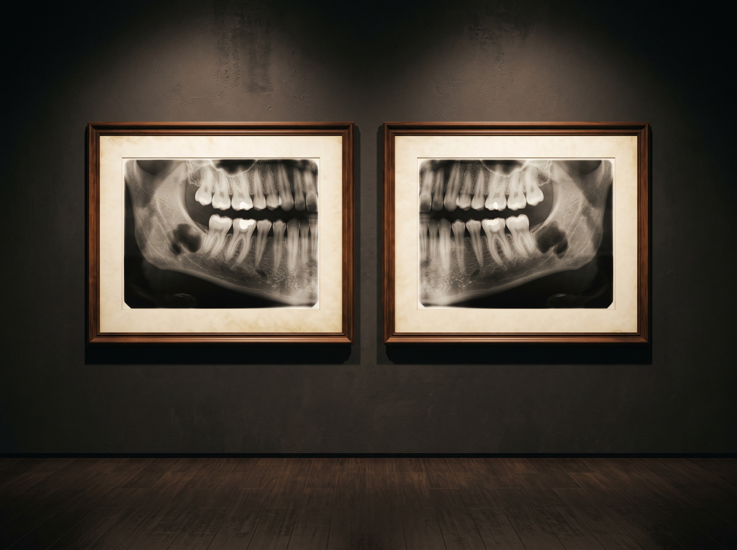
How does a clinician come to tell the pretenders from the real thing? Partly through an atlas of normal anatomy held in memory, and partly through a handful of durable habits. Bilateral symmetry is the first and best of them: a finding mirrored faithfully on the opposite side is usually a shared anatomical feature rather than coincidental disease. Correlation with the clinical picture is the second; a florid radiolucency beneath a vital, painless, fully functional tooth demands scepticism. A second projection at a different vertical or horizontal angle is the third, because true three-dimensional pathology holds its position while a superimposed shadow shifts. And time is the fourth: a short-interval follow-up will betray a growing lesion and exonerate a static landmark. These same disciplines underpin more advanced reading, whether one is interpreting a cone-beam volume for the airway or judging genuine periodontal bone loss against the patient’s record. The catalogue of mimics is simply the negative image of that same expertise.
Future Developments
The study of mimics is quietly being reshaped by automation, and the relationship cuts both ways. On one hand, artificial-intelligence readers trained chiefly to find pathology can be fooled by the same foramina and ghosts that fool a novice, generating confident false positives that a knowledgeable clinician must overrule. On the other, modality is a growing ally: cone-beam imaging dissolves many two-dimensional mimics by separating superimposed structures into distinct slices, so that a shadow which looked like a lesion on a periapical film resolves into an ordinary foramen in three dimensions. The most promising systems will be those that model normal anatomy explicitly, flagging a finding only when it departs from the expected landscape rather than whenever it appears. Until then, and arguably long after, the safeguard remains human and unglamorous: a reader who knows the anatomy so well that its disguises no longer work. In the gallery of the dental record, the most valuable skill is not seeing more, but knowing precisely what one is looking at, and granting the body’s own architecture the recognition it deserves before reaching for the language of disease.
Sources & further reading:
No Comments