


20 Jul Before the Patient Stands Up: Judging a Radiograph at the Moment of Capture
Every clinician knows the sequence. The bitewings were taken an hour ago, the patient is gone, and now — reviewing the series properly — the distal of the second molar is buried under overlap and the premolar apex is cut off. The retake that should happen will not happen, because asking someone to return for a picture is a conversation nobody wants to have. So the image is read anyway, with a quiet asterisk beside it. The interesting development is not that software can see this problem. It is that software can see it in the two seconds after the exposure, while the sensor is still in the patient’s mouth.
The Most Expensive Exposure Is the Second One
Retake rates are among the least examined numbers in a dental practice, and the most consequential. Each repeated periapical is another exposure delivered to a patient who has already absorbed one that produced nothing diagnostic. ALARA is usually discussed as collimation, filtration, receptor speed, and shielding — all worth optimising — but the largest dose reduction available to most operators is simply not needing a second attempt. Technique is a radiation-protection measure.
The cost does not stop at dose. A radiograph is simultaneously a diagnostic document, a legal record, and a claim attachment. Pearl, the dental AI company led by Ophir Tanz, built its rationale for Imagecheck — software giving real-time automated feedback on 2D radiographs at the point of capture — around exactly that overlap: image quality determines diagnosis, treatment and reimbursement at once. An image too flawed to support a diagnosis is often too flawed to support a claim, and a denial arriving weeks later is an expensive way to discover a cone cut.
A Taxonomy of Error and Its Physics
What makes automated quality scoring tractable is that radiographic technique errors are not infinitely varied. They form a short list, each with a specific geometric or exposure cause. Imagecheck targets the common set, worth restating as physical events rather than image defects:
- Cone cut — the central ray and the receptor are not concentric, so part of the sensor sits outside the collimated field and records nothing. A positioning-ring alignment failure, almost always.
- Excessive interproximal overlap — horizontal angulation error. The beam is not directed through the embrasure parallel to the contact, so adjacent surfaces superimpose and the very region you are trying to assess disappears.
- Elongation and foreshortening — vertical angulation error relative to the bisecting angle between the long axis of the tooth and the plane of the receptor. Too little vertical angulation stretches the image; too much compresses it.
- Missing root tips — a framing failure, usually an anterior placement that follows the palate rather than the tooth, truncating exactly the anatomy a periapical exists to show.
- Blurriness and motion artefact — patient movement or tube-head drift during the exposure interval, a window that longer exposure times widen.
- Over- and under-exposure — a mismatch between kVp, mA, and time and the combination of receptor sensitivity and patient anatomy. Digital sensors tolerate wide latitude, which is precisely why exposure drift persists unnoticed until contrast quietly degrades across a whole practice.
Each has a deterministic fix: re-seat the ring, rotate the beam horizontally, increase vertical angulation. This is why capture-time detection is more useful than it first appears — the flag is not merely a verdict, it is an instruction.
Why the Chairside Moment Is Different
Retrospective quality audit is not new. Practices have scored technique from their image libraries for decades, and it changes behaviour slowly if at all, because feedback arriving weeks later lands as criticism rather than correction. The operator no longer remembers what their hands did, the patient is gone, and nothing can be acted on.
Feedback at capture inverts all of those conditions. The patient is still positioned, the holder still assembled, and the operator retains a fresh proprioceptive memory of the angle used. A flag reading horizontal overlap becomes a one-adjustment retake thirty seconds later, and a correction that gets encoded. Repeated across a few dozen exposures, this is a training loop rather than a performance review — a distinction that matters more than the accuracy of any individual detection.
Craft, With an Instant Second Opinion
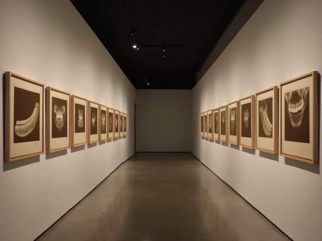
There is a useful analogy in fine-art photography. A print can be beautifully composed and still fail because the exposure was wrong or the frame clipped the subject; no interpretive skill recovers detail that was never recorded. A radiograph is the same object under different physics. Diagnosis is only as good as the image it rests on, and the discipline of capture — alignment, angulation, framing, exposure — is the craft that makes interpretation possible at all.
What has changed is that this craft now has an instant, unsentimental second opinion. Nor is this solely a vendor claim: peer-reviewed work in Nature has evaluated deep-learning assessment of periapical radiographic image quality, indicating a genuine research base beneath the commercial products. Pearl’s partnership with Dentsply Sirona on AI radiology integration shows where this ends up architecturally — quality assessment embedded in the imaging hardware pipeline rather than bolted on afterwards. Related tools go further, flagging when a retake is warranted and enhancing contrast to improve diagnostic visibility.
Calibrating a Practice, Not Correcting a Person
The most immediate practical value is in multi-operator settings. A practice with four hygienists and two assistants taking radiographs has six technique signatures — six habitual vertical angulations, six exposure preferences, six framing tendencies — and a library inconsistent in ways that make longitudinal comparison harder than it should be. A uniform standard applied at capture is the fastest route to a genuinely standardised archive.
For new team members it functions as a calibration instrument: a newly qualified hygienist learning full-mouth series technique gets immediate, specific correction on every exposure, from a system with no tone of voice. Practices adopting it well track aggregate flag categories over time as a quality metric — watching a persistent overlap tendency disappear over a month — rather than treating individual flags as incidents.
Where the Clinician Must Remain the Judge
Two caveats deserve emphasis. First, quality scoring is not diagnosis. A radiograph can be technically imperfect and entirely sufficient for the clinical question in front of you. A slight cone cut flagged on a film that clearly answers whether the lesion crosses the DEJ does not need retaking, and a system implying otherwise is giving bad advice.
Second, and more seriously, threshold calibration is a dose issue. A tool tuned too aggressively produces retake creep — more exposures, not fewer — inverting the benefit it exists to deliver. The operator must remain the decision-maker, the software advising rather than instructing. Validation across receptor types and populations is still maturing: performance on adult digital periapicals does not automatically transfer to paediatric anatomy or an older sensor. Adopt it, but audit its flags as you would audit anything else you delegate.
Future Developments
The trajectory points upstream of the exposure itself. Positioning guidance derived from the same models, offered before the button is pressed, would move quality control from correction to prevention — the difference between a critique and a rehearsal. Integration into sensor firmware rather than the software that receives the image is the logical endpoint of the hardware partnerships now forming. And as these systems accumulate technique data across thousands of operators, the picture of which errors are most persistent and most responsive to feedback will reshape how radiographic technique is taught.
What none of this displaces is the operator’s judgement, or the artisanal nature of the work. The discipline of the perfect exposure — the patient positioned with care, the beam aligned with intent, the frame containing exactly what it must — remains a human craft. The instruments are simply becoming honest enough to tell us, immediately, whether we practised it well. Every image a practice keeps is a piece of its collected work; the aspiration is a library in which none of them carry an asterisk.
Sources & further reading:
- Orthodontic Products — New Pearl Software Flags Dental Radiograph Errors at Time of Capture
- Orthodontic Products — How Pearl’s AI Radiograph Tools Are Reshaping Orthodontic Practices
No Comments